*Corresponding author
Albrektsson T, Department of Biomaterials, University of Gothenburg, Box 412, 405 30, Gothenburg, Sweden, Tel: 46705916607; Email: Submitted: 22 April 2017 Accepted: 21 June 2017 Published: 24 June 2017 Copyright © 2017 Albrektsson et al. ISSN: 2573-1548 OPEN ACCESS Keywords • Osseointegration • Foreign body reaction • Oral implantology • Craniofacial implants • Hip arthroplasties
Review Article
Osseointegration of Implants
– A Biological and Clinical
Overview
Albrektsson T
1,2*, Chrcanovic B
2, JacobssonM
2, and Wennerberg
A
21Department of Biomaterials, University of Gothenburg, Sweden
2Department of Prosthodontics, Malmö University, Sweden
Abstract
Osseointegration was discovered in 1962 and coined as a term in 1977. Original definitions implied direct contact between foreign materials and bone without any interposed soft tissue layers. Today, osseointegration is regarded to be a foreign body response to separate foreign elements from bone. A new definition of the term is suggested in this paper; “Osseointegration is a foreign body reaction where interfacial bone is formed as a defense reaction to shield off the implant from the tissues”. Excellent clinical results of osseointegrated implants have been reported from dentistry and Ear Nose Throat surgery, the latter with the indications of a stable anchorage of hearing aids or facial epistheses in cases of facial trauma. In Orthopaedic surgery a randomized controlled clinical trial has been undertaken demonstrating very good clinical results supported by positive radiostereo-photogrammetical data.
INTRODUCTION
Osseointegration - a historical overview and the
coining of the term
One man was behind osseointegration – Per-Ingvar Brånemark of Sweden. He started placing experimental implants in rabbits and with time in dogs and found them impossible to remove without fracturing the bone. Brånemark immediately saw clinical possibilities with his discovery that was made in 1962 whilst working at the University of Gothenburg. Earlier years claimed for his discovery occasionally quoted, are incorrect and therefore best ignored. Brånemark had presented his ph D thesis about microcirculation in rabbit bone and bone marrow [1], by grinding down the fibula to transparency and in this manner encompassing translucency of bone and marrow capillaries for about 3 weeks before the bone had regrown which made further inspections impossible. His description of the marrow circulation obtained by vital microscopy of ground down bone presented the very first direct observations of marrow circulation ever published [2]. He realized himself one shortcoming of his microscopy; he was dependent on an external light source and realized that if that was changed to trans-illumination straight through his studied tissue specimens this would mean improved microscopic conditions.
When Brånemark moved to the University of Gothenburg by the beginning of the 1960s he had access to a particular workshop that was capable of manufacturing metalic implants.
Based on works by Emnéus [3] Brånemark selected commercially pure (c. p.) titanium as material for his implant to be and he then constructed a threaded implant that was hollow with glass rods glued inside (Figure 1). In fact, two glass rods were separated by a space of about 100 micrometers. When this implant was screwed home through the tibial bone of a rabbit, he was able to decide whether the space would be placed in the marrow zone or in the cortical bone. His ambition was to study blood vessels that he hypothesized would grow straight through his implant at the level of the 100 micrometer wide space. He was proven right, but
Figure 1This is a radiogram of the original chamber, placed in rabbit bone, that lead to the discovery of osseointegration in 1962.
not only blood vessels grew through his implant – so did marrow tissues or bone tissues depending on the precise location inside the bone envelope of the space between the glass rods. In some studies, the top glass rod was replaced by a cover glass that then allowed inspections with higher magnification. Brånemark continued to use similar, if shorter, chambers placed in human skin pedicle tubes to study human microcirculation, thereby obtaining to this day the most high power images of human microcirculation ever published [4]. The original implants were used in other studies of bone tissue and bone grafts [5,6],with the focus of visualization of live bone cells and following cortical bone remodeling by creeping substitution. In further studies, the bone chambers were used for analyzing the impact of heat or irradiation on bone tissue reactions [7,8].
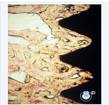
However, experimental studies interesting as those may have been, were soon to be obscured by clinical applications of what was to be called osseointegration. Brånemark´s serendipity based observation that his implant chambers remained most difficult to remove from the bone, immediately made him conclude that the implants were anchored in bone tissue [9]. This was contrary to the concepts of those days that saw a direct bone anchorage of any metallic specimens as impossible [10,11]. Allegedly, a fibrous tissue sheath surrounding titanium implants would be a necessary finding if direct microscopy of the interface would be performed. However, at the time no technique had been developed that made it possible to visualize and verify whether bone or soft tissue actually surrounded titanium implants, and attempts by Brånemark to prove his concept of a bony encapsulation were in very low power (Figure 2) or based on decalcified specimens where the implant had to be removed prior to sectioning of the bone. The inevitable interfacial soft tissues were then accidentally removed together with the implant, claimed those who did not believe in bony anchorage of metals. First with the advent of the cutting and grinding concept through the work by Donath [12], was it possible to analyze intact bone to metal specimens, thereby confirming Brånemark´s ideas of a direct contact between bone and titanium at the resolution level of the light microscope (Figure 3).
However, Brånemark did not wait for this academic controversy to be finally solved – in 1965 he operated his first patient with oral implants based on his conviction that they would prove satisfactory in clinical function due to bone anchorage of the implants [13]. Oral implants were not recognized at all by academic dentistry of those days and his clinical work was to be severely criticized by what then seemed to be united Swedish dentistry. The issue was to be settled first in 1977 following a largely positive clinical report by some Swedish academicians [14]. The term osseointegration to describe the anchorage of the implants was coined by Brånemark in 1976 and first used in 1977. The term was coined in a manner characteristic for P-I Brånemark; he consulted with a University linguist informing this man of his clinical concept and in return had the suggestion of osseintegration to describe it.
Suggested older definitions of osseointegration
The first time that a direct bony anchorage of oral implants was suggested was in 1969 [9], but at the time no specific term was used for the alleged direct bone-to-implant contact. The first time the term “osseointegration” was used in the literature was in the paper by Brånemark et al. [13], where the term was in the headline of the paper but not precisely defined. The first definition of osseointegration that was claimed to be “a direct contact between a loaded implant surface and bone at the light microscopical level of resolution” was published a few years later [15]. An alternative definition“ A direct structural and functional connection between ordered, living bone and the surface of a load-carrying implant” [16], was followed a somewhat more complicated one: ”A continuing, structural and functional co-existence, possibly in a symbiotic manner, between differentiated, adequately remodeling, biologic tissues and strictly defined and controlled synthetic components providing lasting, specific clinical functions without initiating rejection mechanisms”[17]. Figure 2One very early implant design with flanges used by P-I Brånemarkand his team between 1965 and 1970. The radopgram to the right is a 30-year control.
Figure 3According to an old definition, Osseointegration represents a direct (at the light microscopical level of resolution) between bone and implant.
Yet another attempt for definition was“ A process whereby clinically asymptomatic rigid fixation of alloplastic materials is achieved and maintained in bone during functional loading” [18]. These different early definitions of osseointegration either concentrated on an interfacial image of the bone-to-implant contact or on implant function. The functional aspect was further stressed in a biomechanically based definition “A bony attachment with resistance to shear and tensile forces” [19]. However, the question is whether resistance to pure tear off forces really exists over the osseointegrated interface. This topic is further discussed under the heading “osseointegration as a chemical interaction”. The main question is whether the interface is stronger than the surrounding bone; i.e. attempts to remove the implant would cause primarily fracture in the bone, not in the interface. However, in animal experiments it was convincingly demonstrated that attempts to implant removal by torque units, indeed result in interfacial separation between bone and titanium oxide [20].
Osseointegration as a potential chemical interaction
between foreign material and the bone
Osborn and Newesly [21], presented some data indicative of that metals are incorporated into bone by distance osteogenesis, whereas bioinert materials may establish a contact osteogenesis but only so called bioactive implants would be presenting bond osteogenesis. This notion was criticized by the Gothenburg team [22]. In the first years after introducing osseointegrated implants clinically, their chemical composition theoretically was regarded as capable of establishing bonds across the surface. This alleged molecular bonding would then be between the inevitable about 5 nanometer thick oxide layer of the bone and the tissues. It was assumed that bonding at the interface represents a combination of local chemical bonds and long range dipolar or electrostatic interaction [23]. Chemical bonding would assume a bioinert behavior of the titanium implants. This reasoning was further developed by Sul [24], who saw indications of mechanical interlocking as well as biochemical bonding acting over the titanium to bone interface. Sul´s postulate was based on an assumed movement of Ca cations that lead to an electrostatic bonding with proteins. Ca cations stimulate RGD surface receptors and further triggers recruitment of osteoprogenitor cells and osteoblast via calcium signaling pathways [24]. The problem remains to prove such chemical connections, if they exist. In fact, there seems to be no proper methodological approach developed that can verify the theories of Sul [24].
Osseointegration, a foreign body response, motivating
a suggested new definition
The first to suggest an alternative outlook on osseointegrated implants was Donath [25,26]. Donath saw bone development around implants as nothing but a foreign body response. Titanium and other metal implants are not inert, but in reality identified as foreign bodies by the defense mechanisms of the body. The result is to build bone around the foreign body to isolate it from the tissues. Donath [26], described bone attachment to shrapnel found in humans. The bone protection can be regarded similar to soft tissue embedment of foreign bodies – Suska et al. [27], described soft tissue capsule formation developing around materials such
as titanium and copper in a rat model at 28 and 56 days after implant placement. The resultant soft tissue capsule was thicker with more inflammatory cells, particularly macrophages and giant cells, around copper compared to titanium. In another study [28], it was noted that sham recruited cells produced low levels of cytokines in comparison to titanium and particularly to copper. Similar observations were made in a comparative study in bone tissue between sham and titanium (yet unpublished observations). It is not at all surprising that bone tissue seems to react in a similar manner as soft tissues: Bone is precisely like soft tissue belonging to the connective tissues of the body with one main difference between bone and soft tissue being that the ground substance is calcified in bone but not in connective tissue proper cases. Nevertheless, it is not surprising if these two tissues react in a similar manner if provoked by a foreign body such as titanium or copper.
Bos [29], analyzed failed hip joints and found evidence of foreign body reactions, a study further developed with failed hip joints by Christiansen [30]. The latter author found innate and adapted immunology behind aseptic loosening of hip arthroplasties. Thiele et al. [31], reported of foreign body reactions to resorbable polylactic screws. Anderson and Rodriguez [32], have summarized foreign body reactions to biomaterials in an overview. The first part of the immune system to recognize foreign bodies is immune complement [33]. Foreign body giant cells are routinely reported from oral implant interface analyses [25]. Albrektsson et al. [34,35], described foreign body reactions to titanium oral implants and reported that marginal bone loss around titanium implants could be explained by an adverse immunological reaction to the foreign body rather than representing a disease. A detailed analysis of innate immunity to biomaterials is reported in a recent paper [36].
A new definition of osseointegration in the light of this new knowledge would read “Osseointegration is a foreign body reaction where interfacial bone is formed as a defense reaction to shield off the implant from the tissues”.
Osseointegrated implants display long term success
in clinical studies of oral implants
With older machined (turned) implants, one study demonstrated that 334 mandibular implants had 99.1% survival over 5-8 years whereas 106 maxillary implants reported a survival of 84.9% over 5-7 years [37] (Figure 4). Studies with minimally rough turned implants commonly showed about 10 % better outcome in the mandible cf the maxilla [38]. The advent of moderately roughened implants (Figure 5) displayed improved clinical results, particularly for implants placed in compromised situations such as patient smoking, direct loading of implant or placement of implants in the maxilla [38]. An alleged disease entitled peri-implantitis displayed what initially seemed to be implanting threatening marginal bone resorption, but in the light of recent information [39], problems were found much smaller; the quoted authors checked what happened with implants declared to have problems with bone loss in a previous 5-20 year study by Fransson et al. [40]. On an average 9.1 years later, i.e. when the implants had been in situ for about 20 years, the alleged problematic implants showed minimal or no further
bone resorption in more than 91% of cases, whereas 95% of the “problematic” implants still functioned as part of bridges [39]. Such findings went hand in hand with several critical analyses of so called peri-implantitis; in reality it seemed like disease problems were quite minor and that one can expect about 95% or more of implants to function very well over 10 years or more in the patient provided properly trained individuals place controlled oral implant systems [35,41,42]. Implants are not the same as teeth (Figure 6)
Osseointegrated implants in craniofacial surgery
After the positive outcome of osseointegrated dental implants it was a natural step to search for other applications of this technology. Brånemark successfully tried bone anchored implants for retaining facial epistheses in a few patients. However, before this became a routine surgical procedure, the bone anchored hearing aid (BAHA) was developed. The important fact that an implant is securely anchored into the hardtissue means that vibrations can be transmitted. This fact was used in the development of BAHA. Basically, an implant is placed in the skull behind the ear and onto this implant a vibrator is mounted, producing vibrations which are then transmitted into the inner ear. This is ideal for patients with so-called conductive hearing loss, where the outer ear canal and/or ossicles are defective due to congenital malformations or following surgery, but the inner ear is still functioning. Previously, these patients had to wear a metallic spring-like headband over the head with a vibrator applying pressure on the soft tissues of the skull, causing pain and ulcerations. Today, models both with and without skin penetration are commercially available. Tjellström et al. [43], developed this method and presented the first clinical follow-up series.
A surgical protocol for inserting implants into the facial skeleton for the retention of aural and facial epitheses was also developed. Previously, due to congenital malformations or following ablative surgery of auricles or in the maxillofacial region, defects were concealed using epitheses retained with eyeglasses or adhesives, which are liable to make the epistheses come askew or fall off. With the use of implants coupled to retention elements on the episthesis, retention is secure and the margins of the epithesis can be made so thin as to actually, to a large extent, follow the movements of the facial muscles and skin. A first small clinical follow-up was published in 1983 [43], followed by a study of 147 patients over a ten year period in 1990 [44]. This method is also today standard clinical procedure in many centres and implants are commercially available.
Osseointegration in orthopaedics
A novel hip arthroplasty design was developed on the concept of osseointegration. The dual shaped externally bead blasted c.p. titanium acetabular component is dome shaped at the top and cylindrical in the distal part with buttresses and was fitted internally with a HDPE liner affixed by buttresses and using thermal expansion [45]. After resection of the femoral head the femoral neck is preserved. The c.p. titanium threaded and blasted femoral component is rotationally symmetrical and features a Figure 4 A retrieved mandibular, Branemark implant from post mortem
patient.
Figure 5A moderately rough titanium surface.
Figure 6Difference between teeth and implants; Good blood flow and nerves in interface of teeth but not so around titanium that shows condensation of bone instead (republished from Albrektsson et al ClinImplant dent rel res 2014 with Permission).
collar that rests on the proximal part of femoral neck. The implant is screwed in place after preparation of a canal that extends from the top of the femoral neck and out through the calcar.
This hip arthroplasty was developed over a number of years and subjected to randomised clinical trials [46,47] where the Spectron stem and the Trilogy acetabular components were used as controls. RSA (radiostereometric analysis) was employed to detect minute changes in the components location relative to the bone tissue surrounding them and functional scoring was used to assess patients’ perception of the hip arthroplasties. After two years no differences were found between the test and control groups regarding stability and functionality. After five years all implants were still in place but signs of HDPE liner wear were apparent. This was probably due to the use of an older sterilisation technique, which rendered the plastic more susceptible to wear. According to the Swedish hip Registry no hip stems had been revised over the first 10 years of follow up.
DISCUSSION AND CONCLUSIONS
1. Osseointegration was discovered in 1962 and originally defined based on a direct contact between bone and implant at the light microscopical level of resolution 2. Today, Osseointegration is regarded as a foreign body
reaction with bone formed to shield off the foreign material from the tissues
3. Osseointegrated implants have demonstrated excellent clinical outcome in oral, craniofacial and orthopaedic sites.
REFERENCES
1. Brånemark PI. Vital microscopy of bone marrow in rabbit. Scand J Clin Lab Invest. 1959; 38: 1-82.
2. Brånemark PI. Bone marrow, structure and function. Adv in Microcirc. 1964; 1: 1-42.
3. Emnéus H. Some aspects of osteosynthetic materials as a foreign body. Acta Orthop Scand 1967; 38: 368-372.
4. Brånemark PI. Intravascular anatomy of blood cells in man. S Karger Basel. 1971: 1-80.
5. Albrektsson T, Albrektsson B. Microcirculation in grafted bone. A chamber technique for vital microscopy of rabbit bone. Acta Orthop Scand. 1978; 49: 1-7.
6. Albrektsson T. Repair of bone grafts. Scand J Plast Reconstr Surg. 1980; 14: 1-12.
7. Eriksson A, Albrektsson T, Grane B, McQueen D. Thermal injury to bone. Int J Oral Surg. 1982; 11: 115-121.
8. Jacobsson M, Albrektsson T, Turesson I. Dynamics of irradiation injury to bone tissue. Acta Radiologica Oncol. 1985; 24: 343-350.
9. Brånemark PI, Breine U, Adell R, Hansson BO, Lindström J, Ohlsson Å. Intraosseous anchorage of dental prostheses I Experimental findings. Scand J Plast Reconstr Surg. 1969; 3: 81-100.
10. Southam JC, Selwyn P. Structural changes around screws used in the treatment of fractured human mandibles. Brit J Oral Surg. 1970; 8: 211-221.
11. Collins DH. Tissue changes in human femurs containing plastic appliances. J Bone Jt Surg. 1954; 36B: 458-467.
12. Donath K, Breuner G. A method for the study of undecalcified bones and teeth with attached soft tissues. J Oral Pathol. 1982; 11: 318-326. 13. Brånemark PI, Hansson BO, Adell R, Breine U, Lindström J, Hallén O, et
al. Osseointegrated implants in the treatment of the edentulous jaw. Scand J PLast Reconstr Surg. 1977; 16: 1-99.
14. Bergenholtz A, Bergman B, Lundberg M. Implantat i odontologisk praxis.Tandläkartidn.1977; 69: 1197-1199.
15. 15. Albrektsson T, Brånemark PI, Hansson HA, Lindström J. Osseointegrated titanium implants. Requirements for ensuring a long-lasting, direct bone anchorage in man. Acta Orthop Scand. 1981; 52: 155-170.
16. Brånemark PI. Introduction to osseointegration. In: Brånemark PI, Zarb G, Albrektsson T, editors. Tissue integrated prostheses. Osseointegration in clinincal dentistry. Quintessence Co berlin, Chicago and Tokyo.1985; 350.
17. Brånemark PI. Precision, predictability. Information booklet Institute for Apllied Biotechnology, Gothenburg Sweden, 1990; 1-15.
18. Zarb G, Albrektsson T. Osseointegration - a requiem for the periodontal ligament? (editorial). Int J Periodontol rest dent. 1991; 11: 88-89. 19. Steinemann SG, Eulenberger J, Maeusli PA, Schroeder A. Adhesion of
bone to titanium. Adv Biomaterials. 1986; 6: 409-415.
20. Johansson C, Albrektsson T. Integration of screw implants in the rabbit. A 1-year follow up of removal torque of titanium implants. Int J Oral Max fac Implants. 1987; 2: 69-75.
21. Osborn JF, Newesly H. Dynamic aspects of the implant-bone interface. In: Dental Implants, Materials and Systems Heimke G, editor. HanserVerlag, München, Wien. 1980.
22. Albrektsson T. The response of bone to titanium implants. CRC critical reviews in biocompatibility. 1985; 1: 53-84.
23. Albrektsson T, Brånemark PI, Hansson HA, Kasemo B, Larsson K, Lundström I, et al. The interface zone of inorganic implants in bone. Ann Biomed Eng. 1983; 11: 1-41.
24. Sul YT. On the bone response to oxidized titanium implants: The role of microporous structure and chemical composition of the surface oxide in enhanced osseointegration. 2002; 1-184.
25. Donath K, Laas M, Günzl H. The histopathology of different foreign body reactions in oral soft tissue and bone tissue. Virchows Arch A Pathol Anat Histopatol. 1991; 420: 131-137.
26. Donath K. Pathogenesis of bony pocket formation around dental implants. J Dent Assoc S Afr. 1992; 47: 204-208.
27. Suska F, Esposito M, Gretzer C, Källtorp M, Tengvall P, Thomsen P. IL-1α, IL-1β and TNF-α secretion during in vivo/ex vivo cellular interactions with titanium and copper. Biomaterials. 2003; 24: 461-468.
28. Suska F, Emanuelsson L, Johansson A, Tengvall P, Thomsen P. Fibrous capsule formation around titanium and copper. J Biomed Mater Res A. 2008; 85: 888-896.
29. Bos I. Gewebereaktionum gelockerte Hüftgelenk-endoprothesen. Der Orthopäde. 2001; 30: 881-889.
30. Christiansen RJ. Metal release from implants and its effect on the immune system. 2016; 199.
31. Thiele A, Bilkenroth U, Bloching M, Knipping St. Fremderkörperreaktion nach implantation eines biocompatible en OsteosyntheseSystems. HNO. 2008; 56: 545-548.
32. Anderson J, Rodriguez A, Chang D. Foreign body reaction to biomaterials. Semin Immunol. 2008; 20: 86-100.
Albrektsson T, Chrcanovic B, Jacobsson M, Wennerberg A (2017) Osseointegration of Implants – A Biological and Clinical Overview. JSM Dent Surg 2(3): 1022.
Cite this article
33. Hulander M, Lundgren A, Berglin M, Ohrlander M, Lausmaa J, Elwing H. Immune complement activation is attenuated by surface nanotopography. Int J Nanomedicine. 2011; 6: 2653-2666.
34. Albrektsson T, Dahlin C, Jemt T, Sennerby L, Turri A, Wennerberg A. Is marginal bone loss around oral implants the result of a provoked foreign body reaction? Clin Implant Dent Relat Res. 2014; 16: 155-165.
35. Albrektsson T, Canullo L, Cochran D, de Bruyn H “Peri-implantitis”: A complication of a foreign body or a man made “disease”. Facts and fiction. Clin Implant Dent Relat Res. 2016; 18: 840-849.
36. Christo S, Diener K, Bachhuka A, Vasilev K, Hayball J. Innate immunity and biomaterials at the Nexus: Friends or foes. Biomed Res Inter. 2015; 23.
37. Albrektsson T, Dahl E, Enbom L, Engevall S, Engquist B, Eriksson A, et al. Osseointegrated oral implants. A Swedish multicenter study of 9139 consecutively inserted Nobel pharma implants. J Periodontol. 1988; 59: 287-296.
38. Jimbo R, Albrektsson T. Long-term clinical success of minimally and moderately rough oral implants: A review of 71 studies with 5 years or more of follow-up. Implant dentistry. 2015; 24: 62-69.
39. Jemt T, Sundén-Pikner S, Gröndahl K. Changes of marginal bone level in patients with “progressive bone loss” at Brånemark system implants: a radiographic follow-up study over an average of 9 years. Clin Implant dent rel res. 2015; 17: 142-147.
40. Fransson C, Lekholm U, Jemt T, Berglundh T. Prevalence of subjects with progressive bone loss at implants. Clin Oral Impl Res. 2005; 16; 440–446.
41. Albrektsson T, Buser D, Sennerby L. Crestal bone loss and oral implants. Clin Implant Dent Relat Res. 2012; 14: 783-791.
42. Albrektsson T, Chrcanovic B, Östman PO, Sennerby L. Initial and long-erm crestal bone responses to modern dental implants. Perio 2000. 2017; 73: 41-50.
43. Tjellström A, Lindström J, Hallén O, Albrektsson T, Brånemark PI. Direct bone anchorage of external hearing-aids. J Biomed Engin. 1983: 5; 59-63.
44. Hakansson B, Liden G, Jacobsson M, Tjellström A, Carlsson P, Ringdahl A, et al. 10 years of experience with the Swedish bone-anchored hearing system. Ann Otol Rhinol laryngol. 1990; 99: 1-16.
45. Albrektsson T, Carlsson L, Jacobsson M, Macdonald W. The Gothenburg osseointegrated hip arthroplasty. Experience with a novel type of hip design. Clin Ort Rel Res. 1998; 352: 81-94.
46. Carlsson L, Albrektsson BJ, Albrektsson T, Jacobsson M, Macdonald W, Regnér L, et al. Stepwise introduction of a bone- conserving osseointegrated hip arthroplasty using RSA and a randomized study: I Preliminary observations - 52 patients followed for 3 years. Acta Orthop Scand. 2006; 77: 549-558.
47. Carlsson L, Albrektsson T, Albrektsson B J, Jacobsson M, Macdonald W, Regnér L, et al. Stepwise introduction of a bone-conserving osseointegrated hip arthroplasty using RSA and a randomized study II. Clinical proof of concept - 40 patients followed for 2 years. Acta Orthop Scand. 2006; 77: 559-566.