Studies towards a general method for
attachment of a nuclear import signal.
Stabilization of the m
3
G-Cap
Mattias Lindvall
Master Degree Thesis
2010-06-03
Supervisors
Prof. Roger Strömberg
Dr Malgorzata Wenska
Karolinska Institutet
Department of Biosciences and Nutrition
Huddinge, Sweden
Examiner
Assoc. Prof. Simon Dunne
Mälardalen University, Eskilstuna/Västerås
Abstract
A synthetic pathway towards the cap-structure of 2,2,7-trimethylguanosine containing a methylene modified triphosphate bridge have been investigated. The modification to the triphosphate bridge is hoped to slow down cap degradation and give the connected oligunucleotide an increased lifetime. This could result in an better understanding of nuclear transport of oligonucleotides and could thereby helping to develop new treatments for different diseases. The synthesis relies on a coupling reaction between the 2,2,7-trimethylguanosine 5’phosphate and 2’-O-methyladenosine with a 5’-pyrophosphate where the central oxygen has been replaced by a methylene group. The reaction pathway consists of 9 steps of which 8 steps have been successfully performed. The last step, which includes a coupling reaction, was attempted but without successful identification and isolation of the cap-structure, and will need further attention. The reaction has been performed in a milligram scale with various yields.
Table of contents
Abbreviations and definitions ... 4
Introduction ... 5
Project aim ... 5
Background ... 6
Results and Discussion ... 12
Experimental ... 18 Equipment ... 18 Material ... 19 Synthesis ... 19 Acknowledgements ... 21 References ... 22
Abbreviations and definitions
In order of appearance:
HPLC ... High Performance Liquid Chromatography DNA ... Deoxyribonucleic acid
RNA ... Ribonucleic acid
NLS ... Nuclear Localization Signal mRNA ... Messenger-RNA
pre-RNA ... Precursor-mRNA m7G ... 7-methylguanosine
snRNP ... Small nuclear ribonucleo protein snRNA ... Small nuclear ribonucleic acid m3G ... 2,2,7-trimethylguanosine
SPN1 ... Snurportin 1
GTPase ... Guanosinetriphosphate hydrolaze RAN ... RAs-related Nuclear protein DcpS ... Scavenger Dcp
m3GpppA ...
2,2,7-trimethylguanosine-5’-triphosphate-5’(2’-O-methyladenosine)
Dcp1/2 ... Decapping enzymes TEAA ... Triethylammonium acetate NMR ... Nuclear Magnetic Resonance NaCNBH4 ... Sodium cyanoborohydride
TLC ... Thin Layer Chromatography DCM ... Dichloromethane
MeOH ... Methanol
MS ... Mass Spectrometry DMF ... Dimethylformamid AcOH ... Acetic acid
Introduction
Project aim
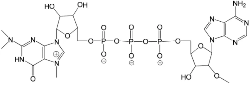
The aim for this project was to stabilize the native triphosphate bridge between the import signal 2,2,7-trimethylguanosine and 2’-O-methyladenosine (Figure 1). The modification was planned to be compared with the native bridge in enzymatic cleavage in cellular extract and 10% human serum, using HPLC to monitor the reaction.
Figure 1 The m3G with the native triphosphate bridge to 2’-O-methyladenosine
The modification was to be achived by separately synthesizing the guanosine-based part and the adenosine-based part of the molecule before coupling them together to give a triphosphate bridge. The bridge bears a methylene group between the α and β phosphate groups. The guanosine-based half is trimethylated and possesses a 5’-phosphate group (Figure 2a). The adenosine-based half will bear the methylene modification. The native molecule is made by substitution in the 5’-position by a pyrophosphate. In the modified half, however, methylenebis(phosphonic dichloride) is used to form a methylenebisphosphonate terminus. (Figure 2b).
Figure 2 a is the trimethylated 5’-phosphateguanosine to be coupled together with the modified (hydroxyphosphoryl)methylphosphonic acid 2’-O-methyladenosine b
Background
The cell nucleus is surrounded by a double membrane. The eukaryotic cell has its DNA localized in one part of the cell instead of spread out as in prokaryotes. This makes transcription more effective. The membrane protects the new, immature RNA from being degraded by enzymes or organelles before it has been processed. On the other side, the nuclear membrane creates transport problems that the cell must overcome; e.g. enzymes and proteins required for replication and transcription must find their way through the nuclear membrane in either direction. This problem is solved by the cell through two different types of transport mechanisms1; The diffusion through nuclear pores is an open channel where small molecules can pass through. As molecular size increases the entry rate to the nucleus decreases. Small molecules (<40 KDa) can diffuse through the cell membrane pore complexes, but larger molecules need help with the transport from a carrier protein2. Larger molecules could be RNA, proteins, protein complexes or a hybrid of either of these required by the nucleus in order to maintain normal function, but the idea of transporting larger molecules into the nucleus can be applied to other desired functions such as drug delivery. In order to be released through the pores into the nucleus, the carrier protein needs to have an identification signal attached to them. This signal (Nuclear localization signal (NLS)) can be an amino acid chain, usually with a positively charged lysine or arginine, that attaches to the carrier protein and the carrier protein is allowed to enter through the nuclear pore complexes. In a similar way, larger molecules can exit the nucleus.1
One of the molecules that uses this transport mechanism to exit the cell is mRNA. mRNA is synthesized within the nucleus in the transcription process, but needs to be transported out to the cytoplasm to undergo translation at the ribosomes. In the translation process, proteins are formed. As opposed to the prokaryotic RNA, the eukaryotic RNA needs processing before it is ready for transport and translation. Immature mRNA, called pre-mRNA, often needs to remove some nucleotide sequences, needs to add a 5’-cap and a 3’-tail. The 3’-tail is a long chain of adenosine molecules, called poly(A) tail. A specific sequence (AAUAAA) gives information to the cell where the poly(A) tail should be added. This 3’-tail protects the mRNA from enzymatic degradation. The longer the tail, the longer is the lifetime of mRNA.3
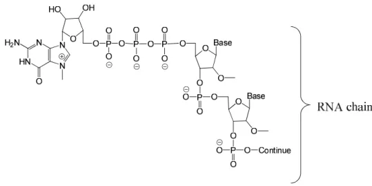
A 5’-cap also prevents degradation of the mRNA and in addition it has an important role in the positioning of the mRNA on the ribosome for correct translation as well as in being a signal for transport out of the nucleus. The 5’-cap consists of a guanosine molecule which is methylated in the 7-position (m7G)and is linked to the mRNA via a 5’-5’-triphosphate bridge (Figure 3)3.
Figure 3 The m7G cap connected to an RNA chain via a 5’-5’ bridge
As earlier stated, pre-mRNA needs to remove some nucleotide sequences in order to become mature mRNA. These sequences, introns, are removed by a protein complex called spliceosome in a procedure called splicing.
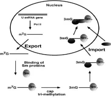
The spliceosome is built up in the cytoplasm by several different proteins, of which the small nuclear ribonucleoproteins (snRNPs) are the major part. These different types of snRNPs contain small nuclear ribonucleoacids (snRNAs). The snRNAs are transcribed by RNA-polymerase II in the nucleus and can be divided into six major classes; U1, U2, U3, U4, U5 and U6.4 The snRNA needs transport to the cytoplasm in order to participate in the assembly of the splicesome complex. Before export an m7G cap is applied to the snRNA. Beside the snRNAs, the snRNPs consist of seven Sm proteins, orientated in a ring, and some proteins that are specific for each snRNA. These Sm proteins are synthezised in the cytoplasm. This complex needs to be assembled correctly to undergo hypermethylation of the 5’-cap and correct processing of the 3’-end. Since the spliceosome is put together in the cytoplasm, outside the nucleus, it needs to be imported into the nucleus. Hypermethylation of the m7G, which previously exported the snRNA, to a 2,2,7-trimethylguanosine (m3G) gives the
spliceosome an import signal5. It is essential to have m3G as a cap to bind into the carrier
The m3G-cap on the spliceosome binds specifically to a protein complex, snurportin-1
(snurportin or SPN1). The 45kDa large snurportin interacts with the m3G-cap, while it ignores
the m7G. This makes the hypermethylation of m7G important7. When m3G interacts with
SPN1, importin β is recruited8. Importin β, with the help of a GTPase called RAN facilitates the entry of the spliceosome into the nucleus where it can start splicing the pre-mRNA. The process of exporting snRNA, assembly of the spliceosome and import to the nucleus can be seen in the simplified figure (Figure 4) by Mattaj and Englmeier9.
The knowledge of how the cell imports snRNA with protein complexes, such as the spliceosome, and the function of the m3G-cap can open the opportunity to import other
proteins or nucleotide sequences as well, opening a new way to treat diseases which have their origin in the nucleus. The problem behind many diseases may be, for example, incorrect transcription, wrong gene expression or failure in splicing. One strategy to affect the DNA and/or the mRNA is if a chain of complementary nucleotides (oligonucleotides) can be imported. These antisense oligonucleotides (AON) are based upon the idea of a sequence-specific recognition complementarity of a certain part of the mRNA molecule10. Using AONs on mRNA involves several strategies, one is to have the AON block the site on the mRNA in the cytoplasm and therefore preventing the gene to express a protein. In some diseases, as for example, Duchenne’s Muscle Dystrophy (DMD) the problems lies within a mutation in the encoding gene which results in an incorrect splicing.
Figure 4 The pathway by which snRNA is synthesized in the nucleus, exported, assembled with Sm protein to form spliceosome, hypermethylated and imported to the nucleus. Mattaj and Englmeier9
DMD causes 1:3 500 newborn males to suffer from a progressive muscular weakness. Before the age of 12 the patients are wheel-chair-bound and as the muscles continue to degrade, which results in death usually before a age of 30. The cause behind the disease is a mutation in the gene coding for the protein dystrophin. The mutation prevents the protein from being expressed. Dystrophin is a part of a complex which keeps the membrane surrounding the muscle fibers intact. Lack of dystrophin causes the muscle cells to die which leads to muscle degradation. There is a milder form, Becker muscular dystrophy (BMD), that hits 1:20 000 and usually allows the patients to live to an age of 40. In BMD the mutation of the gene results in a shorter and less functional protein which partially works, therefore it slows down the degradation. The gene is X-bound which means only men can be affected from DMD and BMD, but females may be carriers. With todays techniques, there is no cure for DMD, but treatment could reduce the symptoms to mimic the milder BMD it would give patients hope of an improved life. Treatment using AOEs that interfere with the mRNA during the splicing process is a promising method11. As mentioned, all pre-RNA’s are spliced in order to become
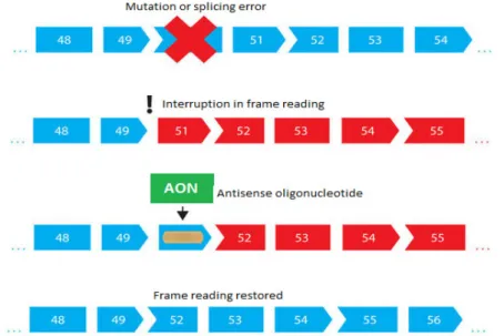
mature mRNA. After splicing the exons remain. The exons can consist of, for example, a coding gene and nonsense RNA. But in the case of DMD the gene mutation results in the splicing mechanism deleting not only the introns but some of the exons as well. The frame-shift may introduce a premature stop codon on the mRNA which prevents dystrophin from being expressed. Using AONs scientists have a chance to partly correct this frame-shift by binding the AONs to one or more exons which lie nearby the deleted exon. By doing so, the exon with the premature stop codon is skipped. The reading frame in the translation process
Figure 5 The illustration of when a mutation or splicing error occur which results in a interruption of the frame reading. Treatment with a Antisense Oligonucleotide (AON) restores the frame reading and gives a partially working protein. Modified from http://www.muscular-dystrophy.org/13
can be corrected, creating a shorter but partly functional dystrophin, making the DMD symptoms appear more like in Becker Muscular Dystrophy (BMD)12 . The exon deletion and exon skipping is illustrated in Figure 513. As stated earlier, one of the roles the 5’-cap fills is protection of the mRNA or AON from degradation. The degradation occurs through two major pathways; 3’-exonucleases which act by degrading the RNA/AON from the 3’-end group (called 3’-5’ degradation) and the 5’-exonucleases, which after an initial breakdown of the 5’-cap by Dcp1 and Dcp2 pyrophosphatases, degrade the RNA/AON from the 5’-end (called 5’-3’ degradation)14-15
In mammals the 3’-5’ degradation is the most occuring. It starts with a deadenylation of the previously mentioned poly(A) tail followed by a 3’-mRNA decay by the exonuclease exosome. The product from this degradation are monophosphates of the RNA and the mRNA cap. The cap is then hydrolyzed by a scavenger enzyme, DcpS16. The DcpS associates with the exosome and is specific for short pieces of nucleotides such as the mRNA cap17. The 5’-3’ degradation is initiated by Dcp1 and Dcp2 which breaks the pyrophosphate bond between the α and β phosphate groups, resulting in a m3G- or m7G-pyrophosphate and a RNA/AON with a
exposed 5’-phosphate. The exoribonuclease Xrn1 then can hydrolyze the RNA/AON from the 5’-end.
The degradation of mRNA is naturally occurring in all eukaryotic cells. mRNA decay is one way to regulate the gene expression and it is important for the mRNA turnover. The exosome, for example, plays an important role in degradation of mRNAs with defect 3’-ends and precluding translation of defective mature mRNAs18. When the cellular mechanisms are used for other purposes than the naturally occurring ones, some of the cell functions must be overcome without interrupting normal functionality. One such purpose is the use of AON’s which have been introduced. A major problem lies within exposure of the AON’s to all the degradation factors. 5’ Degradation could be protected by hiding or protecting the 3’-hydroxy group which normally is the target for degradation after removal of the poly(A)-tail. However, in order to increase the concentration in the nucleus, the 5’m3G-cap structure with
its triphosphate bridge should be present.
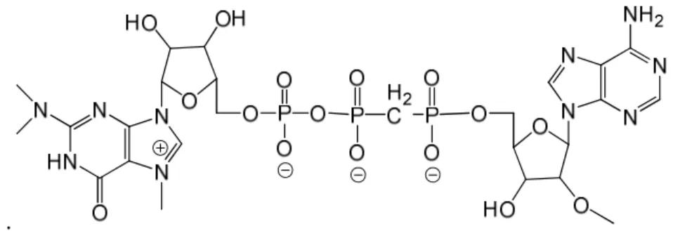
The m3G-cap, linked via a modified triphosphate bridge to a 2’-O-methyladenosine
(m3GpppA) (Figure 6) could protect against the enzymatic cleavage. The modification is a
methylene group between the α and β phosphates (counted from the adenosine). The introduced P-C bond is extremely stable and should prevent any enzymatic Dcp1/Dcp2 cleavage at that specific site, making the degradation of the cap slower 19.
In vitro cleavage studies of the modified m3G-cap will be compared with the non-modified
m3G-cap in cellular extract solution as well as in 10% human serum in order to measure
resistance to similar enzymatic activity that the cap encounters in the body.
.
Figure 6 The 2,2,7-trimethylguanosine cap linked to 2’-O-methyladenosine via a α-β modified triphosphate bridge.
Since the body uses the import mechanism via the m3G-cap, the non-modified cap could be
used to transport some drug/AON into the nucleus and thus it is of interest in getting as little degradation as possible. With less degradation, fewer side-products are left in the cell that can interfere with the natural processes of the cell, and with less degradation lower levels of drug/AON are needed. The bioresponse is therefore increased; one requires lower doses of the drug to get the same effect.
Results and Discussion
As the project aim suggesed intention is to make the cap synthetically. The synthetic pathway can be seen in Scheme 1.
Scheme 1. The synthetic route to give 2’-O-methyladenosine 5’-(β-(N2,N2,N7 -trimethylguanyl-5’-yl)methylenediphosphonate)
In the first step of the synthesis of 2’,3’-di-O-acetyl-N2,N2-dimethylguanosine 2, a shiff base was reduced to give methylation, by using paraformaldehyde and NaCNBH4. In the first
reaction some problems were encountered. The amount of solvent (AcOH) given in the literature was not satisfying in order to fully dissolve compound 1 at room temperature. Before compound 2 had dissolved completely, paraformaldehyde was added. Precipitate was still observed after the addition of paraformaldehyde. When the temperature was increased to 45°C, the starting material fully dissolved and gave a colorless transparent solution. After approximately 1 hr the reducing agent NaCNBH4 was added. The addition resulted in a
vigorous reaction but without any visible change to the reaction mixture. In total, three portions of NaCNBH4 were added over an interval of 1 hr. TLC (DCM:MeOH 90:10) showed
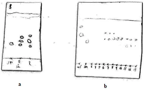
reaction progress (Figure 7a). Two spots plus the starting compound 1 were observed. Doing a preparative TLC with subsequent analysis by MS showed that the spot with lower Rf value
was the dimethylated product. The starting material spot on TLC never vanished before column chromatography nor did it reduce in size. The conclusion was that not all starting material had reacted but the reaction did not go any further under the current conditions. The reaction was performed a second time with more solvent and two more additions of paraformaldehyde and NaCNBH4 in order to try to push the reaction to completion. However
the starting material spot remained. What was not tested was increased reaction temperature. But since nucleosides are thermally vulnerable, it was deemed better to obtain a lower yield due to unreacted starting material than to break down the nucleosides.
Figure 7. TLC analysis for the first step. a the reaction mixture compared to the starting material where SM is starting material and R is reaction mixture. b is results from column chromatography in the second reaction, which shows the pure
Column chromatography was performed on both reaction mixtures and here was the biggest obstacle in the first step of the pathway. In the first reaction it was very difficult to achive any separation between the different products. Chromatography was performed with a gradient (0%-15% MeOH in DCM) as the mobile phase. For the purification of the second reaction mixture a non-gradient mobile phase (DCM:MeOH 85:15) was tested. This eluent gave some separation and yielded 14 mg of pure material (yield 20%). TLC (DCM:MeOH 85:15) showed that even if there were some fractions containing pure material there were many others also containing product (Figure 7b). The separation was thus not optimal but it produced some material to continue working on. In the purification of the first reaction mixture several columns were run but the separation was difficult to achieve. Why the separation partially worked in the purification of the second reaction mixture but not in the first is unknown. But one reason may be inexperience of using a gradient during purification. The following steps included a attachment of a Fluoren-9-ylmethyl-H-phosphonate to the 5’-end of 2 followed by an oxidation with iodine, water and pyridine. The steps were finished with elimination overnight. The pathway to make 2’,3’-di-O-acetyl-N2,N2-dimethylguanosine 5’-phosphate 5 involved these three steps where the product in each step was detected by MS but not isolated before proceeding with the next step. When 2’,3’-di-O-acetyl-N2,N2 -dimethylguanosine 5’-(Fluoren-9-ylmethyl-H-hposphonate) 3 was successfully made, according to the method described by Romanowska et al20, and detected by MS analysis, two interesting peaks were observed. In positive ion detection mode (ES+) the product peak (m/z = 638) was the only peak observed, however in ES- the major peak present was 458. The mass difference between the expected product peak and the observed in ES- is equal to the mass of 3. This gives us an indication that the Fluoren-9-ylmethyl group has been removed. The explanation might lie within the MS spectrometer that bombards the molecule with electrons, tearing apart fragments of various size. One of these fragments happens to be the 9-methylene-9H-fluorene molecule. The conclusion made is that the molecule is still intact but MS makes us see differently. Considering this we went on with the next step.
Oxidation of the 5’-H-phosphonate to 5’-phosphate was a straightforward and fast reaction which was completed after only a few minutes. MS recorded for 2’,3’-di-O-acetyl-N2,N2 -dimethylguanosine 5’-(Fluoren-9-ylmethylphosphate) 4 is similar to the spectrum for compound 3. In ES+ the product peak can be observed at 653.94. The ES+ mode spectra contains another few large peaks, one at 221.59 and one at 675.01. ES- mainly consists of one peak at 473.96 which is the 5’-phosphate without the flourene-group attached. The
explanation for this can be the same as when fragmentation by mass was observed for compound 3. It is difficult to decide what the peaks in ES+ that are not the product represent. Left in the reaction vessel there is material and reagents from the previous steps as well as the current step. Also side products from the previous steps remain and there might be other possible reaction routes for the side products would be difficult to predict. Since we could identify the product in both ES+ and ES- the next step of the reaction was performed.
The original reaction mixture with 3 was split equally into two separate flask. The purpose was to oxidize one half and let the other half react with sulfur in order to produce a thiophosphate. The thiophosphate is also suggested to prevent enzymatic cleavage and if so, we could create a cap-structure containing one methylene modification and a thiophosphate. In theory that cap could resist cleavage in two locations and therefore an even more stable cap could be made. However when analyzed by MS the only product identified was the oxidized product. It is unclear if some contamination has oxidized the compound since the oxidation reaction takes place much faster. The two flasks was therefore added together in order to give more 4.
In the last step of making compound 5 the Fluoren-9-ylmethyl group is removed by a base. In this case, Et3N picks the most acidic proton which leads to removal of the group according to
the mechanism in scheme 2. The reaction is also known as a β-elimination. The step was straightforward and easy to perform; just to evaporate off the solvent in the previous reaction mixture and dissolve the resulting yellow precipitate in DCM:Et3N 2:1 overnight.
Scheme 2 The mechansim for removal of the flourene group. Et3N starts by picking the most acidic proton in the reaction
called a β-elimination.
The final step towards N2,N2-dimethylguanosine 5’-phosphate 6 involves deprotection of 2’- and 3’-acetyl groups to obtain the free hydroxyl groups. After 4 hrs MS was recorded which showed only the expected mass. HPLC purification showed very few impurities which was confirmed by a purity check on HPLC after purification. According to MS, the deprotected
product was the only species both in ES+ and ES-. A yield of 4.8 mg from product 2 was calculated to 20%.
N2,N2,N7-trimethylguanosine 5’-phosphate 7 was first made in a small scale reaction to investigate the experimental method. The main issue was the purification on HPLC where material was lost due to unexpected early retention times. The guanosine-based molecules made in this project usually have a retention time of around 20 minutes. In this run however the elution was much earlier. Already after 5 min a large peak had gone through the column. MS analysis showed that the reagent MeI which was used in the reaction together with m3Gp
was the reason. What probably occurred was that the high concentration of MeI made the solution more non-polar, resulting in the major early peak. When injecting smaller quantities of the reaction mixture into the HPLC, two peaks were visible with retention times of 18 and 24 min. The first of these contained 7 according to MS. To obtain a decent yield three small scale purifications were performed. The yield from these three purifications was 2.1 mg (52%). In all purifications the problem with overloading of the column was encountered. In the reference paper15 the reaction was carried out in DMSO instead of in DMF and the excess of MeI was 12 equivalents. However the working amount was 262 mg of starting material compared to the low quantities used here so workup via extraction and column chromatography was possible in their case. The purification problems therefore gave a lower yield than reported in the paper which was 75% for a 3’-O-methylguanosine 5’-phosphate-. N2,N2,N7-trimethylguanosine 5’-(imidazol-1-yl phosphate) 8 was made from compound 7. Compound 8 was used without any workup due to the small amounts of compound and the possible loss of any compound during purification by HPLC. The goal with the reaction was to activate the compound with imidazolyl phosphate to enhance the formation of the final cap-structure 11. Once 8 was identified by MS, compound 10 was added to the reaction vessel even though no workup had been done.
2’-O-methyladenosine 5’-(methylenediphosphonate) 10 was made from 2’-O-methyladenosine 9 which was commercially available. In a report15 the reaction time of the formation of 10 was 1hr. However after one hour, MS indicated that two products had formed. One of them was the desired 5’-substituted adenosine molecule while the other was a bissubstituted product, possibly 5’,3’-substitution of the molecule since the 2’-position bears a methyl group and is therefore not available to substitution. The retention time of the mono- and bissubstituted forms on HPLC was the same which made the purification difficult. Though Kalek et al.15 discussed the high 5’ regioselectivity we observed otherwise, the
difference being that we used an adenosine as the nucleotide compared to the guanosine nucleotide used by Kalek et al. Even if there are different nucleotides that play different roles in the body there should not be significant differences from a synthetic point of view. After reducing the reaction time to 15 min the reaction seemed to only yield the monosubstituted nucleoside thereby making the workup easier. After two reactions with different reaction times, a third reaction yielded compound 10 (22.2 mg, 20%). Compound 10 is very polar due to the methylenediphosphonate substitution which makes reversed phase HPLC the best separation method (since the molecule binds strongly to silica). The reported yield of 83% was not achieved but considering the reduced reaction time and MS results not all of compound 9 had reacted which could explain the yield of 20%. The reaction must be further optimized in order to increase the yield.
The final step to obtain 2’-O-methyladenosine 5’-(β-(N2,N2,N7-trimethylguanyl-5’-yl)methylenediphosphonate) 11 was performed by adding an excess of compound 10 to the crude reaction mixture from synthesis of 8. To the mixture ZnCl2 was added as a catalyst. The
ZnCl2 did not dissolve but the reaction was carried out at room temperature overnight and
followed by MS analysis. A small amount of unreacted 8 was observed which gave an indication that the reaction was not finished. Therefore the reaction mixture was allowed to react for an additional 18 hrs and this time at 30°C. When the reaction mixture once again was analyzed by MS compound 8 could not be identified, but on the other hand there were a lot of materials and reagents which could interfere with the measurements. HPLC was performed to allow isolation of the different components from the reaction mixture and more easily identify what was obtained from the reaction. MS analysis did not identify the cap-structure and many of the HPLC peaks were identified as the polar compound 10, the excess of Ph3P and dipyridyl disulfide in the activation reaction may have blocked 10 by reacting at the end of the 5’-methylenepyrophosphate bridge and therefore preventing the coupling reaction. As an effort to restore the 5’-end of 10 water was added and was hoped to hydrolyze 10, still 11 could not be identified.
A high yield of 11 was not expected since Kalek15 reporteds a yield for the step of 38%. In the pathway followed in this project one can consider to perform the activation step with imidazole on 10 instead of 7. However lower yields have been reported when activating the 5’-methylenediphosphonate instead of the 5’-phosphate.
Overall, many of the steps has been performed successfully but with various yields, poor separations during chromatography (both silica column chromatography as well as HPLC)
have contributed to yield variance. In certain steps some optimization could be achieved, for example to increase the separation in column chromatography other mobile phases could be explored and, in some reactions, the reaction time and conditions could be altered further. Optimization of a reaction step is time-consuming and since this diploma work involves less than 20 weeks of laboratory work, topic research and report writing the yields can be considered acceptable. Unfortunately the final product, the cap-structure, was not isolated. But I am convinced that with some optimizing and customization of some of the reaction steps in the reaction pathway the cap-structure can be made via this pathway in an acceptable yield.
The project aim was not fully achieved. There were several steps which required more time than expected for purification by HPLC. Several reactions were also run more than one time, either the first reaction did not work or the first reaction was only a pilot reaction.
One thing in this project that personally was a challenge was the scale of the reactions. Previously I have worked on larger scales which have yielded several grams of product. The reactions have mainly consisted of heterocyclic chemistry and have involved substrates and reactions which are not very sensible to high temperatures. The chemistry I had the opportunity to work with in Professor Strömbergs laboratory have been a great experience, not only has it trained me to work with very small amounts (milligram scale) but also it has invited me learn more about the nucleotide chemistry. It have sometimes been challenging and even frustrating but I am happy and satisfied with the work I have done.
Experimental
Equipment
NMR
400 Ultrashield Bruker Avance DRX-400
1H NMR spectra were recorded at 400MHz 13C NMR spectra were recorded at 100.6 MHz.
HPLC
Buffer A (28ml Et3N, 11.5ml AcOH, 4L H2O, pH 6.5)
Buffer B (1L Acetonitrile, 1L H2O, 14ml Et3N, 5.7.5ml AcOH, pH 6.5)
Jasco UV-975 Intelligent UV/VIS Detector Jasco CO-965 Column Oven (27.6°C/22) Jasco PU-986 Intelligent prep. Pump
Jasco LC-net II/ADC (power)
Column: Supelcosil LC18 (25cm*4.6mm, 5µm)
Mass Spectrometer
Micromass LCT, scanning between m/z 100-2500, Kd scientific pump
Freeze dryer
Labconco Freezone plus 6
Balances/Scales
Mettler Toledo AG135 (31 g/191 g ± 0.01 mg/0.1 mg) Mettler Toledo AND HR-60 (60 g ±0.1 mg)
UV-visible equipment
Varian Cary 300 Bio UV-Visible spectrophotometer Vilber Lourmal UV lamp 254 nm
Material
List of chemicals used can be found in Appendix 1.
Synthesis
2’,3’-di-O-acetyl-N2,N2-dimethylguanosine (2) was made by adding
2’,3’-di-O-acetylguanosine 1 (0.17 mmol, 65 mg, 1 eq) to concentrated acetic acid (20 ml). Heating (45°C) and stirring was applied until complete dissolution, resulting in a transparent solution. Paraformaldehyde (0.53 mmol, 15.94 mg, 3 eq) was added followed by NaCNBH4 (0.53
mmol, 33.36 mg, 3 eq) after 1h. This was repeated with a 45 min interval until completion of the reaction (TLC DCM:MeOH 90:10, Rf-values: 1 0.54, 2 0.66). Crude yield: 56%.
Purification by column chromatography (DCM:MeOH 85:15) to gave a yield of 20% or 14 mg. 1H NMR (CDCl3 + 1 drop MeOD); δ 7.92 (s, 1H), 6.00 (t, 1H, J = 5.78 Hz, 2’-H), 5.9 (d,
1H, J = 1.5 Hz, 1’-H), 5.56 (t, 1H, J = 4.6 Hz, 3’-H), 4.205 (q, 1H, J = 2.44, 3.27 Hz, 4’-H), 3.79 (dd, 2H, J = 3.2 Hz, 5’-H), 3.36 (s, 6H, 2-diMe), 2.02 (s, 3H, Ac), 1.99 (s, 3H, Ac)
2’,3’-di-O-acetyl-N2,N2-dimethylguanosine 5’-phosphate (5) was made by adding 2 (0.035
mmol, 14 mg, 1 eq) to a mixture of dry (9H-fluoren-9-yl)methyl phosphonate (dried by coevaporation with 20% Et3N in pyridine solution) (3.89*10-2 mmol, 14 mg, 1.1eq) in
pyridine. The solvent was evaporated and the residue was dissolved in dry DCM:pyridine 9:1 solution (500 µl). To the solution, pivaloyl chloride (5.31*10-5 mmol, 6.3 µl ,1.5 eq) was added. After stirring for 20 min, the 2’,3’-di-O-acetyl-N2,N2-dimethylguanosine 5’(Fluoren-9-ylmethyl-H-phosphonate (3) was identified in the reaction mixture by MS (expected: 637.58)
(ES+; m/z>10%: 637.944 = 100%, 638. 959 = 60%, 1273.73 = 20%, 1274.68 = 10%, ES-; 458,96 = 100%), (see discussion section).
To the solution containing 3, a mixture of 1.1 eq H20 and 1.1 eq I2 in pyridine (approx 100µl)
was added. After a few minutes of mixing the oxidized product, 2’,3’-di-O-acetyl-N2,N2 -dimethylguanosine 5’(Fluoren-9-ylmethyl-H-phosphate (4) was observed by MS (expected: 652.52) (ES+; m/z>10%: 221.59 = 100%, 653.94 = 95%, 675 = 70%, 305 = 50%, 411 = 40%, ES-; 473.96 = 100%, 475.02 = 20%, 557.91 = 20%), (see discussion section).
The solution was evaporated to dryness to give a yellow oil. It was stored in DCM: Et3N 2:1
in a refrigerator overnight to remove the methyl-fluorene substituent. Formation of (5) occured overnight. Yield 0.254 mg according to UV-measurments and calculations. However this must be incorrect since bigger amounts was obtained later as discussed in results and discussion section.
N2,N2-dimethylguanosine 5’-phosphate (6) was made by dissolving 5 in ammonium
hydroxide. The foamy mixture was left for 4 hrs before being analyzed by MS (expected: 390.27)(ES+; m/z>10%: 392.26 = 100%, 414.21 = 30%, ES-; 390.04 = 100%). The deprotection was considered complete. Ammonium hydroxide was evaporated and the resulting residue dissolved in water and extracted with DCM. The aqueous phase was evaporated to yield a yellow solid, which was purified with HPLC (1 ml/min, 30 min, 15% Buffer B, retention times for peaks >5%; 23.33 min) to yield 4.8 mg, percentage yield from compound 2 was 32%.
N2,N2,N7-trimethylguanosine 5’-phosphate (7) was made in a pilot reaction by adding dry
DMF (47µl) to a small amount of 6 (1.1 mg, 2.82 µmol, 1 eq) in an Eppendorf tube. MeI (3.5 µl, 56.3 µmol, 20 eq) was added and the mixture was vortexed overnight. Purification by HPLC (1 ml/min, 30 min, 15% buffer B, rt = 23.33 min). After three identical reactions, all carried on a small scale (with total 3.9 mg of 6) yielded 2.1 mg (52%) of 7.
N2,N2,N7-trimethylguanosine 5’-(imidazol-1-yl phosphate) (8) was prepared from (7) (1.3
mg, 3.2 µmol, 1 eq). To an eppendorf tube containing 7 dry DMF (320 µl) with tri-n-butylamine (20 µl) and tri-n-ethylamine (10 µl) was added followed by an addition of imidazole (26.6 mg, 0.391mmol, 122 eq), triphenylphosphine (10 mg, 38.4 µmol, 12 eq) and 2,2-dithiodipyridine (8.5 mg, 38.4 µmol, 12 eq). The mixture was all for 6 hrs to give a yellow solution. MS (expected: 455.36) (ES-; m/z>10%: 255.38 = 100%, 283.38 = 70%, 453.91 = 75%). No workup was performed before next step due to the low quantities.
2’-O-methyladenosine 5’-(methylenediphosphonate) (10) was prepared by dissolving 2’-O-methyladenosine (9) (50 mg, 0.178 mmol, 1 eq) in trimethylphosphate (2.5 ml) cooled to 0°C. Separately methylenebis(phosphonic dichloride) was dissolved in trimethylphosphate also cooled to 0°C (2.5 ml). The two solutions was added together and stirred at 0°C. After 15 minutes the reaction was checked by MS (expected: 437.25) (ES-; m/z>10%: 437.89 = 100%, 390.90 = 30%, 265.08 = 20%, ES+: 440.23 = 100%, 239.94 = 45%, 541.21 = 45%, 282.49 = 40%). Compound (10) was observed and therefore the reaction was quenched with buffer A followed by evaporation. The yellow oil was put into a refrigerator overnight. Extraction with DCM, evaporation and purification with HPLC (1 ml/min, 30 min, 15% buffer B, rt = 21.8 min) was performed to yield small white crystals. Yield 22.2 mg (20%).
5’-(β-(N2,N2,N7-trimethylguanyl-5’-yl)methylenediphosphonate) (11) was made by an
direct addition of 10 (9.2 mg, 384 mmol) to the reaction vessel where 8 was made. ZnCl2 (5.2
mg, 384 mmol) was added and the reaction left for 6 hrs at room temperature whereby MS was recorded (expected: 827.55) (ES-; m/z>10%: 203.09 = 80%, 306.74 = 30, 419.86 = 90%, 454.87 = 80%, 535.52 = 100%, 673.04 = 30%, ES+: 186.62 = 100%, 279.41 = 30%). Due to the absence of product but the presence of la small amount of starting material 8 was observed the reaction was continued at 30°C overnight. The following day a new MS was recorded (expected: 827.55 ) (ES-; m/z>10%: 437.96 = 100%, 212.31 = 80%, 293.19 = 40%, 535.64 = 90%) followed by HPLC (1 ml/min, 30 min, 15% buffer B). However, the product could not be detected.
Acknowledgements
I want to begin with to thank Professor Roger Strömberg for accepting me to do my Master degree thesis in his lab at Karolinska Institutet. It has been a worthwhile time and a great opportunity for me.
Second I want to thank Dr. Malgorzata Wenska for being my supervisor, her help, knowledge and advices made this diploma work possible.
Thank you Assoc. Prof. Simon Dunne for the fun years, the knowledge you have thought me during my education and for being the examiner of my Master thesis.
I would also like to thank everyone that has been in the laboratory during my work; Erik Partha, Joanna, Stefan, Merita and Mikael. You have all helped and supported me very much and will always be remembered. I will look back at this time with a smile.
Also thank you Mälardalens Högskola for the economical support, which without would have resulted in an expensive time for me.
Sara Andersson, thank you for the last three years of friendship. I am glad I had the opportunity to do this journey with you. You have always supported and cheered me up. Last but not least I want to thank my wonderful girlfriend for always being there for me, my parents and my sister for all support.
References
1Becker, Kleinsmith, Hardin; The World of the Cell, 2006,554-555, Benjamin Cummings,
ISBN: 0-321-31208-2
2 Moreno P, Wenska M, Lundin K, Wrange Ö, Strömberg R, Smith E; Nucleic acid research, 2009, 37 (6), 1925-1935
3 Becker, Kleinsmith, Hardin; The World of the Cell, 2006,673-674, Benjamin Cummings,
ISBN: 0-321-31208-2
4 Kiss T, J. of Cell Science, 2004, 117, 5949-5951
5 Yong J, Wan L, Dreyfuss G; Trends in Cell Biologi, 2004, 15 (5), 226-232
6 Sekine m, Kadokura M, Satoh T, Seio K, Wada T; J. Org. Chem. 1996, 61, 4412-4422 7Huber J, Cronshagen U, Kadokura M, Marshallsay C, Wada T, Sekine M, Lürmann R; The
EMBO J, 1998, 17 (14), 4114-4126
8 Mitrousis G, Olia A, Walker-Kopp N, Cingolani G; J. Bio. Chem. 2008, 238 (12),
7877-7884
9 Mattaj I W, Englmeier L; Annu. Rev. Biochem, 1998, 67, 265-306
10 Isaka Y, Imai E, Takahara S, Rakugi H; Expert Opin. Drug Discov, 2008, 3(9), 991-996 11 Aartsma-Rus A, Janson A.A.M, Kaman, W.E, Bremmer-Bout M, Dunnen Johan T, Baas
Frank, Ommen G-J, Deutekom J.C.T; Human Molecular Genetics, 2003, 12(8), 907-914
12 Toshifumi Y, Qi-long L, Partridge T, Kobayashi M, Nakamura A, Shin’ichi T, Hoffman E;
Annals of Neurology, 2009, 65(6), 667-676
13 Picture from Muscular Dystrophy Campaign, Charity No. 205395, 2010-05-11,
http://www.muscular-dystrophy.org/about_muscular_dystrophy/research_faqs/612_what_is_exon_skipping_and_ho w_does_it_work
14 Houseley J, Tollervey David; Cell, 2009, 136, 763-776
15 Kalek M, Jemielity J, Darzynkiewicz Z M, Bojarska E, Stepinski J, Stolarski R, Davis R E,
Darzynkiewicz E; Mioorg. Med. Chem, 2006, 14, 3223-3230
16 Liu H, Rodgers N D, Jiao X, Kiledjian M; The EMBO J., 2002, 21(17), 4699-4708 17 Van Hoof A, Parker R; Curr. Bio., 2002, 12, R285-R287
18 Tomecki R, Drazkowska K, Dziembowski A; 2010, ChemBioChem, 11, 938-945
19 Kalek M, Jemielity J, Stepinski J, Stolarski R, Darzynkiewicz E; Tetrahedron Lett. 2005,
46, 2417-2421
20 Romanowska J, Szymanska.Michalak A, Pietkiewicz M, Sobkowski M, Boryski J,