ACTA UNIVERSITATIS
Digital Comprehensive Summaries of Uppsala Dissertations
from the Faculty of Medicine
1636
Prognostic signficance of tumor
cell markers in diffuse large B-cell
lymphoma with special emphasis
on lymphoma localization
Dissertation presented at Uppsala University to be publicly examined in Rudbecksalen, C11, Dag Hammarskjölds v 20, Uppsala, Friday, 20 March 2020 at 09:00 for the degree of Doctor of Philosophy (Faculty of Medicine). The examination will be conducted in English. Faculty examiner: Professor Wolfram Klapper (Institut für Pathologie, Sektion Hämatopathologie und Lymphknotenregister Universitätsklinikum Schleswig-Holstein, Campus Kiel).
Abstract
Abdulla, M. 2020. Prognostic signficance of tumor cell markers in diffuse large B-cell lymphoma with special emphasis on lymphoma localization. Digital Comprehensive
Summaries of Uppsala Dissertations from the Faculty of Medicine 1636. 76 pp. Uppsala: Acta
Universitatis Upsaliensis. ISBN 978-91-513-0869-2.
Diffuse large B-cell lymphoma (DLBCL) is the most common type of high-grade B-cell lymphoma with different clinical, morphological, immunophenotypical, and molecular features. DLBCL is curable in 60-70% of patients when treated with standard immunochemotherapy R-CHOP (rituximab, cyclophosphamide, doxorubicin, vincristine and prednisone).
The main aim of this thesis is to identify prognostic factors in DLBCL by studying tumor markers (paper I and II), site of disease (paper III) and tumor microenvironment markers in primary DLBCL of the CNS (PCNSL) (paper IV) in order to better identify different risk groups of DLBCL patients.
In papers I-III, we studied DLBCL patients treated homogeneously with R-CHOP. The
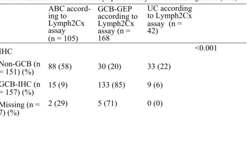
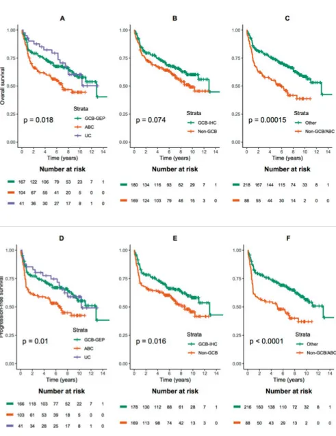
negative prognostic impact of double protein expression of MYC and BCL2 so called “double-expressor lymphoma” (DEL) was a common finding in the three papers. In paper I, we detected DEL in 27% of patients, distributed with no significant difference between the germinal center derived B-cell subgroup (GCB) in 52% of cases and the non-GCB subgroup in 37% of cases. There was no significant difference in survival between GCB and non-GCB patients. The diagnosis in most of the patients with DEL was made on core needle biopsy in this paper. This finding was more thoroughly investigated in paper III with attention paid to the site of biopsy. In paper II, we evaluated the concordance of cell of origin (COO) assignment between gene expression profile (GEP) and immunohistochemistry (IHC) to identify the best predictor of survival in a DLBCL cohort including patients from Sweden and Denmark. The overall concordance between the two methods was 83%. We found that ABC/non-GCB subtype identified by both GEP and IHC is associated with the worst outcome. This finding indicates the importance of precise risk stratification in the era of precision medicine. DEL was more common in ABC patients categorized by GEP. In paper III, we identified abdominal lymph node involvement by radiological examination in 63% of DLBCL patients with an inferior survival, adverse clinical characteristics and significantly more frequent DEL. These findings may indicate a distinct biological behavior in patients with abdominal nodal disease. In paper
IV, we demonstrated a significant association between IDO1 and PD-L1 in PCNSL patients.
This finding indicates the crucial immunosuppressive role of these two molecules. In addition, in PCNSL low frequencies of MYC and BCL2 translocations and high frequency of BCL6 translocation was observed and DEL was detected in 49% of cases. Contrary to our results in systemic DLBCL in papers I-III, there was no significant prognostic impact of DEL in PCNSL.
Maysaa Abdulla, Department of Immunology, Genetics and Pathology, Clinical and experimental pathology, Rudbecklaboratoriet, Uppsala University, SE-751 85 Uppsala, Sweden.
© Maysaa Abdulla 2020 ISSN 1651-6206 ISBN 978-91-513-0869-2
“Nothing in life is to be feared, it is only to be understood. Now is the time to understand more, so that we may fear less”
Marie Curie
Dedicated to all fighters of lymphoma
List of Papers
This thesis is based on the following papers, which are referred to in the text by their Roman numerals.
I. Abdulla M, Laszlo S, Triumf J, Hedström G, Berglund M, En-blad G, Amini RM. A population-based study of cellular markers in R-CHOP treated diffuse large B-cell lymphoma patients. Acta
Oncologica, 2016 Aug 55(9-10):1126-1131.
II. Abdulla M*, Hollander P*, Pandzic T, Mansouri L, Bram Ednersson S, Andersson P-O, Hultdin M, Fors M, Erlanson M, Degerman S, Munch Petersen H, Asmar F, Grønbæk K, Enblad G, Cavelier L, Rosenquist R**, Amini RM**. Cell-of-origin de-termined by both gene expression profiling and immunohisto-chemistry is the strongest predictor of survival in patients with diffuse large B-cell lymphoma. American Journal Hematology, 2019 Nov 95 (1):57-67.
III. Abdulla M, Guglielmo P, Hollander P, Åström G, Ahlström H, Enblad G, Amini RM. Prognostic impact of abdominal lymph node involvement in diffuse large B-cell lymphoma. Accepted for
publication in European Journal of Haematology. Published
online Nov 30th 2019.
IV. Abdulla M, Alexsson A, Sundström C, Ladenvall C, Berglund M, Mansouri L, Lindskog Bergström C, Cavelier L, Enblad G, Hollander P, Amini RM. PD-L1 and IDO1 are important immu-nosuppressive molecules to consider in primary DLBCL of the CNS. Submitted.
*Contributed equally as first author **Contributed equally as last author
Related publications
I. Abdulla M, Laszlo S, Triumf J, Hedström G, Berglund M, En-blad G, Amini RM. Core needle biopsies for the diagnosis of dif-fuse large B-cell lymphoma - a great concern for research. Acta
Oncologica, 2016 Oct 56(1):106-109.
II. Mansouri L, Noerenberg D, Young E, Mylonas E, Abdulla M, Frick M, Asmar F, Ljungström V, Schneider M, Yoshida K, Skaftason A, Pandzic T, Gonzalez B, Tasidou A, Waldhueter N, Rivas-Delgado A, Angelopoulou M, Ziepert M, Arends CM, Couronné L, Lenze D, Baldus CD, Bastard C, Okosun J, Fitzgib-bon J, Dörken B, Drexler HG, Roos-Weil D, Schmitt CA, Munch-Petersen HD, Zenz T, Hansmann ML, Strefford JC, Enblad G, Bernard OA, Ralfkiaer E, Erlanson M, Korkolopoulou P, Hultdin M, Papadaki T, Grønbæk K, Lopez-Guillermo A, Og-awa S, Küppers R, Stamatopoulos K, Stavroyianni N, Kanellis G, Rosenwald A, Campo E, Amini RM, Ott G, Vassilakopoulos TP, Hummel M, Rosenquist R, Damm F. Frequent NFKBIE deletions are associated with poor outcome in primary mediastinal B-cell lymphoma. Blood, 2016 Dec 128 (23):2666-2670.
Contents
Introduction ... 13
Epidemiology ... 13
DLBCL once upon a time ... 14
Before 1970 ... 14
Between 1970-2000 ... 14
After 2000 ... 15
DLBCL in the updated 2016 World Health Organization (WHO) Classification of Tumours of Haematopoietic and Lymphoid Tissues .... 15
Biology and Pathogenesis ... 17
Normal germinal center ... 17
DLBCL ... 17
Primary DLBCL of the CNS (PCNSL) ... 23
The role of immune system in lymphoma ... 25
Tumor microenvironment in DLBCL ... 25
Tumor microenvironment in PCNSL ... 25
Immune checkpoint pathways ... 26
Indoleamine 2, 3 dioxygenase (IDO) ... 28
Clinical aspects and diagnostic approach of DLBCL ... 29
Clinical features ... 29 Radiological examination ... 29 Histopathological examination ... 30 Prognosis ... 31 Treatment ... 32 Frontline treatment... 32
Treatment of patients with refractory/ relapse disease ... 32
Precision treatment with novel therapies in DLBCL ... 33
Immune checkpoint blockade ... 34
Future visions ... 35 Diagnosis ... 35 Treatment ... 36 Aims ... 37 Overall aim ... 37 Specific aims ... 37
Patients in papers I-IV ... 38
Tissue microarray (TMA) ... 39
Immunohistochemical stainings ... 40
Evaluation of immunohistochemical stainings ... 40
GEP in paper II ... 40
RNA extraction and NanoString assay ... 40
Radiological examination in paper III ... 41
In situ hybridization for Epstein-Barr virus-encoded RNA in paper IV .. 41
FISH for MYC, BCL2 and BCL6 in paper IV ... 41
RNA extraction and quality assessment in paper IV ... 42
Library construction and RNA sequencing ... 42
Statistical analysis ... 43
Results and discussions ... 44
Paper I ... 44
Paper II ... 46
Paper III ... 49
Paper IV ... 51
Strengths and limitations ... 53
Summary of results ... 54
Comprehensive discussion and future perspectives ... 55
Populärvetenskaplig sammanfattning på svenska ... 57
Delarbete I ... 58 Delarbete II ... 58 Delarbete III ... 59 Delarbete IV ... 59 Acknowledgements ... 60 References ... 62
Abbreviations
aa-IPI Age-adjusted International Prognostic Index ABC Activated B-cell
AID Activation-induced cytidine deaminase APC Antigen presenting cell
AS Alternative splicing
ASCT Autologous stem cell transplantation
BCLU B-cell lymphoma, unclassifiable, with features intermediate between DLBCL and BL
BCL2 B-cell lymphoma 2 protein BCL6 B-cell lymphoma 6 protein
BL Burkitt lymphoma
CAR Chimeric antigen receptor
CARD11 Caspase Recruitment Domain Family Member 11 CHL Classical Hodgkin Lymphoma
CHOP Cyclophosphamide, doxorubicin, vincristine and prednisone
CNB Core-needle biopsy COO Cell-of-origin CR Complete remission
CT Computer tomography
CTLA-4 Cytotoxic T lymphocyte-associated antigen 4 DEL Double expressor lymphoma
DHL Double-hit lymphoma
DLBCL Diffuse large B-cell lymphoma DNA Deoxyribonucleic acid
EBV Epstein-Barr virus
ESMO European Society for Medical Oncology EZH2 Enhancer of zeste homolog 2
FDA U.S. Food and Drug Administration FFPE Formalin-fixed paraffin-embedded tissues FISH Fluorescent in Situ Hybridization
FNA Fine needle aspiration GCB Germinal centre B-cell GEP Gene expression profile HGBL High-grade B-cell lymphoma HCV Hepatitis C virus
IDO Indoleamine 2, 3 dioxygenase IHC Immunohistochemistry IPI International prognostic index IRF4 Regulatory factor 4 protein LMO-2 LIM domain only-2
LSS Lymphoma-specific survival MAG Myelin Associated Glycoprotein
MSKCC Memorial Sloan-Kettering Cancer Center MYD88 Myeloid differentiation primary response 88 NGS Next-generation sequencing
NHL Non-Hodgkin lymphoma
OS Overall survival PAX5 Paired Box protein 5
PCNSL Primary DLBCL of the CNS PD-1 Programmed Death receptor 1 PD-L Programmed Death Ligand PET Positron Emission Tomography PFS Progression-free survival
PMBCL Primary mediastinal large B-cell lymphoma PRDM1 PR domain containing 1
PTEN The phosphatase and tensin homolog gene R-CHOP Rituximab-Cyclophosphamide combined with
doxorubicin, vincristine and prednisone RNA Ribonucleic acid
SEB Surgical excisional biopsy SHM Somatic hypermutation SSP1 Secreted Phosphoprotein 1 TAM Tumor associated macrophages TCR T-cell receptor
THL Triple-hit lymphomas UC Unclassifiable
WHO World Health Organization XBP1 X-box binding protein 1
Introduction
Malignant lymphomas affect about 2,000 people in Sweden every year. The-se lymphoid malignancies compriThe-se a heterogeneous group of different dis-ease entities with different features, where some patients are diagnosed with high-grade lymphomas with an aggressive clinical course that require imme-diate treatment, while others can live for decades without the need for any treatment. The process for lymphoid stem cells in the bone marrow to be-come highly specialized B- and T-lymphocytes requires several steps of differentiation. The process to become a mature and immunocompetent B- or T-cell is therefore a delicate process, where the lymphocytes are vulnerable to genetic aberrations that may develop along this long pathway of differen-tiation. Lymphomas arise from genetically transformed and clonally expand-ed lymphoid cells at different stages of differentiation. B-cells mature and differentiate in the secondary lymphoid organs like lymph nodes, whereas immature T-cells develop in the thymus. The vast majority of lymphomas are derived from transformed B-cells, whereas T-cell lymphomas constitute only about 5% of all lymphomas. All lymphomas are today classified in the updated 2016 World Health Organization (WHO) Classification of Tumours of Haematopoietic and Lymphoid Tissues, and the different entities are clas-sified mainly based on cell-of-origin (COO) in order to better understand and relate to its normal counterpart for the transformed malignant cells [1].
Epidemiology
In Sweden, the incidence of DLBCL is approximately 5.5 per 100,000, with slight male predominance [2]. DLBCL is predominant in elderly with a me-dian age of 65 year, although it also occurs in young patients and rarely in children [3].
The etiology of DLBCL is unclear in the majority of patients. However, certain factors may influence the risk of developing lymphoma as genetics, comorbid diseases or their treatments (i.e. immunosuppression), environ-mental factors such as ultraviolet radiation, pesticides, hair dyes, and diet. Several infectious organisms have been linked to the risk of lymphomas. Certain subtypes of DLBCL have been highly associated with Epstein-Barr virus (EBV) namely the immunoblastic variant and primary DLBCL of the
CNS [4]. A correlation between DLBCL and hepatitis C (HCV) has been shown [5].
An increased risk of DLBCL has been associated with autoimmune diseases such as rheumatoid arthritis, Sjogren syndrome, and autoimmune hemolytic anemia [6].
DLBCL once upon a time
Before 1970
The history of malignant lymphoma classification can be traced to the time when malignant lymphoma was divided into four types; Hodgkin’s disease, lymphosarcoma, reticulum cell sarcoma, and follicular lymphoblastoma [7]. Hodgkin’s disease was named after Thomas Hodgkin, who first described the disease in 1832 [8]. Subsequently, malignant lymphoma was found to be a heterogeneous group of neoplastic diseases of lymph nodes and to differen-tiate these conditions from Hodgkin’s disease, these were named as non-Hodgkin lymphoma (NHL).
Malignant lymphoma was first mentioned by Theodore Billroth in 1871, to describe neoplastic, infectious and miscellaneous causes of lymphadenopa-thy [9]. However, the disease was first known as “lymphosarcoma” and was recognised by Rudolf Virchow in 1865 [10].
The first attempt to classify malignant lymphoma in modern times was per-formed by Gall and Mallory in 1942 [11], however, the first well-organized classification of malignant lymphomas was proposed by Rappaport in 1956 and modified in 1966 [12]. The Rappaport classification was based on mor-phology; cell size and architecture and also included growth pattern such as diffuse, nodular and histiocytic patterns; each with subtypes [12]. DLBCL was recognized as “diffuse histiocytic lymphoma” by Rappaport [13].
Between 1970-2000
The advances in the science of immunology with the identification of lym-phocytes to be B-, T- and natural killer lymlym-phocytes have provided new insights into the classification of lymphoma. The efforts on both sides of the Atlantic Ocean took a step forward and classification of lymphoma based on morphology and immunology was developed in 1974 by Karl Lennart in Europe (Kiel classification) and Lukes and Collin in USA [14, 15]. DLBCL synonyms were known as centroblastic, B-immunoblastic, large cell anaplas-tic (B-cell) in Kiel classification and large cleaved or large non-cleaved fol-licular center cell (FCC), B-immunoblastic in Lukes-Collins classification.
The different terms used by different classifications and the questionable clinical relevance between the clinicians led to the creation of a working formulation in 1982 in order to interpret these classifications for clinical usage [16]. The working formulation was favored in the USA while in Eu-rope, the Kiel classification was still in use [17]. DLBCL was known as dif-fuse large cell cleaved, non-cleaved or immunoblastic; occasionally difdif-fuse mixed small and large cell in the Working Formulation.
The International Lymphoma Study Group Founded in 1990 by Stein and Isaacson, tried to review and unify the Kiel, Lukes and Collins and the Working Formula classifications. These collaborated efforts led to the de-velopment of the revised European-American classification of lymphoid neoplasms (REAL) classification of lymphoma that was published in 1994. The meeting concluded that lymphomas should be classified according to their normal counterpart and include clinical presentation, morphology, im-munophenotype and genetic information [18]. Certain clinically relevant subtypes of DLBCL were identified including Large B-Cell Lymphoma Subtype; Primary Mediastinal (Thymic) Large B-Cell Lymphoma, Burkitt's Lymphoma and a Provisional Entity: High-Grade B-Cell Lymphoma, Bur-kitt-Like.
After 2000
The World Health Organization (WHO) decided to update the classifications of haematopoietic and lymphoid tissue tumors shortly after the publication of REAL classification. This project was adopted by the Society for Hematopathology and European Association of Hematopathology. The WHO classification was developed over a period of seven years and pub-lished in 2001 and updated in 2008 and 2016 [1, 19]. Different entities were established and provisional categories of NHL based on cell lineage and differentiation were described. At least 80 different entities of NHL are described in the WHO classification, each with specific clinical features, immunophenotype, molecular and genetic alterations [1].
DLBCL in the updated 2016 World Health Organization
(WHO) Classification of Tumours of Haematopoietic
and Lymphoid Tissues
The 2016 WHO classification defines DLBCL as the most common type of high-grade B-cell lymphoma with heterogeneous clinical, morphologic, im-munophenotypic, cytogenetic and molecular features [20, 21]. Biological and clinical studies have subdivided DLBCLs into specific subtypes and/or variants (Table 1) [22]. Some of these defined subtypes or variants of large
B-cell lymphomas represent tumors that arise in a particular site, such as primary mediastinal B-cell lymphoma (PMBCL), primary DLBCL of the CNS (PCNSL), intravascular large B-cell lymphoma and DLBCL, leg type [20, 22].
However, there is still a substantial number of DLBCL cases that have no clearly accepted criteria for subdivision, and these are classified as DLBCL, not otherwise specified (NOS) [3, 22, 23]; DLBCL, NOS account for about 30% of adult non-Hodgkin lymphoma [23].
Few changes of the categories of DLBCL were made in the 2016 WHO clas-sification from 2008 WHO clasclas-sification; mainly including the COO classi-fication in the pathology report i.e. germinal center B-cell type (GCB-DLBCL) versus activated B-cell type (ABC-(GCB-DLBCL), and the identification of the double expression of MYC and BCL2 assessed by immunohistochem-istry (IHC) as a poor prognostic indicator. Another major change is the emergence of two new categories to replace the provisional category “B-cell lymphoma, unclassifiable, with features between DLBCL and Burkitt lym-phoma” (BCLU). These are high grade B-cell lymphoma, with MYC and
BCL2 and/or BCL6 translocations (double-hit or triple-hit lymphoma) and
high grade B-cell lymphoma, not otherwise specified.
Table 1. High-grade B-cell lymphomas (2016 WHO) Classification of Tu-mours of Haematopoietic and Lymphoid Tissues)
DLBCL, NOS
T-cell/histiocyte rich large B cell lymphoma Primary DLBCL of the CNS
Primary cutaneous DLBCL leg-type EBV-positive DLBCL, NOS
Large B cell lymphoma with IRF4 rearrangement Primary mediastinal (thymic) large B cell lymphoma Intravascular large B cell lymphoma
DLBCL associated with chronic inflammation HHV8 positive diffuse large B cell lymphoma Primary effusion lymphoma
High-grade B-cell lymphoma
High-grade B-cell lymphoma, with MYC and BCL2 and/or BCL6 rearrangements
High-grade B-cell lymphoma, NOS
B-cell lymphoma, unclassifiable, with features intermediate between DLBCL and classical Hodgkin lymphoma
Plasmablastic lymphoma
Biology and Pathogenesis
Normal germinal center
Normal B-cells arise in the bone marrow and undergo primary rearrange-ment of their immunoglobulin genes before antigen exposure. They migrate from the bone marrow carrying surface immunoglobulin receptors toward the peripheral lymphoid organs where they encounter antigens and produce high-affinity antibodies [24]. The formation of germinal center in the lym-phoid organs occurs when the antigen induces a T-dependent antibody re-sponse. Two genetic processes take place in the germinal center. These are the somatic hypermutation and switch recombination and both need double-stranded DNA breaks and the presence of activation-induced cytidine deam-inase (AID). B-cell differentiation through the germinal center is controlled by a number of key transcription factors, including BCL6, PRDM1, IRF4, and XBP1. B-cells that have high-affinity antibodies on their surface survive, they re-express BCL2 and exit the germinal center and differentiate into either mature plasma cells or long-lived memory B-cells to be part of the humoral immunity [24]. However, this complex biology is not perfect and errors can happen. Unfortunately, some of these errors may lead to the de-velopment of B-cell lymphomas [24].
DLBCL
Morphology
Morphologically, the 2016 WHO Classification of Tumours of Haematopoi-etic and Lymphoid Tissues defines DLBCL as a diffuse growth of neoplastic large lymphoid cells of B-cell origin with a nuclear size equal to or exceed-ing normal macrophage nuclei cells that totally or partially efface the normal architecture of lymph node or extranodal tissue (Figure 1) [22]. Variable numbers of reactive T-cells and histiocytes are present. Sclerosis and or geo-graphical necrosis may be present. Apoptosis and increased mitotic rates are high, and about 10% of DLBCL may be associated with starry sky pattern. The WHO classification describes a number of morphological variants of DLBCL: centroblastic, immunoblastic, and anaplastic variants. In addition, rare morphological variants have been described in the literature: lymphoma cells may have multilobated nuclei, small size lymphoma cells (small cen-troblastic), signet ring appearance (like gastric carcinoma), spindle-cell ap-pearance (like sarcoma), or lymphoma cells may have cytoplasmic granules or microvillous projections or intracellular junctions. Inferior prognosis has been reported by some studies in immunoblastic variant [25, 26] and also in certain subsets of anaplastic variants that carry TP53 [27].
Figure 1. DLBCL characterized by diffuse infiltration of large neoplastic B-cells; A: Haematoxylin and eosin (HE), B: CD20.
Gene expression profile (GEP)
Alizadeh and colleagues were among the first to use the gene expression profile (GEP) to identify two distinct molecular subtypes of DLBCL, namely GCB, and ABC subtypes, which expressed genes characteristic of their re-spective COO, with 15% remaining unclassifiable group (UC) [28]. This molecular distinction provides prognostic and predictive information with the GCB subtype exhibiting a better outcome than ABC in DLBCL patients treated with CHOP. This observation was confirmed subsequently in patients treated with R-CHOP [29]. In addition, using the GEP to understand the pathogenesis of DLBCL plays a promising role in the selection of targeted therapies [22, 28-30].
GEP is a reliable method for the identification of the GCB and ABC molecu-lar subtypes of DLBCL; however, it is not a routinely available test for clini-cal use due to the lack of a standardized commercially available test and the requirement for fresh-frozen tissue specimens.
Several studies assessed the use of IHC as an alternative and practical tool to determine the COO, and different algorithms have been developed with dif-ferent levels of concordance with GEP [31, 32]. The most commonly used algorithm developed by Hans et al. defines cases as GCB and non-GCB based on three IHC markers (CD10, BCL6 and MUM1) [33].
It is acknowledged that the IHC algorithms do not recognize the UC subtype that represents 10 to 15% of tumors according to GEP, thus they do not cor-relate exactly with the molecular categories, and are not uniformly reported to have prognostic utility and nor do they determine therapy. However, since GEP is not available as a routine clinical test, the use of IHC algorithms is considered to be acceptable [20].
In the recent years new methods based on the quantification of RNA tran-scripts extracted from formalin-fixed paraffin-embedded tissues (FFPE) have been developed and provide results concordant with conventional microarray GEP, are reproducible between laboratories, and capture the prognostic im-pact of the COO classification. The Lymph2X is one of these methods that is based on NanoString technology and includes 20 genes in the panel to de-termine COO. These methods are still not accessible to most laboratories but may represent a promising alternative to current IHC-based algorithms [30, 34-38].
GCB-DLBCL
GCB-DLBCLs are believed to originate from lymphoid cells residing in the germinal center and express CD10, BCL6 and LMO-2 [39]. BCL6 is charac-teristically upregulated in GCB-DLBCL. BCL6 represses the transcription of several genes including TP53 tumor suppressor, which controls DNA dam-age-induced apoptotic response, allowing germinal-center cells to tolerate the physiological DNA breaks required for immunoglobulin class switch recombination and somatic hypermutations [40]. Chromosomal transloca-tions involving BCL6 are found in 40% of GCB-DLBCL cases, but other mechanisms leading to BCL6 upregulation have been reported, such as so-matic mutations [41, 42]. The GCB subtype has more often translocations of
BCL2, MYC, mutations involving histone methylation or acetylation like EZH2, EP300, CREBBP, KMT2D (figure 2) [39, 43, 44], and mutations
in-volving B-cell homing like GNA13, GNA12, SIPR2 [45], mutations involv-ing PI3K pathway signalinvolv-ing, and JAK-STAT pathway [46].
ABC-DLBCL
ABC-DLBCLs are considered to originate from B-cells at a plasmablastic stage, just prior to germinal center exit [39] and express a plasma-cell like transcription program.
The ABC subtype has more often genetic abnormalities that activate the B-cell receptor signaling and the Toll-like receptor signaling pathways, result-ing in activation of NF-κB (nuclear factor kappa-light-chain-enhancer of activated B-cells) signaling pathway like mutations of TNFAIP3, CARD11,
CD79B and MYD88 (figure 2) [47-49].
Upon activation, the NF-kB pathway promotes cell survival, proliferation and inhibition of apoptosis, as well as driving the cell towards plasma cell differentiation [47, 50, 51].
Both subtypes of DLBCL share overexpression of antiapoptotic protein BCL2, although due to different mechanisms. In GCB-DLBCL, overexpres-sion of BCL2 is largely due to presence of the t(14;18), whereas in ABC DLBCL it is caused by other mechanisms, such as transcriptional upregula-tion and gene amplificaupregula-tion [52].
Unclassifiable (UC)
The GEP of UC subtype is still obscure. While some studies presented UC as having a similar outcome to ABC-DLBCL [29, 53], a recent study demonstrated that a concomitant NOTCH2 mutations and BCL6 translocations is common in this subtype and is associated with a favorable outcome [54].
Figure 2. Key oncogenic pathways in DLBCL. Figure adapted from Sehn et al, Blood 2015 [39] with permission.
Monti and colleagues subdivided DLBCL into three groups by using GEP data. These are: oxidative phosphorylation, B-cell receptor/proliferation, and host response [55]. The oxidative phosphorylation group includes tumors carrying t(14; 18) and tumors with apoptotic pathway defects. The B-cell receptor/proliferation includes tumors carrying BCL6 translocation and may overlap with ABC subtype. The host response group has a T-cell and den-dritic cell signature and probably includes cases of T-cell/histiocyte rich large B-cell lymphoma.
Dybkaer and colleagues assigned B-cell-associated gene signature (BAGS) based on normal B-cell subset phenotypes and identified five subtypes: na-ive, centroblast, centrocyte, memory, and plasmablast B-cell subtypes [56]. These signatures provided additional prognostic significance to the molecu-lar ABC/GCB subtypes where the GCB-centroblast subtype had inferior prognosis compared to the GCB-centrocyte subtyp [56].
Next generation sequencing (NGS)
The next generation sequencing (NGS) enables our understanding of DLBCL heterogeneous genetic pathogenesis as well as provides potential targets for therapeutic agents. The whole genome sequencing allows high throughput DNA sequencing and provides the opportunity to determine the pattern, frequency, and location of somatic point mutations across the entire genome.
An average of 30 to > 100 genetic mutations per neoplasm have been identi-fied in DLBCL [57-59]. Data indicate that DLBCL cells undergo multiple rounds of clonal expansion and that gene mutations can happen at any time point during this process [45]. These mutations are divided into driver and passenger mutations. The driver mutations occur early in DLBCL pathogen-esis and aid in progression of the disease, and they appear in most or all sub-sequent clones and have therefore the potential to be the best therapeutic targets [60]. On the other hand, the role of passenger gene mutations in DLBCL pathogenesis is not known yet. A challenge that makes highly mu-tated alleles appear to be drivers when they are in fact passengers mumu-tated by aberrant somatic hypermutation is due to the effect of the enzyme AID [61-65]. The most well-defined AID induced gene mutations by aberrant somatic hypermutation include BCL2, BCL6, MYC, RHOH/TTF, PIM1, PAX5, IRF4,
ST6GAL1, BCL7A, CIITA, LRMP, and SOCS1 [66].
Chapuy and colleagues have proposed five distinct DLBCL subsets with different pathological mechanisms and outcome (clusters 1-5) [46]. Three of these have previously been undescribed, a low-risk ABC-DLBCL group (C1); poor risk GCB-DLBCLs with BCL2 SVs and alterations of PTEN and epigenetic enzymes (C3), a newly defined group of good-risk GCB-DLBCLs with distinct alterations in BCR/PI3K, JAK/STAT, and
BRAF pathway components and multiple histones (C4). In addition, a
COO-independent group of tumors with biallelic inactivation of TP53, 9p21.3/CDKN2A and associated genomic instability (C2), and a group in which the genetic signature is associated with extranodal tropism and with frequent BCL2 gain, concordant MYD88CD79B mutations and additional mutations of ETV6, PIM1, GRHPR, TBL1XR1 and BTG1 (C5). Schmitz and colleagues identified four distinct genetic subtypes in DLBCL. These are: MCD with co-occurrence of MYD88 and CD79B mutations, BN2
with BCL6 fusions and NOTCH2 mutations, N1 with NOTCH1 mutations, and EZB with EZH2 mutations and BCL2 translocations. These groups have different gene-expression signatures and respond differently to immu-nochemotherapy, where the BN2 and EZB subtypes have favorable outcome and the MCD and N1 subtypes are associated with inferior outcomes [54]. Rearrangement and expression of MYC, BCL2, and BCL6
MYC is a transcriptional factor located at 8q24 and is essential for normal B-cell growth, proliferation, and apoptosis [67, 68]. The dysregulation of MYC is essential to induce lymphomagenesis in B-cell lymphomas. MYC rear-rangement is found in 5 to 15% of DLBCL. BCL2 is an apoptosis regulator factor that inhibits the apoptotic death of cells and is located at 18q21. BCL2 rearrangement is observed in nearly 30% of DLBCL with GCB subgroup and around 5% in ABC subgroup. BCL6 is a sequence-specific repressor of transcription, and has been shown to modulate the transcription of STAT-dependent IL-4 responses of B-cells. The BCL6 gene is located at 3q27.
Double hit lymphoma
MYC translocation can occur in association with BCL2 translocation and/or,
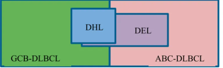
to a lesser extent, BCL6 translocation, in the so called double-hit lymphoma (DHL) or triple-hit lymphomas (THL), which represent a new category of high-grade B-cell lymphoma in the updated 2016 WHO Classification (High-grade B-cell lymphoma with MYC and BCL2 and/or BCL6 rearrange-ments) [69]. These lymphomas which were first defined by Aukema and colleagues in 2011 [70] represent about 10% of high grade B-cell lympho-mas and are highly aggressive, with advanced stage disease, extranodal in-volvement, high serum lactate dehydrogenase (LDH) level, high-intermediate to high International prognostic index (IPI) score and with high failure rate with most treatment protocols [71, 72]. Interestingly, DHL and THL with rearrangement of MYC/ BCL2 respective MYC/BCL2/ BCL6 be-long almost always to the favorable GCB subtype (figure 3) [73].
DHL with rearrangement of MYC and BCL2 is the most common type of DHL and represents about 65% of all cases, while triple-hit lymphoma (THL) with rearrangement of MYC, BCL2 and BCL6 represents about 20% and DHL with rearrangement of MYC and BCL6 represents about 15% of DHL cases [71]. There are other types of DHL that have been proposed, of these double rearrangement of BCL2 and BCL6 [74], MYC and CCND1 [1], and MYC and TP53 [75, 76].
In addition to rearrangement, extra copies of MYC/BCL2 have been demon-strated in DLBCL. Poor prognosis is reported in DLBCL with double extra copies of MYC/BCL2 without rearrangement or when there is rearrangement of MYC and extra copies of BCL2 or the reverse i.e. MYC extra copies with rearrangement of BCL2 and those named atypical double-hit lymphomas to
differentiate them from the typical double-hit lymphomas with rearrange-ment of MYC and BCL2 [77, 78].
Double expressor lymphoma
The use of reliable antibodies to assess MYC and BCL2 over-expression by IHC with cut-offs of 40% for MYC and 50-70% for BCL2 has identified double expression of these two proteins in 20 to 35% of DLBCL cases and is more common in ABC subtype (figure 3) [79]. Most of these tumors do not carry MYC/BCL2 chromosomal alterations and have been named double-expressor lymphoma (DEL) in contrast to double-hit lymphoma. DELs have inferior outcomes compared to other DLBCL, NOS, but they are not as ag-gressive as the HGBL, with rearrangements of MYC and BCL2 and/or BCL6 [80]. In the WHO 2016 Classification, it is suggested that double expression of MYC and BCL2 proteins without gene aberrations should be considered a prognostic indicator in DLBCL, NOS but not as a separate category [20].
Figure 3. The distribution of DHL and DEL in GCB and ABC subtypes of DLBCL.
Primary DLBCL of the CNS (PCNSL)
PCNSL is a distinct entity of DLBCL that predominantly affects elderly patients [20]. It accounts for 2-3% of all NHL with increasing incidence in the last three decades [81-83]. Despite improved treatment results for sys-temic DLBCL, PCNSL still has a dismal outcome [82, 84, 85].
Pathogenesis
In contrast to systemic DLBCL in which GEP has identified two main sub-types, the GCB and the ABC, most of the cases of PCNSL belong to the ABC subtype, which may explain the poor prognosis of this distinct entity [86].
Our understanding of the pathogenesis of PCNSL is slowly growing and is challenging due to the rarity of the disease and also on account of the sparse tissue sampling which may limit the powerful work up of most of the single cohort studies and indicates the importance of multicenter collaboration.
ABC-DLBCL GCB-DLBCL
A “CNS signature” that can replace the predictive and prognostic role of COO is still largely unclear. Molecular investigations could identify high frequency of mutations in specific genes involved in important pathways that could be the driver mutations responsible in PCNSL tumorigenesis such as
MYD88, CD79A and the CARD11 [87-90], dysregulation of factors involved
in JAK/STAT pathways such as IL-4, IL-10, and intratumoural JAK1[91-94], recurrent chromosomal losses affecting the 6q, 6p21.32 (HLA locus) and copy number alterations (CNAs) such as loss of 9p21.3 (CDKN2A) or gain of 9p24.3 [95, 96]and copy number alterations of 9p.24/PD-L1/PD-2 [97].
However, there are still pitfalls that need to be overcome like the mystery why this lymphoma entity develops and is confined to the CNS where very few B-cells, if any, are found under normal circumstances [98], and also whether the B cells home to the CNS in a benign or malignant state.
Malignant transformation may occur in the CNS or outside it. Three hypoth-esized mechanisms have been suggested by Deckert and colleagues [99]. First, a transformation of B-cells happens outside the CNS and is dissemi-nated but may be elimidissemi-nated outside the CNS by a specific antitumor im-mune response, whereas they escape the imim-mune response in the CNS due to the immunoprivileged status of the CNS. Second, transformation of B-cells happens outside the CNS and malignant B-cells acquired a high affinity and tropism for the CNS, which make them home to the CNS. Third, non-malignant B-cells enter the CNS either due to an immune response such as a pathogen inducing inflammation or accidently when the blood-brain barrier permeability is increased. The B-cells may persist in the brain and they can transform in the CNS leading to the development of lymphoma. The molecu-lar clues of tropism and selective dissemination of DLBCL within the brain have been studied in recent years and probably gene alterations of SPP1 and
MAG are two of the most prominent gene alterations that have been shown
to be involved in the pathogenesis of PCNSL [100].
SPP1 is a secreted non-collagenous, chemokine-like glycoprotein and has numerous cellular functions, including cell communication, focal adhesion, immune cell activation, and immune cell migration [91]. SPP1 is thought to be involved in CNS tropism, B-cell migration, proliferation and aggressive clinical behavior [100]. MAG is a cell membrane glycoprotein and member of the immunoglobulin superfamily. MAG regulates the interaction of mye-lin and axons, such as the initiation of myemye-lination and the maintenance of myelin integrity, by expression on periaxonal myelin membrane of the CNS and peripheral nervous system [101, 102]. MAG is suggested to play an im-portant role in perineural cancer invasion [100, 103].
The role of immune system in lymphoma
Tumor microenvironment in DLBCL
The tumor microenvironment of B-cell lymphomas is characterized by vari-ably robust numbers of immune cells, stromal cells, blood vessels and extra-cellular matrix [104]. The interaction of tumor cells with their microenvi-ronment is fundamental for tumor cell survival in B-cell lymphoma [105, 106]. The GEP of non-malignant cell populations in DLBCL revealed mo-lecular signatures that correlated significantly with survival after treatment [105]. A stromal-1 gene expression with favorable prognosis reflects extra-cellular matrix deposition and infiltration of the tumor by macrophages and a non-favorable stromal-2 gene expression was associated with endothelial cells and angiogenesis [105]. These stromal signatures are independent of COO, as they can be seen in both GCB and ABC subtypes [39]. This may explain the inhomogeneous outcome in homogenous cases of GCB and ABC subtypes of DLBCL [28].
Numerous aberrations have been recognized in tumor cells in B-cell lym-phoma, which affect interaction between tumor cells and the tumor microen-vironment. Recurrent gene mutations, copy number aberrations and translo-cations in the lymphoma cells are examples of aberrations that reflect the significance of these interactions as important mechanisms of oncogenesis in many of the lymphoma subtypes [104].
Tumor microenvironment in PCNSL
Tumor cells in PCNSL infiltrate brain parenchyma with a variably heavy inflammatory response with tumor-associated macrophages (TAM) consti-tute a major stromal component as do reactive T-cells [107]. An interaction between the enhanced production of signal transducer and the activator of transcription 3 (STAT3) by malignant B-cells and dense infiltration by CD163-positive macrophages (M2) in the CNS microenvironment has been demonstrated in PCNSL and proposed to have an important role in patho-genesis of PCNSL [107]. STAT3 plays an important role in tumor progres-sion by inducing anti-apoptotic proteins such as BCL2 [108] and also by allowing tumor cells to escape the immune system by inducing immunosup-pressive molecules such as IL-6, IL-10 and transforming growth factor β [109, 110]. It has been reported that higher expression of STAT3 on lym-phoma cells predicts an inferior prognosis in DLBCL [111].
Reactive T-cell infiltrates are present to varying degrees either in the form of scattered lymphocytes or perivascular cuffing occurring alone or in between the vessel wall and malignant cells (reactive perivascular T-cell infiltrate that is shown to be a predictive of favorable outcome (RPVI) [112, 113].
Immune checkpoint pathways
The immune checkpoint pathways are critical for the maintenance of self-tolerance and also for the protection of tissues from damage when the im-mune system responds to pathogens under normal physiological conditions [114]. There are different immune checkpoint pathways, of these, Cytotoxic T lymphocyte-associated antigen 4 (CTLA-4) and Programed Death-1 (PD-1) pathways that block the T cell-mediated immune system at different lev-els. CTLA-4 essentially regulates initial T-cell activation, whereas PD-1 mainly regulates the immune attack in peripheral tissues at the site of im-mune effector response [115].
The T-cell is part of the acquired immune system that provides lifelong pro-tection against pathogens. The activation of T-cell reaction is triggered by the recognition of an antigen presented on the major histocompatibility com-plex (MHC) by the T-cell antigen receptor (TCR). Recognition at the site of inflammation results in an attack against infected tissue. However, in the steady state, all the nucleated cells express self-antigens on MHC and can potentially activate self-reactive T-cells. Most self-reactive T-cells are elimi-nated in the thymus through a mechanism called “negative selection”; though, many of them escape selection and are present in the periphery and can lead to an attack on self-tissue, leading to autoimmunity. Thus, the im-portance of immune checkpoint pathways that prevent activation of self-reactive T-cells in the periphery.
To complete the T-cell activation, another set of signals is required, called costimulatory molecules which are mainly provided by activated antigen-presenting cells (APCs). The best-characterized costimulatory system is the CD28 receptor on T-cells and its ligands, namely CD80 and CD86 on acti-vated professional APCs (dendritic cells, macrophages, and B-cells). Upon exposure to antigen, the APCs are activated and CD80 and CD86 are up-regulated which in turn trigger CD28 receptor on T-cells leading to T-cell activation, proliferation, cytokine production, and development of effector functions [116]. The CTLA-4, which has similar structure to CD28, binds to CD80 and CD86 with greater avidity than CD28 and this leads to suppres-sion of T-cell responses [117].
The CTLA-4 is a protein receptor for the APCs protein ligands CD80 and CD86 and it belongs to the immunoglobin superfamily of proteins. CTLA-4 is encoded by the CTLA-4 gene on chromosome 2 and expressed mainly in T-cells upon activation [118]. The dual physiological role of CTLA-4 by down modulation of helper T-cell activity and enhancement of regulatory T- cell immunosuppressive activity has been a subject of interest for many re-search studies, to investigate the antitumor effect of CTLA-4 blockade, mainly in solid cancer [119, 120]. Few studies have shown that CTLA-4
may have an essential role for B-cell lymphoma proliferation and survival [121, 122].
PD-1 (CD279) is a protein receptor for the APCs ligands PD-L1 (CD274) and PD-L2 (CD273) and it belongs to the immunoglobin superfamily of proteins CD28/CTLA-4 [123]. PD-1 is encoded by the PCCD1 gene on chromosome 2 and is expressed on activated CD4+ and CD8+ T-cells, NK cells, B-cells, macrophages and some types of dendritic cells (DCs) [123, 124].
Upon activation, PD-1 interacts with its ligands; PD-L1 and/or PD-L2 result-ing in inhibition of cell proliferation and production of cytokines, namely IL-2, IL-4, interferon (IFN)-γ, and IL-10 [125].
As previously mentioned in this section, the role of PD-1 as an immune checkpoint pathway is mainly to regulate the immune attack in peripheral tissues at the site of immune effector response. However, it is assumed that PD-1 modulates the immune response in initial phases of T-cell activation too. It is believed that T-cells that are stimulated in the absence of the CD28 mediate an incomplete T-cell activation and shift to an unresponsive state called “clonal anergy” and become refractory to further stimulation by the same antigen [126]. However, other studies have demonstrated that the in-teraction of PD-1 with its ligand PD-L1 results in the so-called “clonal aner-gy” and maintenance of this phase [127-129]. In addition, PD-1 is suggested to be involved in the regulation of innate immune cells [130].
The PD-1 ligands: PD-L1 and PD-L2 are encoded by CD274 gene and CD273 gene respectively, both located on chromosome 9. PD-L1 is ex-pressed on a wide range of cell types including epithelium, muscle, mesen-chymal stem cells, T- and B-cells, DCs, macrophages, and cancer cells, while PD-L2 expression is more restricted and expressed on immune-related cells such as DCs, macrophages, and mast cells [131].
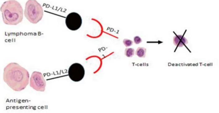
In cancer, the binding of PD-L1 or PD-L2 to its receptor PD-1 inhibits the proliferation of activated T-cells and allows the tumor cells to escape the antitumor adaptive immune response (figure 4) [132].
High expression of PD-1 on leukocytes and PD-L1 on tumor cells is demon-strated to be associated with inferior outcome in solid malignancies [133]. However, the prognostic impact of PD-1 and its ligands PD-L1 and PD-L2 is still unclear in hematological malignances and several studies with small and large cohorts have been published in the recent years with variable results. Increased numbers of PD-1 positive leukocytes in DLBCL were shown to be associated with superior outcome in two studies [134, 135] whereas in one study on PCNSL, PD-1 was an indicator of poor survival [136]. High ex-pression of PD-L1 on tumor cells was reported to be associated with inferior
outcome in DLBCL [137, 138], while high expression of PD-L1 on leuko-cytes had no prognostic impact in DLBCL and in PCNSL [137, 139]. Other studies reported favorable prognostic impact associated with high PD-L1 expression on leukocytes [140, 141]. A recent study on DLBCL reported variable results with adverse prognosis associated with increased numbers of PD-1 positive leukocytes and with PD-L1 on leukocytes, while high PD-L2 expression on tumor cells was reported to be associated with better outcome [142]. The diverse results between the above different studies may be partly explained by the use of different antibodies and different methods for eval-uation of PD-1, PD-L1 and PD-L2 expression.
In addition to the PD-1 and CTLA-4 pathways, there are numerous other immune checkpoint pathways that can be targeted clinically with monother-apies and in treatment combinations like Lymphocyte activation gene 3 (LAG3), T-cell immunoglobulin-3 (TIM-3) and B- and T- lymphocyte at-tenuator (BTLA) [131, 143, 144].
Figure 4. The activated T-cells are deactivated via the PD-1 pathway by interaction of PD-1 with its ligands PD-L1/ PD-L2 on antigen presenting cells (APC) and lym-phoma B-cells in DLBCL.
Indoleamine 2, 3 dioxygenase (IDO)
IDO is an intracellular cytosolic enzyme that is encoded by the IDO1 gene, located on chromosome 8 [145]. The IDO enzyme is expressed by different types of cells such as endothelial cells, APC, fibroblasts, macrophages and DCs [146, 147]. There are two types of IDO enzyme in the human body, IDO1 and IDO2. IDO2 is more restrictedly expressed than IDO1 and has only 3-5% the enzymatic activity of IDO1 [148, 149] and it is not as well investigated as IDO1.
The role of IDO in immune tolerance was demonstrated by Munn, Mellor and their colleagues who presented IDO expression by placenta cells to vent maternal T-cells from destroying the fetus during pregnancy, thus
pre-venting fetus rejection in utero [150-152]. It is assumed that the same con-cept might explain the role of IDO in the immune escape of tumor cells based on the preferential sensitivity of T-cells to tryptophan deprivation [153].
The initial observation of IDO association with cancer was first reported in 1956 when catabolism of tryptophan was found to be elevated in bladder cancer [154], but the impact of this association was obscure. IDO is involved in the kynurenine pathway and is responsible for degrading the essential amino acid tryptophan into L-kynurenine [155]. Tryptophan deprivation results in blockade of cell proliferation by inducing cell cycle arrest of T-cells and increases their apoptosis [156]. In addition, increasing amounts of tryptophan metabolites, especially Kynurenine, are found to be toxic to lym-phocytes and this activates the transcription of the aryl hydrocarbon receptor (AHR)[157] which in turn induces the CD4+ T-cells differentiation to immu-nosuppressive regulatory T-cells [158].
Clinical aspects and diagnostic approach of DLBCL
Clinical features
Most DLBCL patients present with a rapidly growing mass involving lymph node and or extranodal site [159]. Approximately 40% of cases manifest as extranodal disease [160] and 50% present with stage III-IV disease. About one-third of patients present with at least one of the B-symptoms: night sweats, fever, weight loss. Serum LDH and beta-2-microglobulin are often increased above normal level.
Radiological examination
Positron emission tomography (PET) in combination with contrast enhanced computer tomography (CT) is superior to either PET or CT alone [161]. PET/CT is a highly sensitive and specific imaging study for the assessment of extent of disease for staging and assessment of response to treatment [162]. However, a contrast enhanced CT scan is also recommended if meas-uring size of nodes is important [163].
Numerous studies have raised the superior benefit of radiological examina-tion compared to bone marrow biopsy (BMB) for staging of DLBCL [164-166]. However, the histology of bone marrow involvement is of prognostic significance, in which concordant involvement i.e. bone marrow involve-ment by DLBCL predicts a worse overall survival while discordant in-volvement i.e. bone marrow inin-volvement by low grade B-cell lymphoma has no prognostic impact [167-169], hence BMB remains to be a standard test for staging of DLBCL particularly when PET is negative or not available.
Recent studies have demonstrated that PET is a prognostic indicator irre-spective of IPI and COO and can predict survival and response to immu-nochemotherapy in DLBCL patients [170, 171].
Histopathological examination
The primary aim of histopathological classification of lymphoma is to identi-fy morphological, immunophenotypical and genetic characteristics that can identify lymphoma subtypes with different clinical behaviors in order to facilitate risk-adapted stratification of treatment [172, 173].
According to the WHO, accurate diagnosis of lymphoma is established on morphology, IHC, and flow cytometry reviewed by an experienced hemato-pathologist [1]. According to European society for medical oncology (ESMO) guidelines a surgical excisional biopsy (SEB) is preferred to pro-vide adequate tissue for these examinations and for further molecular studies to accurately categorize the lymphoma. However, a core-needle biopsy (CNB) can be considered when excisional biopsy is not possible [174] such in patients with deeply located lymph node or extranodal mass as in the ab-dominal cavity or mediastinum in which SEB is difficult to perform or in patients with comorbidity and where surgery would entail excessive risk. A fine-needle aspirate (FNA) should not be used as the only primary assess-ment analysis for diagnosis of DLBCL [174]. In a multi-institution clinical study very poor accuracy between FNA and SEB was shown with only 12% accuracy suggesting that FNA is neither useful for primary diagnosis of lymphoma, nor cost effective, and in addition it may misguide treatment [175]. SEB allows evaluation of lymph node architecture, growth pattern and cellular composition which are mandatory for accurate diagnosis and classi-fication of lymphoma [1].
CNB and FNA are said to be safe, fast, and cost-effective and are regarded by many studies to be an appropriate alternative to SEB to evaluate lym-phoma patients. However, the safety discussed in these studies was regard-ing the procedure itself rather than the safety of acquirregard-ing correct diagnosis and establishing appropriate treatment. The cost-effectiveness is also a ques-tionable matter, it is important to evaluate the quality of material obtained by CNB and how often these biopsies are retaken to achieve an accurate judg-ment. Repeated procedures and delay in diagnosis and treatment are not cost-effective.
Frederiksen and colleagues reviewed 42 publications between 1989 and 2012 which were aimed at studying the effectiveness of CNB and FNA in the diagnosis of lymphoma. The review concluded that FNA and CNB need-ed be followneed-ed by a SEB to achieve definitive diagnosis and classification of lymphoma in 25% to 35% of cases and that the role of FNA and CNB in
lymphoma should be reserved for the staging and assessment of recurrent disease rather than primary diagnostic process [176].
The main goal of the accurate primary diagnosis of lymphoma is to establish appropriate treatment which is often urgent. CNB and/or FNA are fast but the more important question is whether they are correct. It is alarming to see that there are hundreds of studies that suggest CNB and FNA as appropriate tools for the diagnosis of lymphoma and they have become the frontline diagnostic procedures in the recent years [177, 178]. However, most of these studies are single-center studies and they lack the discussion of the follow up and outcome of patients included in these studies.
Prognosis
DLBCL is a heterogeneous group of aggressive B-cell lymphomas with var-iable clinical course and outcome. In the era of rituximab, the disease is cured in about 60%-70% of patients. The remaining 30-40% patients are either refractory or relapse shortly after remission. The recognition of pa-tients within the curative or refractory group at time of primary diagnosis is of paramount importance for the choice of accurate treatment. There are different clinical and biological prognostic parameters that have been devel-oped to distinguish patients in either group.
The IPI is the most commonly used score since it was designed in 1993 [179]. The IPI score includes five variables: age (>60), Eastern Cooperative Oncology Group performance status (ECOG) (0-1, 2-4), serum LDH level (>normal level), number of extranodal sites (0-1, ≥2), and Ann Arbor stage (stage I-II, III-IV). DLBCL patients were divided into four risk groups ac-cording to their IPI score: low, low-intermediate, high-intermediate, and high risk. However, in the era of rituximab, the outcome of the patients im-proved and the utility of IPI needed to be justified to maintain its powerful stratification. An age adjusted IPI has been suggested (aa-IPI), which in-cludes three parameters for each age category (over or under 60 years): LDH (≤ the upper normal limit (UNL) vs > UNL; Ann Arbor stage (≤ 2 vs >2) and ECOG (≤ 1 vs > 1).
The National Cancer Center Network (NCCN) demonstrated that a more appropriate split of age into four categories (< 40, 40–60, 61–75 and > 75-years-old) and LDH into three categories (≤ UNL, UNL-3xUNL and > 3xUNL) could better stratify risk DLBCL patients receiving chemoimmuno-therapy [180]. In the NCCN-IPI, specific extranodal sites are of more prog-nostic significance than the number of extranodal sites. Disease in bone mar-row, central nervous system, liver, gastrointestinal tract or lung were shown to be relevant parameters associated with prognosis [180].
In PCNSL, Memorial Sloan-Kettering Cancer Center (MSKCC) is used to identify three prognostic groups based on age and Karnofsky performance status (KPS): class 1 includes patients <50 years; class 2 includes patients ≥50 years and KPS≥70; and class 3 includes patients ≥50 years KPS<70 [181].
These score systems are based solely on clinical parameters and new prog-nostic parameters that reflect tumor heterogeneity need to be incorporated for the stratification of accurate therapy.
Treatment
Frontline treatment
Although rapidly fatal if left untreated, DLBCL is potentially curable. Intro-duced in the 1970s, chemotherapy with (CHOP) definitely changed the treatment options for NHL, prolonging patients’ lives and providing better quality of life [182, 183].
In the era of the monoclonal anti-CD20 antibody rituximab, which was in-troduced in the 1990s, about 60–70% of DLBCL patients treated with a combination of Rituximab and chemotherapy regime (R-CHOP) are cured [184].
Treatment of patients with refractory/ relapse disease
Despite the significant improvements obtained with R-CHOP, 10 to 15% of patients exhibit primary refractory disease (non-response or relapse within 3 months of therapy) and an additional 20 to 25% relapse following initial response to therapy. Most relapses occur within the first two years, however, about 10% of all progressions occur >5 years after treatment [39].
Among patients who progress during initial immunochemotherapy or soon after a brief complete remission (CR), only 30 to 40% will respond to sal-vage chemotherapy and may subsequently undergo consolidation with autol-ogous stem cell transplantation (ASCT) [185-187].
Dose-adjusted etoposide, prednisone, vincristine, cyclophosphamide and doxorubicin with rituximab (DA-EPOCH-R) is another dose-intensive regi-men that has shown encouraging results in DLBCL [188, 189].
Chimeric antigen receptor modified T-cell (CAR T-cells) is a revolutionary method for the treatment of malignant tumors. Immune cells (T-cells) from the patient are collected from the blood, engineered in the laboratory and genetically modified to express a chimeric antigen receptor (CAR) that mod-ifies antigen specificity and activates T-cells to better fight the tumor cells,
to then be reinfused into the patient [190]. Several clinical studies have shown that B-cell antigen CD19-targeted CAR T-cells form an excellent and potent target with satisfactory achievement in refractory/ relapse DLBCL [191]. CD20-targeted CAR T-cells have been developed and small studies have reported promising results among patients with refractory/relapse DLBCL [192, 193]. However, this type of treatment is highly toxic, expen-sive and is not available in most of the oncology centers.
Polatuzumab vedotin is a monoclonal antibody against CD79b covalently conjugated to the anti-mitotic cytotoxic agent monomethyl auristatin (MMAE) via a cleavable linker. After infusion, polatuzumab vedotin binds to CD79b on the tumor cell (B-cell surface), is internalized inside the tumor cell and the linker is cleaved to release MMAE, where it inhibits division and induces apoptosis of the tumor cell [194]. Recent studies have presented the beneficial effects of Polatuzumab in combination with chemotherapy and rituximab in DLBCL patients with refractroy/ relapse disease [195, 196].
Precision treatment with novel therapies in DLBCL
As stated, DLBCL is a heterogeneous disease and the standard immu-nochemotherapy is not suitable for all patients, which is confirmed by the refractory/relapse rates in about 30 to 40% of patients. Recently, novel ther-apies have been developed targeting different specific molecular pathways involved in the pathogenesis of DLBCL with the aid of GEP and NGS [197]. Targeted therapy for the GCB-DLBCL
The GCB-DLBCL subtype is critically dependent on the intrinsic pro-oncogene BCL6, making the BCL6 protein an ideal target for specific thera-py in this subset of lymphomas. Pre-clinical studies of BCL6 inhibitors are encouraging. A small BCL6 inhibitor, 79-6, was designed with potent inhibi-tory activity against DLBCL [198]. The combination of this BCL6 inhibitor and R-CHOP has been demonstrated. Other target agents investigated in clinical trails are EZH2 inhibitors since 20 to 25% of GCB-DLBCLs display
EZH2 mutations [199, 200].
Targeted therapy for the ABC-DLBCL
Multiple therapies targeting the NF-kB pathway or the B-cell receptor sig-naling pathway under evaluation are promising for ABC-DLBCL [39]. Bortezomib, a proteasome inhibitor that blocks the degradation of IkBα (an inactivating protein for NF-kB), has demonstrated selective benefit within ABC-DLBCL when combined with DA-EPOCH-R in patients with relapsed DLBCL [201]. Drugs targeting various components of the B–cell receptor cascade (including BTK, SYK, PKCβ and PI3K) are under evaluation. Sin-gle agent ibrutinib, a potent BTK inhibitor, showed remarkable activity in
patients with relapsed ABC-DLBCL with a response rate of 41% compared to 5% in GCB-DLBCL [197, 202].
Idelaisib, a PI3K δ inhibitor, recently showed potent clinical efficacy in in-dolent refractory or relapsed B-cell lymphoma but has not been fully investi-gated in DLBCL [203, 204]. However, based on the frequent activation of PI3K/ACT/MTOR pathway in GCB-DLBCL, this drug could be used in the treatment of that subtype [39, 202].
Spleen tyrosine kinase (SYK) is an adaptor that initiates and amplifies the BCR pathway. Fostamatinib disodium (prodrug of R406), a SYK inhibitor, has initially shown promising activity in recurrent lymphoma with various histology subtypes and has demonstrated a response rate of 22% in refracto-ry DLBCL [205, 206].
MYD88 mutation is found in approximately one-third of ABC-DLBCL,
ini-tiating a constitutive NF-kB activation through another pathway, other than that of the BCR signaling cascade. IRAK4 is one adapter constituently acti-vated by the mutation responsible for NF-kB activation. Currently, IRAK4 inhibitors have been successfully tested in vitro in DLBCL cell lines and might constitute promising treatment in ABC-DLBCL [207].
Another type of drugs showing promising efficacy in the treatment of DLBCL both as a single agent and in combination with immunotherapy or immunochemotherapy is Lenalidomide, which is an immunomodulatory drug with antiangiogenic activity and an inhibitory effect on NF-kB pathway [208]. Interestingly, a preferential activity has been reported in re-lapsed/refractory ABC-DLBCL as compared to GCB-DLBCL with a 52% vs 9% response rate [209].
Immune checkpoint blockade
T-cells are regulated in both the priming phase where CTLA-4 has an im-portant impact on regulating the activation of T-cells and the effector phase where the PD-1 present on the T-cell interacts with the ligands PD-L1 and PD-L2 on APC and/or tumor cell. Immune checkpoint molecules on the T-cell surface upon interaction with their ligands can shut down the activity of the T-cell and help the tumor cell to escape the immune response. The main principle of immune checkpoint inhibitors is to block the receptor or their ligands and thus prevent the negative signal and restore the activity of the T-cells.
Both CTLA-4 and PD-1 pathways are being targeted in an increasing num-ber of solid tumors and hematological malignancies [210].
In 2011, blockade of CTLA-4 using the monoclonal antibody ipilimumab was the first immune checkpoint inhibitor approved by the U.S. Food and Drug Administration (FDA) for the treatment of patients with advanced ma-lignant melanoma [211]. A phase I clinical trial of ipilimumab in 18 patients with relapsed/refractory B-cell lymphomasincluded 3 patients with DLBCL. Two of these patients had clinical responses and one achieved a complete response that lasted more than 31 months [212]. However, the role of the CTLA-4 pathway in DLBCL is still largely unexplained and further research needs to be evaluated. About 25% of DLBCL express PD-L1 and PD-L2 that correlate with amplification of 9p24.1 making them potential targets for PD-1 inhibitors [2PD-13].
High response rate has been demonstrated in relapsed and refractory CHL and encouraging results in PMBCL. The U.S. FDA approved monoclonal anti-PD-1 antibody (nivolumab) for treatment of refractory or relapsed CHL in 2016 [214], and monoclonal anti-PD-1 antibody (pembrolizumab) for the treatment of adult and pediatric patients with refractory or relapsed PMBCL in 2018.
Another study demonstrated that PD-1 blockade restores T-cell function in vitro in EBV-positive DLBCL patients [215]. One study on five patients with PCNSL showed response in all patients when treated with PD-1 block-ade [216]. In a phase 1 trial testing nivolumab as monotherapy in R/R lym-phoid malignancies, 18% of 11 patients with DLBCL achieved CR and 18% achieved partial remission (PR) [217].
Despite the essential role of the PD-1/PD-L1 pathway and the promising results of PD-1 blockade in DLBCL, clinical trials have demonstrated that there is a subset of patients who did not respond or progress after short re-sponse [217]. Another challenging complication is the immune-related ad-verse events that may affect almost all tissues. Additional research is there-fore needed and further clinical trials are required to overcome the disease resistance to the PD-1 blockade and the immune-related adverse effects.
Future visions
Diagnosis
“The future is here”
The heterogeneity of DLBCL may represent the key to improving outcome for patients in the refractory/relapse group. Upon primary diagnosis of DLBCL, the use of GEP and NGS is a future model of diagnostic work and precision therapy can be designed which can suit the patients accordingly. At the present time an important consideration is to ensure adequate tissue sam-ples at the time of primary diagnosis to ensure highly qualified diagnosis and
![Figure 2. Key oncogenic pathways in DLBCL. Figure adapted from Sehn et al, Blood 2015 [39] with permission](https://thumb-eu.123doks.com/thumbv2/5dokorg/4267857.94592/20.727.128.539.230.687/figure-oncogenic-pathways-dlbcl-figure-adapted-blood-permission.webp)