D
igital holographic cytometry (DHC) is a state-of-the-art quantitative phase imaging (QPI) method that permits time-lapse imaging of cells with-out induced cellular toxicity. DHC plat-forms equipped with semi-automated image segmentation and analysis software packages for assessing cell behavior are commercially available. In this study we investigate the possible uptake of nano-probes in macrophages in vitro over time. Quantitative phase imaging (QPI) is a promising label-free technique for live-cell imaging [1-6]. The general technique for QPI is the use of interference to convert the phase information into pixel intensity, and thereby directly record the quantita-tive phase delay [7]. Digital holographic cytometry (DHC) builds on red coherent laser light from a diode laser, which is split into two beams, a sample beam and a reference beam (fig. 1). The sample beam passes through living cells, and merge with the reference beam, thus creating an inter-ference pattern as it hits a digital sensorDigital Holographic Cytometry
Macrophage Uptake of Nanoprobes
Louise Sternbæk1,2, Martha Wamaitha Kimani3, Kornelia Gawlitza3, Birgit Janicke1, Kersti Alm1, and Anette Gjörloff Wingren2
©
Juan Gärtner - A
dobe-Stock.com
(e.g. CDD-sensor) [8]. The sensor captures the hologram and converts the signal into a holographic image based on the light phase shifting properties of the cells. Sev-eral DHCs can be housed in a mammalian cell incubator and image cells in a large assortment of common culture plates or media, which enables non-invasive and non-interruptive cell analysis. Cellular behavior as well as morphology can be quantified and evaluated using analysis software packages.
Nanoprobes can be quite diverse in design and have substantial clinical potential, such as drug-delivery or detection by artificial antibodies. One of the main issues plaguing nanoprobes circulation in humans, is that it can lead to induction of host inflamma-tory responses due to non-specific recog-nition and uptake by macrophages in vivo [9]. Macrophages are leukocytic cells capa-ble of phagocytosing cellular debris, bacte-ria, and particles through energy-consum-ing membrane-engulfenergy-consum-ing. Their essential role is in early response to foreign material/ substances and its clearance due to release
of cytokines or endotoxins, which activate the macrophages. Phagocytosis occur within minutes of recognition, increasing their rates of phagocytosis [10, 11].
In this study we used molecularly imprinted polymers (MIPs) targeted to sialic acid (SA). SA is a monosaccharide that plays an important role in the regulation of the innate immunity [12]. The amount of SA has been found to correlate with cancer, with an upregulation on more aggressive cancers [13-15]. A SA-imprinted polymer shell was grafted from a silica core particle using deprotonated SA as template for MIP growth [16]. Here we demonstrate how DHC can be used to analyze the possible uptake of SA-MIPs in macrophage cells in in vitro cultures.
Material and Methods Cell Culturing and MIPs
RAW 264.7 cells (ATCC TIB-71, ATCC LGC Standards, Teddington, UK) were cultured in RMPI-1640 medium (Gibco, ThermoFisher
and object identification are the basis for morphological values and cell count.
As the laser intensity is approximately 2W/m2 during imaging, and exposure time is
maximum 10 ms it is assumed that the laser irradiation has only minimal effects on the physiological functions of the cells [17-20]. Result and Discussion
Macrophage Uptake of SA-MIPs
RAW 264.7 cells were analyzed with and without SA-MIP treatment in vitro by ho-lographic monitoring of the macrophages over time. Tracking several cell populations over time, which are analyzed at a given time point and average representations of cell morphological parameters area, thick-ness, and volume are presented (fig. 2A-C). These parameters help monitoring the cel-lular processes and their response to treat-ments. In response to SA-MIP treatment, macrophage cell size increased (fig. 2A-C). The role of macrophages is to phagocytose foreign substances, therefore we assume that the increase in cell size is a response to SA-MIP treatment [9-11]. The increase in cell area and cell volume is substantial during the first 24h, which could be caused by an increased phagocytosis after recognition of SA-MIPs (fig. 2A and 2C), as seen in the hu-man body with foreign substances [9-11].
Interestingly, the phagocytosis of the nanoprobes does not affect the macrophages’ ability to keep proliferating (fig. 2). The num-ber of cells increased equally over time for untreated and treated cells, meaning that nei-ther the SA-MIP treatment nor DHC seemed to affect the overall physiological functions of the macrophages (fig. 2D) [17-20].
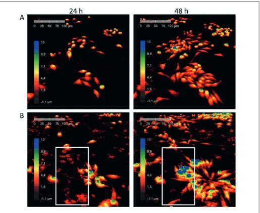
In the holographic images, the SA-MIPs are represented by red dots and clusters that are present only in images showing treated cells (fig. 3B) and not in the untreated cells (fig. 3A). The quantity of SA-MIPs decreased over time, which is due to an uptake and ingestion by the macrophages (see video online). Moreover, as the nanoprobes will not dissolve due to their chemical construc-tion, it would be assumed that they have been ingested by macrophages. Several clus-ters of SA-MIPs are seen in the framed area of Fig. 3B after 24h incubation. After 48h they have disappeared from the images (fig 3B). The time-lapse movie clearly shows that the macrophages ingested the nanoprobes as would be expected (see video online). Conclusion
There are several advantages with DHC and other QPI platforms. Since they can be Scientific, Grand Island, USA) with
addi-tion of 10% fetal calf serum (FBS, Invitro-gen, San Diego, CA, USA) and 1% penicil-lin-streptomycin (Invitrogen) and incubated in 37 ˚C with 5% CO2.
The SA-MIPs were prepared according to a previously described protocol, with depro-tonated SA being used as template [16]. Deprotonation was achieved by addition of an equimolar amount of tetrabutylammo-nium hydroxide to the SA stock solution prior to use in the polymerization. The dried SA-MIPs were resuspended in phosphate saline buffer (PBS, Invitrogen), to a stock solution of 1.0 mg/ml. The SA-MIP had an average size of 200 nm in diameter. Digital Holographic Cytometry
RAW 264.7 cells were seeded in a 6-well polystyrene plate (Sarstedt, Germany)
con-Fig. 1: Schematic illustration of the HoloMonitor M4 and image reconstruction (PHIAB, Lund, Sweden).
Fig. 2: RAW 264.7 cells were seeded at 6 x 10⁴ cells per well and monitored for 48 h. A-C)
SA-MIPs increased the average cell area, average cell thickness, and average cell volume, respec-tively. D) The number of cells was not affected by the SA-MIP treatment. All parameters were calculated using digital holographic cytometry. Error bars are based on the standard deviation.
taining 60,000 cells per well and incubated overnight for cells to adhere at 37 ˚C and 5% CO2 (day 0). The RAW 264.7 cells were
treated with 0.04 mg/ml SA-MIPs in media (day 1). The lid was changed to Hololids (PHIAB, Lund, Sweden), which enable cell imaging by HoloMonitor M4 DHC (PHIAB, Lund, Sweden). Time-lapse phase imaging as well as image processing, segmentation and analysis were conducted with Hstudio software package (PHIAB) with preset mean values of refractive indexes for cells and culture medium. To monitor SA-MIP uptake, cells were imaged every 15 min after addi-tion for a total of 48 h, at eight posiaddi-tions in each well. Images were segmented followed by object identification for every time point, for further investigation of the uptake of SA-MIPs by RAW 264.7 cells. Segmentation
Fig. 3: Raw 264.7 macrophage cell were untreated (A) or treated with SA-MIPs (0.04 mg/ml) (B). Images of the cells were captured by digital holographic cytometer during 48 h. Framed area in the images show a disappearance of SA-MIPs after 48 h, due to an uptake by the cells (see video online). All colors are pseudocolors related to the thickness of the cells as shown by the color bar.
housed in mammalian cell incubators, they can monitor the behavior of unlabeled cells in real time and calculate the number of cells easily. In this work, we demonstrated that neither the DHC or the SA-MIPs did affect the physiological functions of macrophages as they phagocytosed the nanoprobes over time and still proliferated in a similar man-ner as the control macrophages. The utility of this method is that it is quick, quantita-tive, and qualitaquantita-tive, which is essential for future cell-based research.
Acknowledgement
The authors would like to thank the Eu-ropean Union’s Horizon 2020 research
and innovation program under the Marie Sklodowska-Curie grant agreement No 721297 for financial support and Malmö University.
Affiliations
1Phase Holographic Imaging AB, Lund,
Sweden,
2Department of Biomedical Sciences,
Facul-ty of Health and SocieFacul-ty, Malmö UniversiFacul-ty, Malmö, Sweden
3Chemical and Optical Sensing Division,
Bundesanstalt für Materialforschung und -prüfung (BAM), Berlin, Germany
Contact Louise Sternbæk
Phase Holographic Imaging AB Lund, Sweden
Department of Biomedical Sciences, Faculty of Health and Society Malmö University
Malmö, Sweden louise.sternbaek@phiab.se