Interobserver variability
when measuring the
abdominal aorta with
ultrasound
MAIN AREA: Biomedical Laboratory science, clinical physiology, 6th term
AUTHORS: Emma Filipsson & Cecilia Olsson SUPERVISOR: Emma Kramer & Ida Åström Malm
EXAMINER: Anna Bjällmark JÖNKÖPING: 2018-05
Summary
A rupturing abdominal aortic aneurysm (AAA) has a mortality rate of 50% while an elective repair has a mortality rate of 3-8%. Screening programmes with ultrasound are used in some countries to detect AAA at an early stage. Ultrasound is however very observer bias and dependent on the observer's experience. The study was performed in Vietnam, a country that have experienced exceptional economic growth with increasing risk factors for cardiovascular diseases but has no national screening programme. The aim of this study was to examine the interobserver variability when measuring the abdominal aorta with ultrasound on young adults in Vietnam. The abdominal aorta was measured in the longitudinal and the transverse axis by two different observers using the leading edge to leading edge method. Participants in this study were 31 voluntary students, 16 men and 15 women in the ages 18-26 years, from the Da Nang university of medical technology and pharmacy and the design was a cross-sectional study with a quantitative approach. The study showed a significant difference between the longitudinal measurements but not between the transverse measurements. Despite this statistical significance, the observers' differences were within the recommended limit of 5 mm.
Sammanfattning
Observartörsvariation vid mätning av bukaortan med ultraljud – en jämförelse mellan mätvärden observerade i de longitudinella och transversella snitten
Ett rupturerande bukaortaaneurysm har en dödlighet på 50% medan förebyggande vaskulär kirurgi har en dödlighet på 3-8%. Screeingprogram med ultraljud används i vissa länder för att upptäcka bukaortaaneurysm i ett tidigt skede innan de rupterar. Ultraljudsundersökningen är dock väldigt beroende på utövarens erfarenhet och utbildning. Studien genomfördes i Vietnam, ett land som genomgått stor ekonomisk tillväxt men även en ökning av riskfaktorer för kardiovaskulära sjukdomar. Vietnam har dock inget nationellt screeningprogram för bukaortaaneurysm.Syftet med studien var att undersöka observatörsvariationenvid mätning av bukaortan med ultraljud på unga vuxna i Vietnam. Populationen var ung för att minska patologiska fynd. Bukaortan mättes med metoden leading edge to leading edge, både i det longitudinella och i det transversella snittet av två olika utövare. Observatörerna var två studenter från Biomedicinska analytikerprogrammet med inriktning klinisk fysiologi, Jönköpings Universitet. Deltagare i studien var 31 frivilliga studenter, 16 män och 15 kvinnor i åldrarna 18-26 år, från Da Nang University of medical technology and pharmacy och designen var en tvärsnittsstudie med kvantitativ ansats. Studien visade en signifikant skillnad i de longitudinella mätningarna men inte i de transversella mätningarna. Trots denna statistiska signifikans var observatörernas differenser inom den rekommenderade gränsen på 5 mm.
Content
Introduction ... 5
Background ... 5
Abdominal aortic aneurysm ... 5
Ultrasound ... 7
Screening worldwide ... 8
Challenges with ultrasound ... 9
Aim ... 10
Research methodology ... 11
Design ... 11
Equipment and measurement method ... 11
Imaging acquisition ... 12 Statistical analysis ... 13 Ethical considerations ... 13
Results ... 14
Discussion ... 17
Results ... 17 Method ... 19Conclusion ... 21
Acknowledgements ... 21
References ... 22
Appendix 1
Appendix 2
5
Introduction
Abdominal aortic aneurysm (AAA) is a vascular disease that can be fatal if it ruptures. It is common that patients with AAA also suffers from other cardiovascular diseases (1). In 2014, 506 people died because of an abdominal aortic aneurysm in Sweden. Due to a low level of autopsy the unrecorded number is supposedly higher. To decrease the mortality of AAA, Sweden has a screening program that includes an abdominal ultrasound of all men over 65 years of age (2).
In the year of 2015, the population of Vietnam was 93 448 000. During that year, approximately 126 300 people died of cardiovascular diseases. There is no statistics on how many people died
because of an AAA, however AAA is a part of the category cardiovascular diseases and this
category has increased in recent years (3). Vietnams economy has developed rapidly and due to this globalisation the population is getting older, smoking more, there is an increase in obesity and high blood pressure is more common (4). Smoking, overweight, old age and male sex are all risk factors for AAA (5).
Background
Abdominal aortic aneurysm
The largest blood vessel in the human body is the aorta that originates from the left chamber of the heart. The aorta is divided into different sections, the first being the aortic root, followed by the ascending aorta, the aortic arch and then the descending aorta. The descending aorta is divided into the thoracic and abdominal aorta. The abdominal aorta begins at the diaphragm and ends at the bifurcation where the abdominal aorta divides into the right and left common iliac arteries (6). The wall of the vessel is built up by three layers, from inner to outer: intima, media and adventitia (7). A permanent dilation of these three layers is called an aneurysm if the dilation expands over 30 mm or by a 50% increase compared to a normal segment (Figure 1). An aneurysm of this size is not harmful and are in most cases asymptomatic, it can however expand and increase the risk of a rupture (2, 6, 8). A rupture is defined as a fracture of all three layers with a bleeding outside of the adventitia and is without acute treatment fatal (9). When
6
the aneurysm grows to a certain size the patient may eventually feel discomfort, pressure and pain if the aneurysm is growing fast (5). In most cases however, the symptoms occur first when the aneurysm rupture (6).
Figure 1: Illustration of a normal abdominal aorta and an abdominal aortic aneurysm. 1
The dilation of the abdominal aorta is caused by loss of elastin, the death of smooth muscle cells and the accumulation of balancing collagen. It was preciously claimed that atherosclerosis was the cause of AAA and that an AAA was a local phenomenon in the arterial wall. Recent studies now claim that atherosclerosis is the result of the changed blood flow in the AAA rather the cause of it. Today's evidence suggest that AAA is a systematic disease including the complete vasculature and involves a complicated pathological process with the key aspects being inflammation, reduction of the cell matrix, breakdown of proteins and the death of smooth muscle cells (6). Small aneurysms of the popliteal artery that are uncommon in a normal population are found in approximately 70% of patients with AAA. Furthermore, the carotid artery in patients with AAA have also shown slight dilation and loss of compliance, supporting the statement that the whole vascular system is involved (6).
Abdominal aortic aneurysm is more common in an older population and incidence increases with age, AAA related mortality is very low under the age of 65. Gender is also an important factor since the incidence in men is six times greater compared to women. The cause of this gender difference is unknown but is supposedly due to exposure of risk factors, genetics and hormonal factors (6). Smoking, hypertension, family history, male sex and obesity are all
1 Received from:
7
factors that increase the risk of an aneurysm (5, 6). Even though diabetes is related to atherosclerosis, data suggest a lower incidence of AAA in patients with diabetes and formed AAA's also grew slower in this patient group. Smoking is yet the only factor that can be related to all processes of an AAA; developing AAA, growth and rupture (6).
Ultrasound
Ultrasound is used to examine the abdominal aorta since it is a non-invasive and cost-effective method that requires no patient preparation. The transducer can send out and receive soundwaves because of the properties of piezoelectric crystal inside it. The piezoelectric crystal can transform electric energy into mechanical energy, sound waves, and contrariwise. For abdominal aortic ultrasound a frequency of 2-5 Megahertz (MHz) is used (10). By adjusting the frequency of the soundwaves, the penetration depth and resolution can be altered. A low frequency will improve the penetration of the soundwaves but will also impair the resolution. Consequently, a high frequency will improve the resolution but decrease the penetration depth. To achieve a good visualisation of the abdominal aorta, a balance between these two components are vital (8, 11). In addition to frequency, a certain pressure needs to be applied with the transducer to achieve a good visualization of the aorta. Applied pressure decreases the intra-abdominal air and additionally reduces the distance between the transducer and aorta (10). Flatus can obstruct visualization and fasting 4-8 hours before the examination is therefore recommended (8).
The abdominal aorta can be measure in both the longitudinal and the transverse axes. One axis can sometimes be easier to visualize than the other and an image of good quality is needed for optimal measurements (5). In the longitudinal axis the diameter is measured from the anterior wall to the posterior wall (AP-diameter). In the transverse axes, aside from the AP-diameter, the transverse diameter can also be measured. Using the ultrasound image there are three different methods for measuring the abdominal aorta; inner to inner (ITI) leading edge to leading edge (LELE) and outer to outer (OTO) (figure 2). There is no international accepted method and observers therefore measures differently, making the reproducibility low. LELE is the method with less variability compared to the other methods and is recommended when measuring the abdominal aorta (12). LELE is also the method used in Sweden's screening program for AAA (13).
8
Figure 2: An ultrasound image demonstrating the three methods to measure the abdominal aortic
diameter, inner to inner (ITI), leading edge to leading edge (LELE) and outer to outer (OTO) (12).
Screening worldwide
Screening programs using ultrasound are a cost-effective system to reduce the aneurysm related mortality. Ultrasound also has a specificity and sensitivity of almost 100% (14). A rupture with expensive acute surgery and care has a mortality risk of 50% while an elective AAA repair has a mortality risk of 3-8% (5, 15). Even though, only a few developed countries have begun with national screening programmes. Between the countries that screen there are some differences on who to screen and how often. All the programs defined AAA as ≥ 30mm but differed in minimum diameter required for surgery. Western Australia, Italy and USA will consider surgery when the aneurysm is ≥ 50mm while Denmark, Norway, England, New Zealand, Scotland, Sweden, Wales and Northern Ireland consider surgery when the AAA is ≥ 55mm. Most countries would only screen men over 65 but a few countries would also include females. New Zeeland and USA would screen if the patient had a high cardiovascular risk. USA would also recommend screening for patients who have ever smoked more than 100 cigarettes and individuals over 50 years of age with a family history of AAA. The time between follow-ups, how often and at what size to do the follow-ups also varied between the countries (15).
Unlike Sweden and a few other countries, there is today no screening of the abdominal aorta in Vietnam. It is a long process to set up a national AAA-screening program and it requires educated personal since operating the ultrasound machine and perform the measurements necessitate a certain level of skills. Since AAA is most often discovered as an incidental finding
9
during different pathology examinations, screening is needed to discover the AAA at an early stage, eliminate mortality and decrease the need for expensive surgery (5).
A lot of studies have been made on AAA in western countries, but studies of AAA in Asian countries are scarce. In other Asian countries the incidence of AAA is lower than in western countries, however an increasing trend of the AAA incidence, prevalence and mortality have also been observed during the years 2005 to 2011 (1). Alternative data suggest comparable incidence as in western countries (16). Furthermore, it was also discussed that the incidence was hard to examine in Asian countries since the incidence was calculated on patients where AAA was discovered due to symptoms or as an accidental finding. Patients with asymptomatic AAAs, which is most common, is therefore not included in the data since there is no national screening for the disease (1). The incidence also varied depending on which criteria for AAA was used. Incidence was higher with the criteria 50% dilation compared to normal aortic diameter and lower with the criteria above 30 mm (14). Similar to western countries, the incidence was higher in an older population as well as in a male population compared to a female population. Women generally also had a smaller aortic diameter. Common comorbidities were hypertension, smoking, chronic obstructive pulmonary disease and other cardiovascular diseases (1, 16).
Challenges with ultrasound
Despite being cost effective and non-invasive, ultrasound is however very dependent on the observer’s skill, experience, medical discipline and level of training which creates a disadvantage for the method. Studies have shown an inter-observer difference of 10 millimetres that indicates observer bias. This is problematic since the diameter of the aneurysm is a direct indicator for either operation or follow up (5, 10). According to the National Abdominal Aortic Aneurysm Screening Program (NAAASP), the variation between two observers should not exceed 5 millimeters. Variability in measurements can depend on the observer’s education, scanning technology, the patient and the anatomical localization on the abdominal aorta where the measurements are obtained. A high interobserver reproducibility is important since it should not matter which examiner the patient receives (5). Another disadvantage with ultrasound that can affect the measurements of the abdominal aorta is the angulation of the transducer. The angulation of the transducer affects which part of the aorta that is visualized, and it can result in false measurements of the diameter (10).
10
The quality of the image is also dependent on the patient’s stature since the frequency needs to be decreased to penetrate the increased distance which results in inferior resolution (11). Measurements are more dependent on the patient's stature, if the measurement are taken superior to the renal arteries compared to if the measurements are taken inferior to the renal arteries. AAA is also more common proximal to the bifurcation due to the hemodynamic strains prior to the first major division (5, 6). The measurements are therefore most commonly taken infrarenal but a complete scanning of the aorta, from the diaphragm down to the aortic bifurcation is needed (10, 17).
For AAA screening to be successful, formal quality controls, standardized measurement methods and standardized clinical training are significant (5, 10). It is very important with a correct measurement method and a precise estimation when measuring the abdominal aorta since diagnosis and treatment are directly related to the diameter of the aneurysm. Small variations when measuring the diameter could therefore make a great difference on how to proceed with treatment, affecting the patient safety (18, 19). Interobserver variability between observers is problematic since all patients should receive equal care and diagnosis and treatment should not depend on the examiner (20).
Aim
The aim of this thesis is to evaluate the interobserver variability when measuring the abdominal aorta with ultrasound on young adults in Vietnam.
11
Research methodology
Design
The study was a cross-sectional study with a quantitative approach and was performed in Vietnam, at the Da Nang University of Medical Technology and Pharmacy. The observers were two students studying the Biomedical Laboratory programme focusing on clinical physiology 6th at Jönköping University with no previous experience of abdominal aortic ultrasound. The
population was a convenience sample selected by Dr. Phuong, a teacher at the university and the Vietnamese supervisor of the study. All participants had to be at least 18 years old to be allowed to participate in the study. The number of participants was 35 and consisted of voluntary students from the university, both males and females. All 35 individuals were examined with ultrasound, but one participant was excluded from the study since that individual was not a student. The data from three other participants were also excluded due to a change of ultrasound equipment during the beginning of data collection. Thus, the final statistical analyses were performed on 31 participants in the longitudinal measurements and on 30 participants in the transverse measurements since one transverse diameter was not obtained by one observer due to poor visualisation. An ethical approach has been applied and an ethical examination prior of this study has been performed according to Jönköpings University's guidelines. According to which, a permission from an ethical committee was not needed (19).
Equipment and measurement method
The ultrasound equipment was provided by the Da Nang University of Medical Technology and Pharmacy in Vietnam. The brand of the machine was an ALOKA SSD-1100 Flexus (ALOKA, Tokyo, Japan) and the brand of the transducer was a curved ALOKA VST-934N-3.5 with the frequency of 3,5 MHz (Figure 3). To eliminate the air between the transducer and the participant, ultrasound gel was also used.
The measurements of the abdominal aortic diameter were taken in the long axis (longitudinal) and in the short axis (transverse). The measurements were taken with the tool caliper, 2-4 centimetres proximal of the bifurcation, inferior to the renal branches. Measurement method used was LELE and the caliper markers were placed at the anterior adventitia and at the posterior intima in the still ultrasound image (12, 13).
12
Imaging acquisition
Before the ultrasound examination, all participants received an informative letter translated to Vietnamese informing about the purpose of the study and the procedure of the ultrasound examination. The participants had to sign an informed consent prior to participating. The informed consent contained questions concerning gender, age, height and weight (appendices 1 and 2). After signing the form, the participant entered the examination room and was asked to lay down during the examination. The abdominal aorta and the anatomical localization was first identified in the longitudinal axis and the optimal image, clearly visible walls, was obtained. The transducer was then turned 90 degrees anti clockwise to visualise the transverse axis and obtain the optimal image in the second axis. The images were paused in systole, when the abdominal aorta was at is largest due to the pulsation. The measurements were taken with the images abreast in the ultrasound frame (Figure 4) and the markers of the caliper tool were placed at the first reflection of the anterior wall and at the second reflection of the posterior wall in both images. The longitudinal and the transverse diameter were documented by the observer and the images were deleted before the other observer started the next examination. Both observers separately performed the ultrasound examination for each participant, measuring in separate images unaware of previous results. All participants therefor underwent the same ultrasound procedure twice performed by two different observers.
Figure 3: Ultrasound equipment, ALOKA SSD-1100 Flexus.
13 Statistical analysis
The agreement between the two sets of data collected by the two observers, the body mass index (BMI) of the participants and descriptive statistics of the measurements were calculated and analysed using the software program International Business Machines Corporation, Statistical Package for the Social Sciences 25 – IBM, SPSS 25 (IBM, Armonk, USA). Microsoft Excel 2016, version 1803 was used to create the tables and the plots (Microsoft, Redmond, USA). Bland-Altman plots were used to determine the agreement between the observers, comparing the longitudinal and transverse measurements (21, 22). A paired sample T-test was performed to calculate if the mean differences between the two observers' measurements were significantly different from zero (23). The level of significance was α = 0.05.
Ethical considerations
The ethical aspect has been examined according to Jönköping university guidelines. A permission from an ethical committee was not needed since this study was not a research with the purpose to be published in a scientific journal. Neither does this study treat sensitive personal data, require any physical intervention, do not purpose to affect the participants mentally nor physically. The ultrasound examination is harmless and does not use biological material (24).
Figure 4: An ultrasound image of the abdominal aorta
visualizing the longitudinal and transverse axis. The arrows demonstrate the placement of the caliper markers.
14
Before participating the students received written information through a form regarding the aim of the study, that the study is anonymous, that they can withdraw at any time without reason and if wanted, receive a copy on the results of the study via email (25). The participants also receive information about the ultrasound procedure. The information was translated into Vietnamese to avoid misunderstandings and the students that wanted to participate signed their form, giving their informed consent that they had understood the information given and that they wanted to participate (26, 27). A risk during this study was a pathological finding, even though the risk was low. A younger population minimized the risk of pathology findings related to the abdominal aorta. A physician was therefore present or nearby during the data collection. The physician also helped with translation when the participants did not understand what the observer was saying. The informed consents and the collected data have been handled confidentially and anonymously by identifying the participants as a number during the compilation of data. The data has also been presented as a group and not individually (24, 25). Some of the images were printed out before they were deleted but the images did not contain any personal information about the participants. During the collection of data, the authors got the impression that some of the participants had not read the informed consent since they seemed confused about the procedure of the examination. It could also be that the complied information failed to give a clear perception of the study procedure.
Results
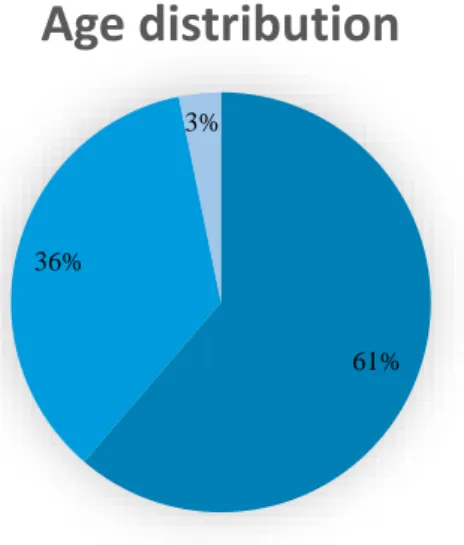
The population consisted of 31 individuals, 16 males and 15 females in the ages 18 to 26 years. The majority of the population had a normal BMI (Figure 5) and were between 18-20 years old (Figure 6). Table 1 and 2 demonstrates the paired sample -test for the longitudinal and transvers measurements obtained by two observers. Mean difference being 0.6 mm for the longitudinal axis and 0.05 mm for the transverse axis. The paired sample T-test showed a statistical significant difference in the longitudinal measurements between the two observers (p = 0.004). The difference between the observers´ transverse measurements did not show a statistical difference (p = 0.802) (Tabell 1-2). The interobserver variability is statistical significant between the observers’ longitudinal measurements but not between the transverse measurements.
15
Table 1: Paired sample T-test for longitudinal measurements of the abdominal aorta obtained by
observer 1 and 2
Paired sample statistics Mean N Std. Deviation Std. Error mean
Longitudinal, observer 1 11,2 31 1,3 0,3
Longitudinal, observer 2 10,6 31 1,4 0,4
Paired sample t-test Mean Std. Deviation Std. Error mean Sig. (2-tailed)
Longitudinal, observer 1
- longitudinal, observer 2 0.6 1.1 0.2 0.004
Table 2: Paired sample T-test for transverse measurements of the abdominal aorta obtained by observer
1 and 2.
Paired sample statistics Mean N Std. Deviation Std. Error mean
Transverse, observer 1 11.3 30 1.2 0.2
Transverse, observer 2 11.2 30 1.3 0.2
Paired sample t-test Mean Std. Deviation Std. Error mean Sig. (2-tailed)
Transverse, observer 1
-transverse, observer 2 0.1 1.2 0.2 0.802
Figure 5: A pie chart demonstrating
the populations weight distribution. Underweight (BMI < 18,5), normal (18,5-25) and overweight (25-30) according to the body mass index scale. N = 31.
Figure 6: A pie chart demonstrating the
participants age (years) distribution. 18-20 years, 21-23 years and 24-26 years. N = 31. 26% 71% 3%
BMI
<18,4 18,5-24,9 25-30 61% 36% 3%Age distribution
18-20 år 21-23 år 24-26 år16
The Bland-Altman plots showed that the differences vary across the mean and that there is no systematic measurement error made by the observers, meaning that the differences are not proportional to the mean. Both plots also have over 95% of the differences within the limits of agreement and the range of their limits off agreement are all below 5 mm (Figure 7-8).
Figure 7: The mean difference of 0,6 mm is represented by the middle line while the dotted lines
represent the limits of agreement and over 95% (30/31=97%) of the differences are within these limits. The lower limit of agreement = -1,6 mm and the upper limit of agreement = 2,8 mm. N = 31.
Figure 8: The mean difference of 0,05 mm is represented by the middle line while the dotted lines
represent the limits of agreement and over 95% (30/30=100%) of the differences are within these limits. The lower limit of agreement = -2,2 mm and the upper limit of agreement = 2,4 mm. N=30.
-2,5 -1,5 -0,5 0,5 1,5 2,5 3,5 9 10 11 12 13 14 15 L ongit ud inal di ff ere nc e ( m m )
Longitudinal mean diameter (mm)
-3 -2 -1 0 1 2 3 9 10 11 12 13 14 15 T ransve rse d if fer ence ( m m )
17
Discussion
Results
The aim of this thesis was to examine if it were any differences between the two observers when measuring the abdominal aorta with ultrasound in the longitudinal and the transverse axes. The purpose has been achieved for this thesis since the differences between the observers were determined, considering there was a statistical significant difference between the longitudinal measurements but not between the transverse measurements. According to NAAASP, it should not differ more than 5 mm between two observer’s measurements. In this study, above 95 % of the differences for longitudinal and transverse measurements were within the limits of agreement. Since the range of the limits of agreement were smaller than 5 mm, the differences has therefore no clinical relevance (5, 22). The measurements with the lowest mean difference between the two observers (0.05 mm) were the ones taken in the transverse axis. This might be explained by studies that have shown higher repeatability on measurements taken in this axis (18). The transverse axis is also the most common plane to obtain measurements in studies (5). Another possible explanation for the transverse measurements lower mean difference compared to longitudinal could be because the longitudinal measurements are more dependent of the angle placement of the caliper marker against the angle of the abdominal aorta. As seen in figure 3, the abdominal aorta in the longitudinal axis is angled down in the ultrasound image towards the patient's head (left side of the image) and more angled up towards the patient's lower body (right side of the image). Using the caliper tool and performing a vertical measurement, anterior to posterior in the ultrasound image of the abdominal aorta would result in a larger diameter. The caliper tool should be placed orthogonally, at a 90 degrees angle to the abdominal aortic wall (10).
This angulation problem has also been discussed when ultrasound is compared to the gold standard, computed tomography (CT). When the two methods are compared, ultrasound repeatedly underestimated the diameter. But when measuring with orthogonal plane and then comparing with CT the underestimation for ultrasound decreased and resulted in a mean difference of 0.8 mm (10). The observers focus was to obtain the optimal image and a clearly visible aorta, more effort could have been addressed the angulation of the caliper tool but also the angulation of the transducer. The increased mean difference within the longitudinal measurements could therefore depend on the observer’s different angulation of the caliper tool and the angulation of the transducer.
18
In a study comparing the reproducibility of ultrasound in nine different studies the maximum variability detected was 10 mm. All studies used different ultrasound machines, sonographers had different medical background and seven different measurements methods or combination of methods were used. Despite the variations between the studies, most of the studies did however show an inter-observer variability below the recommended limit of 5 mm and that an abdominal aortic scan can be performed by operators with varying experience (5). Like this study, the observers in eight out of nine studies performed real time ultrasound examinations and obtained the images in which the diameter would be measured. In the ninth study the interobserver variability was compared in still images. Even though the observers were inexperience in this study, the limits of agreement (Figure 7-8, -2.2 mm to + 2.4 mm for the transverse and -1.6 mm to + 2.8 mm for the longitudinal measurements, ) are closer to the lower range of the limits of agreement in the study (− 1.9 to + 1.9 mm and − 10.5 to + 10.4 mm) (5).
It was also discussed that the patient's waist circumference could affect the inter-observer variability since increased depth limits visibility (5). Since only 3 % of the study subjects in this study had a BMI above normal (Figure 5), the variations in measurements between the two observers are more likely to be explained by other factors than BMI. Despite that the observers had no previous experience of abdominal aortic ultrasound scanning the measurements were still within the recommended limit of 5 mm. However, the true diameters of the study subjects are still unknown since the measured diameters by the two observers were never controlled by experienced observers or compared to a CT examination which is the gold standard for abdominal aortic measurements (10).
The population examined in this study was chosen to improve image quality and might have been a component for the low variability since they were young adults with a normal BMI, making the ultrasound examination less challenging for the observers. If the study would have been performed on men over 65 years of age, the target group for screening programmes, the data collection could have been more demanding and the outcome another. Two different studies investigated the effect of ultrasound training with novice operators. The studies concluded that education to perform abdominal aortic scans with ultrasound can make novice operators perform ultrasound examinations on the abdominal aorta. But one study also claims that despite the training they failed in more demanding cases (28). When screening for AAA, challenging cases will appear, and it is therefore important that the operators have enough experience to achieve correct results. The differences between these studies are the medical
19
backgrounds of the novice operators and how they were trained (28, 29). Apart from the patient's physical state, the treatment of an AAA is dependent on its diameter. A difference of 10 mm will therefore have significant clinical relevance since it affects the decision whether the AAA should be treated surgically or not. Since the diameter also conclude the existence of an AAA and if the patients should be followed over time, a difference of 10 mm is not acceptable (5).
The average mean diameter in this study, both male and female, was 11.1 ± 1.2 mm which is smaller compared to another study performed on a Chinese population, 12.9 ± 2 mm (14). The smaller mean could depend on the younger population, 18-26 years compared to 45-80 years. The average mean diameter is however much smaller compared to previous studies performed on a western population, 19.1 ± 2.9 mm and 18.6 ± 2.2 (14). Since AAA is more common in countries with a Western lifestyle, most studies have been performed on a Caucasian population. Not many studies have been performed on an Asian population and the few published demonstrates various results regarding the incidence of AAA (1, 14, 16). They do however conclude that the average diameter of the abdominal aorta is smaller compared to the diameter of a western population, despite adjusting for variations in body size. Because of this, a different criterion for AAA may be needed for an Asian population since the abdominal aorta is smaller from the beginning. The criteria 30 mm used may not be suitable and the criteria of an 50% increase compared to a normal segment may therefore be preferable (14).
All factors discussed highlight the importance for the training and experience needed to become a professional within ultrasound. Besides the hours of training, great knowledge regarding the anatomy and physiology of the abdominal aorta is required (28, 29).
Method
One limitation with the method was the observers´ lack of experience. The inexperience could have resulted in variations of the identification of the anatomic location of the abdominal aorta. Meaning that the measurements were obtained at different segments of the aorta. Although limited experience within ultrasound, the observers did only have minimal training connected to abdominal aortic examination and the LELE method prior to the study. The ultrasound equipment was changed during the data collection since the images from the first machine were of poor quality. A newer ultrasound machine might have improved the image quality providing
20
more optimal conditions for the measurements. The equipment was also unfamiliar to the observers. The data was collected for one day and due to this limited time, only one measurement was obtained in each axis. Three measurements taken in each axis and a calculated mean for each observer and axis could have made the results more reliable instead of one single measurement. Since the observations were performed by inexperienced observers the results of the interobserver variability cannot be generalised to trained sonographers but could apply to other students with the same level of training.
A second limitation with the method is that the observers, instead of doing a visual estimation of the maximum diameter, could have used electrocardiography (ECG) to make sure the measurements were taken at the same time in the heart cycle considering that the arterial wall fluctuates 2-4 mm during the heart cycle. The fluctuation occurs independent of the presence of an AAA, but ECG is not required in screening programmes despite that the measurements could be made more accurate (10). Even though it might not be relevant for screening in clinical practise it could have been applicable in this study to improve reliability of the measurements since without a connected ECG a difference of 2-4 mm could simply depend on the fluctuation of the arterial wall between diastole and systole if the measurements are not taken in the same cycle (10).
A curved transducer with the frequency of 3,5 MHz was used since it provided the best visualisation of the abdominal aorta and therefore optimised the conditions for the measurements taken with the LELE method (10). This study uses the LELE method since it is the method used in the Swedish national screening programme but also because it measures at points that are easier to distinguish in the ultrasound image due to increased difference in acoustic impedance resulting in stronger echoes (11, 12). The three different methods, ITI, LELE and OTO, that can be used to measure the abdominal aorta with ultrasound are all well discussed in different articles but differ as to which method is preferable. In two different studies, OTO and LELE were deemed the better, however the same method that was suggested as the best was also the method that the sonographers of the studies use in everyday practise (12, 18). This also demonstrates that experienced sonographers, practising the method for obtaining the measurement, will improve the reliability of the results. However, considering that LELE was assessed as the preferable method in more than one study it became the method of choice in this study (10, 12).
21
In contempt of the previous, an additional study suggests that LELE and OTO are two different variations of the same method since computed tomography (CT) have showed that the aortic wall is 2 mm thick. The general difference between OTO and LELE is also 2 mm and the difference between the OTO and LELE could simply depend on the aortic wall thickness. The difference between the two methods are that LELE measurements are calculated from anterior outer wall to inner posterior wall while OTO measures from anterior outer wall to posterior outer wall. Between the inner posterior wall to posterior outer wall the thickness is approximately 2 mm. The study therefore suggests that LELE and OTO are two versions of the same method (10). Further research with a larger sample, ECG and experienced observers would be needed to increase reliability and generalizability of the results. It would also be interesting to examine the average diameter of both Swedish and Vietnamese students due to the small mean aortic diameter in this study. More research is also needed regarding abdominal aortic aneurysm in Asia as well as an international standardised method for abdominal aortic measurements with ultrasound to improve interobserver variability.
Conclusion
This study showed no statistical significant difference between the observers' transverse measurements. There was a statistical significant difference between the two observers’ longitudinal measurements, however, its clinical relevance can be considered as low since the differences were within the recommended limit of 5 mm.
Acknowledgements
The authors would like to express their gratitude towards the staff and students at the Da Nang University of Medical Technology and Pharmacy in Vietnam for their help and warm welcome. A special thanks to our supervisor in Vietnam, Dr. Phuong, whom without the collection of data would have been considerably more challenging. The authors would also like to thank the program of Minor Field Studies (MFS), The School of Health and Welfare and Jönköping University's International Relations for the scholarship funding and opportunity to perform our thesis in Da Nang, Vietnam. A final thanks to our Swedish supervisors Emma Kramer and Ida Åström Malm.
22
References
1. Wang SW, Huang YB, Huang JW, Chiu CC, Lai WT, Chen CY. Epidemiology, Clinical Features, and Prescribing Patterns of Aortic Aneurysm in Asian Population From 2005 to 2011. Medicine (Baltimore). 2015;94(41):e1716.
2. Socialstyrelsen, SBU. Screening för bukaortaaneurysm Vetenskapligt underlag Bilaga 1
socialstyrelsen2016 [cited 2018 April 3]. Available from:
https://www.socialstyrelsen.se/SiteCollectionDocuments/2016-6-5-Bilaga-1-Vetenskapligt-underlag.pdf.
3. WHO. Viet Nam [Fact sheet]. World Health organization; 2018 [cited 2018 April 3]. Available from: http://www.who.int/countries/vnm/en/
4. WHO. UN Interagency Task Force on NCDs joint country mission to Viet Nam World Health organization2016 [cited 2018 April 3]. Fact sheet]. Available from: http://www.who.int/ncds/un-task-force/vietnam-mission-september-2016/en/
5. Beales L, Wolstenhulme S, Evans JA, West R, Scott DJ. Reproducibility of ultrasound measurement of the abdominal aorta. Br J Surg. 2011;98(11):1517-25.
6. Nordon IM, Hinchliffe RJ, Loftus IM, Thompson MM. Pathophysiology and epidemiology of abdominal aortic aneurysms. Nat Rev Cardiol. 2011;8(2):92-102.
7. Haug E, Bjålie JG, Sand O, Sjaastad ØV. Människokroppen2007-08.
8. Jogestrand T, Rosfors S. Klinisk fysiologisk kärldiagnostik: Studentlitteratur AB; 2002.
9. Bergqvist D. Artärsjukdomar : utredning, klinisk bedömning och behandling: Studentlitteratur AB; 2013.
10. Schäberle W, Leyerer L, Schierling W, Pfister K. Ultrasound diagnostics of the abdominal aorta: English version. Gefasschirurgie. 2015;20(Suppl 1):22-7.
23
11. Jonson B, Wollmer P. Klinisk fysiologi med nuklearmedicin och klinisk neurofysiologi: Liber; 2011.
12. Gürtelschmid M, Björck M, Wanhainen A. Comparison of three ultrasound methods of measuring the diameter of the abdominal aorta. Br J Surg. 2014;101(6):633-6.
13. Wanhainen A, Björck M. The Swedish experience of screening for abdominal aortic aneurysm. J Vasc Surg. 2011;53(4):1164-5.
14. Guo W, Zhang T. Abdominal Aortic Aneurysm Prevalence in China. Endovascular today. 2014.
15. Stather PW, Dattani N, Bown MJ, Earnshaw JJ, Lees TA. International variations in AAA screening. Eur J Vasc Endovasc Surg. 2013;45(3):231-4.
16. Yii MK. Epidemiology of abdominal aortic aneurysm in an Asian population. ANZ J Surg. 2003;73(6):393-5.
17. Wanhainen A, Hultgren R, Linné A, Holst J, Gottsäter A, Langenskiöld M, et al. Outcome of the Swedish Nationwide Abdominal Aortic Aneurysm Screening Program. Circulation. 2016;134(16):1141-8.
18. Chiu KW, Ling L, Tripathi V, Ahmed M, Shrivastava V. Ultrasound measurement for abdominal aortic aneurysm screening: a direct comparison of the three leading methods. Eur J Vasc Endovasc Surg. 2014;47(4):367-73.
19. Patientsäkerhetslag. Patientsäkerhetslag (2010:659). In: Socialdepartementet, editor. Sveriges riksdag2010.
20. sjukvårdslag H-o. Hälso- och sjukvårdslag (2017:30). In: Socialdepartementet, editor. Sveriges riksdag2017.
24
22. Bland JM, Altman DG. Statistical methods for assessing agreement between two methods of clinical measurement. Lancet. 1986;1(8476):307-10.
23. Petrie A, Sabin C. Medical Statistics at a Glance: Wiley-Blackwell (an imprint of John Wiley & Sons Ltd); 2009.
24. 2003:460 E. Lag (2003:460) om etikprövning av forskning som avser människor. In: Utbildningsdepartementet, editor. Sveriges riksdag2003.
25. 2009:400 Sf. Offentlighets- och sekretesslag (2009:400). In: L6 J, editor. Sveriges riksdag2009.
26. Jönköping Hi. Dokument [Fact sheet]. Studentwebben, Jönköping University2018 [cited 2018 April 3]. Available from: http://ju.se/student/studier/blanketter--dokument/halsohogskolan.html.
27. Sandman L, Kjellström S. Etikboken : etik för vårdande yrken: Studentlitteratur AB; 2013.
28. Nguyen AT, Hill GB, Versteeg MP, Thomson IA, van Rij AM. Novices may be trained to screen for abdominal aortic aneurysms using ultrasound. Cardiovasc Ultrasound. 2013;11:42.
29. Bonnafy T, Lacroix P, Desormais I, Labrunie A, Marin B, Leclerc A, et al. Reliability of the measurement of the abdominal aortic diameter by novice operators using a pocket-sized ultrasound system. Arch Cardiovasc Dis. 2013;106(12):644-50.
Appendix 1
Information regarding participation in student bachelor thesis data
collection using ultrasound to measure the abdominal aorta.
We are two students from Sweden who are going to perform our bachelor thesis here in Vietnam. We are studying Biomedical Science, focusing on clinical physiology and are very interested in medical ultrasound. When using ultrasound to measure the abdominal aortic diameter, experience of the operator and measurement accuracy are of great importance. The purpose of our study is to examine the inter-observer reliability between two different observers when measuring the abdominal aorta with ultrasound.
The method that will be used in this study is ultrasound. Ultrasound is painless and non-invasive. Since two different observers will obtain measurements the exact same examination will be performed twice. The examination will in total take a maximum of 30 minutes.
The study is completely anonymous and name, social code will not be presented in the study. Participation is completely voluntary. If you decide to participate but change your mind you have the right to withdraw without any explanation. Finally, if you would like to receive a copy of the completed bachelor thesis please fill in your email address.
If you would like to participate you need to be over 18 years old and sign the following form on the next page. Fill in your height (in centimetres), weight (in kilograms) and circle your gender and appropriate age interval. If you are not sure about your exact height and weight an estimation is okay.
People responsible for this study:
Supervisor in Sweden Ida Åström Malm
Lecturer, Department of Natural Science and Biomedicine ida.astrom-malm@ju.se
School of health and welfare, Sweden
Supervisor in Vietnam Dr Phuong
Teacher at the I.M depature hoangphuong149@gmail.com
Cecilia Olsson Student
Olce15cc@student.ju.se
School of Health and Welfare, Sweden
Emma Filipsson Student
Fiem15fi@student.ju.se
Informed consent
I have read the information above and I have understood the purpose of the study and I hereby choose to participate in this study with the knowledge that I can withdraw at any time without reason. Name………. Date………
Yes, I would like to receive a copy of the final results of this study to the following email (optional). Email……….
Height (in centimetres) ………
Weight (in kilograms) ……….
Gender (circle your gender)
MALE FEMALE
Age (circle the interval)
Appendix 2
THÔNG TIN CHO SINH VIÊN THAM GIA THU THẬP DỮ LIỆU CHO
ĐỀ TÀI SỬ DỤNG SIÊU ÂM ĐO ĐỘNG MẠCH CHỦ BỤNG
Hai chúng tôi là sinh viên Thụy Điển sẽ thực hiện đề tài ở Việt Nam. Chúng tôi là sinh viên Khoa Sinh lý học lâm sàng, tập trung nghiên cứu sinh lý lâm sàng và yêu thích siêu âm. Khi sử dụng siêu âm đo đường kính động mạch chủ bụng, kinh nghiệm của người thực hiện và việc đo đạc chính xác là rất quan trọng. Mục đích của nghiên cứu của chúng tôi là kiểm tra độ tin cậy giữa hai lần quan sát (đo) được thực hiện bởi hai người khác nhau khi đo động mạch chủ bụng trên siêu âm.
Phương pháp được sử dụng trong nghiên cứu là siêu âm. Siêu âm là phương pháp không can thiệp và không đau. Từ hai lần quan sát khác nhau sẽ có được số liệu chính xác trên cùng một người bệnh được thực hiện hai lần . Việc thăm khám này được thực hiện tối đa trong 30 phút. Nghiên cứu sẽ được bảo mật về tên và mã xã hội. Người tham gia hoàn toàn tự nguyện. Khi bạn quyết định tham gia bạn không nên bỏ cuộc giữa chừng. Cuối cùng, nếu bạn muốn nhận bản sao hoàn thiện của đề tài thì vui lòng điền địa chỉ email vào đây.
Điều kiện tham gia bạn phải trên 18 tuổi và ký tên vào thông tin trang sau. Điền chiều cao, cân nặng và giới tính và độ tuổi của bạn.
Người tham gia nghiên cứu
1. Sinh viên Cecilia Olsson – Olce15cc@student.ju.se
Trường sức khỏe và phúc lợi Thụy Điển
2. Sinh viên Emma Filipsson – Fiem15fi@student.ju.se
Trường sức khỏe và phúc lợi Thụy Điển
Người hướng dẫn tại Thụy Điển
Ida Astrom Malm – ida.astrom-malm@ju.se
Giảng viên khoa khoa học tự nhiên và sinh y học, Trường sức khỏe và phúc lợi Thụy Điển
Người hướng dẫn tại Việt Nam
Trương Thị Hoàng Phương – hoangphuong149@gmail.com
CAM KẾT
Tôi đã đọc những thông tin trên và hiểu những mục đích của nghiên cứu và tôi được chọn tham gia nghiên cứu .
Tên:……… Ngày:……… Tôi muốn nhận bản sao kết quả cuối cùng của nghiên cứu bằng email. Email: ………. Chiều cao: ………..(cm) Cân nặng: ………..(kg) Giới tính (khoanh tròn) NAM NỮ Tuổi (khoanh tròn) 18 – 20 21 – 23 24 – 26 >26