ACL reconstruction with a hamstring graft,
can a forced foot dorsiflexion alter
isokinetic knee flexion performance
a pilot study
Kristoffer Lindbäck
Physiotherapy, bachelor's level 2019
Luleå University of Technology Department of Health Sciences
LULEÅ TEKNISKA UNIVERSITET Institutionen för hälsovetenskap Fysioterapeutprogrammet, 180hp
ACL reconstruction with a hamstring graft, can a forced foot dorsiflexion alter
isokinetic knee flexion performance
A pilot study Kristoffer Lindbäck
Kurs: S0090H Termin: VT19 Datum: 190530
Handledare: Ulrik Röijezon; biträd proffessor Examinator: Irene Vikman, Universitetslekto
Abstract
Introduction: The prevalence of anterior cruciate ligament tear is around 7000 every year
within the swedish population and 95% of reconstructive surgery was with a hamstring tendon graft. Six months post surgery a 13% decrease in knee flexion strength compared to non injured side. The need for a study that determine the role of gastrocnemius and foot position in knee flexion is lacking in the litterature and could benefit Physiotheraphy ACL rehabilitation. Purpose: ofhis study was to investigate M gastrocnemius medialis (MGM) activation in the ipsilateral knee after removal of hamstring tendon for ACLR, and se if ankle position alter knee flexion outcomes. Method: 3 women, mean age 24 years who underwent ACL reconstruction with hamstring graft on one leg was tested. The time since injury was 3, 6 and 7 years respectively with an mean ,5 years wait until surgery. Isokinetic knee flexion and extension testing was done in the Biodex system 3 pro. EMG electrodes measured medial gastrocnemius activation with and without ankle in dorsiflexion. Results: Knee flexion peak strength was higher by a mean of 3 Nm (3,5%) with dorsiflexed foot. Mean (MGM) activation remained the same with or without dorsiflexion. Overall knee flexion strength was higher by a mean of 5 Nm compared to the injured side. Conclusion: Knee flexion in the biodex with foot in dorsiflexion could allow for isometric plantarflexion and increas power output. Certain changes in inclusion criteria and methodology as well more studies is needed to come to a given conclusion.
Table of contents
1. Introduction...4
1.1 Anterior cruciate ligament tear...4
1.2 Biomechanics and muscles in the knee...4
1.3 Electromyography (EMG)...5
1.4 Rehabilitation and clinical relevance...5
2. Purpose...6
2.1 Questions at issue...6
3. Methods...6
3.1 Design and selection...6
3.2 Data collection instruments...7
3.3 Testing procedure...7
3.3.1 Warm up...7
3.3.2 EMG placement...7
3.3.3 Specification of test procedure...8
3.4 Materials and data processing...9
3.5 Statistical analysis...10
3.6 Ethical considerations...10
4. Results...11
4.1 Knee flexion torque...11
4.2 EMG data on M. Gastrocnemius medialis...12
5. Discussion...13
6. Conclusion...14
Introduction
1.1 Anterior cruciate ligament tear
The reported prevalence of anterior cruciate ligament tear in Sweden is between 7000 cases every year. An injury to the ACL (Anteriror cruciate ligament) is either followed by reconstructive surgery (ACLR) & physiotherapy or only physiotherapy, depending on the need of the individual to return to sports or daily activities (1). In 2012, 95% of the primary ACL reconstructions in Sweden were with a hamstring tendon graft (2). Injury to the ACL can result in abnormal stiffness in the joints and a decrease ROM (Range of motion) but also can affect the muscles ability generate power at various speeds (3). Post operative treatment after ACLR include sessions and training with a Physiotherapist to reduce swelling, regain full limited ROM, increasing strenght, endurance, balance, proprioception, joint control & agility in the knee joint (1). An ACL injury is serious in the sense that it takes up to 6 months at best to go through a full rehabilitation and the individual might need to stop their current sport for good to protect from reinjury due to the high risk of osteoarthriis. This can result in changes not only in activity level but in social life in general (4). From a study of danish handball players who underwent ACLR “I still have to be careful all the time, and I'm always a little scared that something might happen.” Joanne, 23 years old; (4). Restoring the lower extremity muscle function is regarded as important after an ACL injury. This includes restoring neuromuscular function which can be important to prevent further damage to the knee. (5). Limb symmetry index (LSI) is often used to determine muscle performance progress in rehabilitation. This is calculated of % in relation to the uninjured sides performance in Isokinetic knee flexion-extension and hop tests (5, 6). After 6 months a hamstring tendon graft group had a 87% LSI in knee flexion power, in contrast to the patellar tendon graft group who had 102%. Also after 24 months all subjects saw a significant
improvement in leg muscle function and hop tests except for knee flexion power which had dropped by a mean of 6% for all subjects post ACLR and persisted after the 24 month follow up, no data was found each graft group. It’s likley that knee flexion deficit was higher in the hamstring group (5).
1.2 Biomechanics and muscles in the knee
M. Gastrocnemius originates from the posterior medial and lateral femur condyles and attaches posterior on the calcaneus via the achilles tendon. This muscle is a two joint muscle meaning it can plantar flex and supinate the foot as well perform flexion in the knee joint. Together with the hamstrings and quadriceps they control motions, resist external moments and help to actively stabalize the knee joint. Popliteus initiates knee flexion by unlocking the knee from a straight leg (7). By viewing the biomechanics in the knee the Gastrocnemius muscle seems to be overlooked in the current litterature. The muscle could play an important role in knee flexion, especially when hamstring strength is decreased post ACLR surgery.
1.3 Surface electromyography (EMG)
Electromyography (EMG) is a method used in researach, rehabilitation, ergonomics & sport science to evaluate neuromuscular activation. It measures the electrical signals in the
membrane of muscle fibers and changes depending on the muscle activation (8).
1.4 Rehabilitation and clinical relevance
The goal of the rehabilitation process after ACLR is to restore the patient to full or even better capacity than before the injury. Limb Symmetry Index (LSI) is the ratio between the injured and the non-injured side in percentage. Recovery of muscle function was defined as achieving an LSI of 90% or higher in muscle function test (6). Hamstring to quadriceps ratio is often mentioned, without taking into consideration of other knee flexion muscles. A ratio with a hamstring strength of 90% of the quadriceps on the same side is a prefered preference for rehabilitation goals as an indicator for patient percieved knee function (9). Furthermore rehabilitation should limit anterior tibial translation developed by a quadriceps co-contraction close to full knee extension in order to avoid strain on healing tissue (10). The antagonist role of quadriceps and agonist role of hamstrings in loading and unloading ACL during flexion, however gastrocnemius role in ACL loading remains controversial. (11) Another study suggested that an increase in hamstring strength is, in theory desirable in patients with ACL tear because activation can reduce the anterior glide of the tibia, therefore providing a decreasing load on passive structures as well increasing the joint compression (12). Reconstructive surgery to the ACL with a hamstring graft could alter muscle activation pattern and make the patients experience an altered kinetic pattern, in this case an elevated
gastrocnemius activation for knee flexion because of inhibited strength in the hamstring musculature due to removal of the tendon for reconstruction. Upon conventional testing in the biodex machine an altered muscle strategy from hamstring to gastrocnemius muscle activation is impossible to determine without use of EMG. Putting the foot in dorsiflexion stretches the Musculus gastrocnemius medialis (MGM) and could therefore alter knee flexion power. The need for a study that determine if the foot position in which the subject is tested with, can be of benefit in ACL rehabilitation protocols for Physiotherapists is lacking in the current litterature.
2. Purpose
The purpose of this study was to investigate if an increased gastrocnemius activation was present in the injured knee of women due to ACLR with hamstring graft. As well to
investigate what effect forced foot dorsiflexion has on isokinetic knee flexion test when using the biodex machine to determine if an altered muscle strategy has been adapted.
2.1 Questions at issue
1. Do ACL reconstruction with a hamstring graft alter the muscle activation pattern in the affected leg for isokinetic knee flexion in the biodex?
2. Does a forced foot dorsiflexion alter outcome measures on isokinetic knee flexion and muscle activation?
3. Are there differences in the injured leg in comparison to the unaffected?
3. Methods
3.1 Design and selection
To answer the study´s purpose and questions an empiric anatomic (quantitative) approach with a cross section and controlled experimental design was chosen. This design aims to see phenomenons that are studied in isolation and context free. In this approach variables shall be well defined and the result should be reproducible. The author chose this approach to see what impact the MGM can have on knee flexion in ACLR with hamstring graft. A pilot cross sectional study design was chosen to see the connection between different variables to answer the purpose, a pilot study is a small scale study which aims to investigate whether crucial components of a main study will be feasible (13). In order to recruit participants,
EMG Placement
Test persons lying on the belly, knee extended and with the foot projecting over the end of the table, a Noraxon DTS (Noraxon, USA) 16 channel wireless EMG was attached to the most prominent part of the MGM with the orientation of the leg after shaving and scrubbing with an alcohol swab. Both placements were secured with double sided tape. Electrode placement was confirmed by assessing EMG prior to testing using a unilateral standing plantar flexion contraction to increase validity (16).
Picture 1. Electrode placement on the bulk of the medial gastrocnemius muscle (MGM) for EMG measurements.
3.3.3 Specification of test procedure
Quasi random sampling assignment for left and right leg was used by selecting starting side from the previous test in the biodex. For example a subject started left side and finished right, then the next subject started on the right side and finished left. The quasi random assignment also saved time during tests because armature was allready placed. The subjects was then seated in the Biodex system 3 pro for isokinetic knee flexion and extension testing, strapped fast in a seated position and were told to hold hands crossed on shoulders when performing the test. A warm up at 8 reps 50% of max followed by 1 minutes rest then followed by 8 reps on 75% then 1 minute rest then followed by 3 reps of 90% then 3 minutes’ rest and then recording of EMG data for 4 tries of 1 rep max with 2 minutes rests in between. First Repetition Maximum (RM) the leg was tested with foot hanging free followed by the next RM in dorsiflexion. A Dictus band from the company Erimed with a non flexible cord was used to put the foot in a fixed dorsiflexed position (Picture 1).
Picture 2: Dictus band for dorsiflexion fixtion.
The degree of dorsiflexion was set by putting the person in 0° knee position with maximum voulentary dorsiflexion of the foot then strapping them with an orthosis to keep the grade of dorsiflexion (picture 2). Isokinetic extension and flexion was repeated a total of 4 RM, two on each method. Then a change of sides and a repeat of the above mentioned procedure on the opposite side.
3.4 Materials and data processing
Data was captured and thereafter processed in the Noraxon MR3.10.64 software (Noraxon, USA). In order to comprise raw data of the EMG amplitudes a bandwidth filtering on 20-500hz and root mean square (RMS) with 100ms window was chosen. This was done due to possible conflicting frequencies, which was taken away in this process, as well as turning negative amplitudes to positives to gain a mean value of activation. Marks were placed out in the start and finish of knee flexion, a total window of 1.5 seconds.
Picture 3. Placing of markers for MGM activation during knee extension and flexion in the Noraxon
MR3.10.64 software. Right marker with the highest amplitude shows MGM activation.
3.5 Statistical analysis
Comprise of raw data of the EMG amplitudes a bandwidth filtering on 20-500hz and root mean square (RMS) with 100ms window to determine mean EMG amplitude. Data for each individual was then inserted in excel sheet to calculate the mean and standard deviation for right and left side knee flexion peak strength in the biodex with the foot free and with foot in dorsiflexion. Also left and right side EMG activation in the MGM with the foot free and with forced foot dorsiflexion was calculated. LSI in percent was calculated by the formula
(injured/non injured x 100 = LSI).
3.6 Ethical considerations
The autonomity principle was used and participation was voluntary and a written form of consent was taken before testing. Participants in this study could at any moment decide to not participate without need to provide an explanation (13). The risks involving maximum testing would be an injury to the muscles tear surrounding the knee. Risks was minimised with a proper warmup similar to “Projekt Korsband” (6) . Also the fact that participants having done knee rehabilitation and completed the surgical year (1 year, since surgery) limited the risks of reinjury or injury (2). The benefits of potentially increasing our understanding of knee rehabilitation & return to sports far outweigh the small risk of injury when testing. This study could improve the intense and time consuming physiotherapy period. following a ACL reconstruction and improve the rehabilitation process on 7000 Swedish people every year.
4. Results
Four participated in the study, but one of them could not attend on testing day. The
participants descriptive data was presented in mean value. Age 24 +- 0.6, Years since injury 5.3 +- 2.3 Years since surgery 3.8 +- 2.3, All were women who played fotball, one was still active. One had underwent ACLR twice, once with a hamstring and once a patella graft.
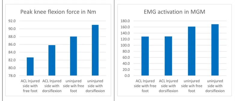
Figure 1. Left: peak knee flexion force in the biodex. Right side: EMG activation in MGM
4.1 Knee flexion torque
Knee flexion peak strength was higher by a mean of 3 Nm with the foot dorsiflexed on both the injured and uninjured side, 3,5% ACLR side and 3,4% uninjured (Figure 1 left side). Mean M. Gastrocnemius medialis (MGM) activation remained the same with both testing methods on the ACL injured side (Figure 1, right side). On the non injured side the mean knee flexion peak strength was also 3 Nm higher with the foot in dorsiflexion and overall knee flexion strength was higher by a mean of 5 Nm compared to injured side (Figure 1 left).
4.2 EMG data on M. Gastrocnemius medialis
EMG recordings showed no major difference between foot dorsiflexion and free foot during knee flexion, except in trials with the foot in dorsiflexion on the non injured side. Here EMG activation was 5% higher than on the ACLR side (Figure 3).
78.0 80.0 82.0 84.0 86.0 88.0 90.0 92.0 ACL Injured side with free foot ACL Injured side with dorsiflexion uninjured side wih free
foot
uninjured side with dorsiflexion
Peak knee flexion force in Nm
0.0 20.0 40.0 60.0 80.0 100.0 120.0 140.0 160.0 180.0 ACL Injured side with free
foot
ACL Injured side with dorsiflexion
uninjured side wih free
foot
uninjured side with dorsiflexion
Table 1. Individual peak knee flexion value (Nm) on all 4 Rep maximum (all participants)
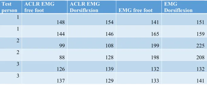
Table 2. Individual muscle activation of MGM during knee flexion moment in the biodex on
all 4 Rep maximum for all 3 participants.
Test
person ACLR Free foot ACLR Dorsiflex Free foot Dorsiflexion
1 76 79 75 78 1 82 82 79 79 2 89 97 92 109 2 86 91 100 106 3 80 85 91 87 3 83 81 91 87 Test person ACLR EMG free foot ACLR EMG
Dorsiflexion EMG free foot
EMG Dorsiflexion 1 148 154 141 151 1 144 146 165 159 2 99 108 199 225 2 88 128 198 208 3 126 139 132 132 3 137 129 133 141
Discussion
The study was a quantitative experimental pilot study choosen for the purpose to study the connection in muscle activation in a healthy vs injured knee in the biodex knee flexion and extension test. This study investigated what effect a forced foot dorsiflexion has on isokinetic knee flexion test when using the biodex machine and if there could be a way to improve the method for testing. But also to se if an increased gastrocnemius activation was present in the injured knee of women due to ACLR with hamstring graft.
The knee flexion strength was significantly lower on the ACLR side even several years since surgery, which is in accordance with data from previous studies (5, 12, 17). Results of EMG recordings showed no major difference between foot dorsiflexion and free foot during knee flexion. Comparing the peak knee flexion force data from the biodex results showed an increase by a mean 3,5% on ACLR side and 3,4% on uninjured with the foot in dorsiflexion for all subjects. Knee flexion strength was lower on the ACLR side even several years since surgery, which is in accordance with data from previous studies (5, 12, 17).No higher EMG activation in MGM on the ACLR side was seen. This could be because tested subjects had a long time since surgery (4-5 years). It would be feasible that subjects with lesser time since ACLR with hamstring graft would have more inhibition in the hamstring musculature and higher activation in other muscles performing knee flexion such M. gastrocnemius. This was the case in a study from Thomeé et al, where knee flexion muscle power LSI in the ACLR with hamstring group, power dropped by 13% post surgery at 6 month follow up (5). The LSI at 6 months was at 10% lower compared to the non injured side indicating that knee flexion power also dropped on the non injured side, likely due to inactivity.
Limitations: A more sportlike position to perform the tests for a specific sport or activity, for example. Laying on the back at prone position in the biodex machine would mean that knee extension and flexion would be performed with the hip in a more neutral position this would represent more the range of motion a fotball player performs in. Sitting in the biodex places the hip at 90° flexion which could put different muscles in more or less optimal working positions. Adouni et al reported that the peak working capacity for hamstring was at 90° flexion (11). Possible bias upon testing was limited by a standard testing procedure,
commands and instructions when testing. A possible weakness could be that subjects had a sports background as well as being physiotherapist students who had great experience in musculoskeletal testing which would have altered test results. This was managed by giving as
little information as possible about the different testing procedures. We were able to follow the study procedure done in project korsband with only minor alterations such as EMG
attached and the ankle in a different position. Also, one extra repetition maximum was used in order to get 4 rep maximum reps split in two reps with the foot in dorsiflexion and two reps without. The testing procedure is widely applied and has little risk for injury and a high reproducibility (18)
A study by Koutras et al concluded that the position of the subject upon testing in the biodex machine had a role in knee flexion activation. They found that the gastrocnemius muscle had a bigger role in knee flexion when testing in prone (lying on the back) as opposed to a seated position (17). Testing in this pilot study was performed in a seated position. It’s likely to yield a higher activation in the EMG in the MGM by testing the subjects in the prone position. The results from this study showed that when subjects had the foot in dorsiflexion the peak knee flexion strength increased on both sides, contrary to the hypothesis that lengthening of the MGM would put it in a less favourable position to perform knee flexion. Increase could be explained by that the M gastrocnemius muscle can pre-tense (isometric plantar flex), thus putting the muscles in the leg in a more favourable position for activation. The Biodex test then suddenly goes from an open kinetic chain to a closed one by putting the foot into a locked dorsiflexed position. A study by Wilk et al questioned the isokinetic testing due to its limitations to functional activity, and the possibility of it to have deleterious effects because it’s an open kinetic chain exercise (18).
6. Conclusion
This pilot study points towards that the follow implications could be beneficial. Making the Isokinetic biodex testing a closed kinetic chain by adding a steel plate for the foot to push against could be a better way to test as realistically as possible. Not to mention early stage rehabilitation exercises, in which many knee flexion exercises in the authors experience are open chain exercises not involving active plantar flexion. Thus, not mimicking ground
reaction force moments. The results suggest that isokinetic knee flexion in the biodex with the foot in forced dorsiflexion increases the power output on young healthy females. Certain changes in inclusion criteria and methodology and more studies in this area is needed to come to a given conclusion.
Reference list
(1) Bahr R, McCrory P, LaPrade R, Meeuwisse W, Engebretsen L. Idrottsskador en illustrerad guide. 2 upplagan. Stockholm. Sisu idrottsböcker. 2015.
(2) Kvist J, Kartus J, Karlsson J, Forssblad M. Results From the Swedish National Anterior Cruciate Ligament Register. Division of Physiotherapy, Erratum in Arthroscopy. 2014 Aug;30(8):1044.
(3) Shumway-Cook A, Woollacott, M. H. Motor control: Translating research into clinical practice. Fifth edition . Philadelphia. Wolters Kulwer (2017).
(4) Thing, LF. “Voices of the broken body”: the resumption of non-professional female players’ careers after anterior cruciate ligament injury. The female player’s dilemma: is she willing to run the risk? Scand J Med Sci Sports. 2006;16: page 369.
(5) Thomeé, R., Neeter, C., Gustavsson, A., Thomeé, P., Augustsson, J., Eriksson, B., Karlsson, J. Variability in leg muscle power and hop performance after anterior cruciate ligament reconstruction. Knee Surg Sports Traumatol Artrosc. Springer-Verlag. 2012. (6) Beischer S, Senorski E-H , Thomeé C, Samuelsson K, Thomeé R. Young athletes return too early to knee-strenuous sport, without acceptable knee function after anterior cruciate ligament reconstruction. Knee Surg Sports Traumatol Arthrosc. 2018 Jul;26(7):1966-1974 (7) Kendall F-P. Muscles, Testing and function with posture and pain. 5e upplagan. Lippincott Williams & Wilkins. 2005.
(8) Konrad, P. The ABC of EMG: A Practical Introduction to Kinesiological Electromyography (phamplet). Arizona. Noraxon Usa inc. Version 1,4. 2006.
(9) Hohmann, E. Tetsworth & K. Glatt, V. The hamstring/quadriceps ratio is an indicator of function in ACL-deficient, but not in ACL-reconstructed knees. Arch Orthop Trauma Surg. 2019 Jan;139(1):91-98.
(10) Biscarini A, Contemori S, Busti, D, Botti F, Enrico Pettorossi V. Knee flexion with quadriceps cocontraction: A new therapeutic exercise for the early stage of ACL
rehabilitation. Journal of Biomechanics. 49. 10.1016/j.jbiomech.2016.10.026.
(11) Adouni MBA, Marouane H, Shirazi-Adl A (2015). Role of Gastrocnemius Activation in Knee Joint Biomechanics: Gastrocnemius Acts as an ACL Antagonist. Computer Methods in Biomechanics and Biomedical Engineering. Comput Methods Biomech Biomed Engin. 2016;19(4):376-85
(12) Kim, H. J., Lee, J. H., Ahn, S. E., Park, M. J., & Lee, D. H. (2016). Influence of Anterior Cruciate Ligament Tear on Thigh Muscle Strength and Hamstring-to-Quadriceps Ratio: A Meta-Analysis. PLoS One. 2016 Jan 8;11(1):e0146234
(13) Olsson, H. & Sörensen, S. Forskningsprocessen: Kvalitativa och kvantitativa perspektiv. 3. Uppl. Stockholm. Liber. (2011).
(14) Drouin J, Valovich McLeod T, Shultz S, Gansneder B, H Perrin D. Reliability and validity of the Biodex System 3 Pro isokinetic dynamometer velocity, torque and position measurements. Eur J Appl Physiol. 2004 Jan;91(1):22-9
(15) Khoddami S, Talebian S, Izadi F, nakhosin Ansari N. Validity and Reliability of Surface Electromyography in the Assessment of Primary Muscle Tension Dysphonia . J Voice. 2017 May;31(3):386.e9-386.e17
(16) Seniam (wy) Noraxon EMG placement Gastrocnemius medialis [Internet] : Hämtad 24-03-19 från http://seniam.org
(17) Koutras G, Bernard M, Terzidis IP, Papadopoulos P, Georgoulis A, Pappas E.
Comparison of knee exion isokinetic de cits between seated and prone positions after ACL reconstruction with hamstrings graft: Implications for rehabilitation and return to sports decisions. J Sci Med Sport. 2016;19:559–62.
(18) Wilk KE, Romaniello WT, Soscia SM, Arrigo CA, & Andrews JR. The relationship between subjective knee scores, isokinetic testing, and functional testing in the ACL-reconstructed knee. Journal of Orthopaedic and Sports Physical Therapy (JOSPT). 1994; 20(2), 60-73.
Bilaga 1 informationbrev
Hej! Har du genomfört en korsbands operation eller känner någon som gjort det?
Jag går sista terminen på Fysioterapiprogrammet på Luleå tekniska universitet och skriver nu mitt Examensarbete.
Rehabilitering efter en korsbandskada är omfattande och tidskrävande. Målet med den här studien är att se hur olika musklerna jobbar i knäet & resultaten kan hjälpa till att förbättra rehabiiteringsprocessen för opererade korsband.
Vi kommer testa styrka och muskelaktivering i benböj & sträck sittandes. Uppvärming på stationär cykel följt av knätester där elektroder kommer mäta muskelaktivering, tidsåtgång ca 35-40 minuter. Resultat av testera kommer hanteras konfidentiellt och inga namn kommer nämnas i uppsatsen. Dina testresultat kan på begäran skickas till din mail om du är intresserad av din knästyrka. Deltagare måste vara mellan 18-55 år.
Hör av er till mig via sms till 0706870810 eller mail för frågor och mer information Kristoffer Lindbäck – Mail :Krilio-5@student.ltu.se