JONAS ANDERUD
ON GUIDED BONE
REGENERATION USING
CERAMIC MEMBRANES
JON AS ANDERUD MALMÖ UNIVERSIT Y 20 ON GUIDED BONE REGENER A TION USIN G CER AMIC MEMBR ANES DOCT OR AL DISSERT A TION IN ODONT OL OG YO N G U I D E D B O N E R E G E N E R A T I O N U S I N G C E R A M I C M E M B R A N E S
Malmö University
Faculty of Odontology Doctoral Dissertation 2016
© Jonas Anderud 2016 ISBN 978-91-7104-676-5 (print) ISBN 978-91-7104-677-2 (pdf) Holmbergs, Malmö 2016
JONAS ANDERUD
ON GUIDED BONE
REGENERATION USING
CERAMIC MEMBRANES
Malmö University, 2016
Faculty of Odontology
Department of Prosthodontics
This publication is available in electronic format at: http://dspace.mah.se/handle/2043/20132
To Malin, August and Linn To my Mother and Father
This thesis is number 48 in a series of investigations on implants, hard tissue, and the locomotor apparatus originating from the Department of Biomaterials, University of Gothenburg and the Department of Prosthodontics/Material Sciences, Malmö University, Sweden. 1. Anders R Eriksson DDS, 1984. Heat-induced Bone Tissue
Injury. An in vivo investigation of heat tolerance of bone tissue and temperature rise in the drilling of cortical bone. Thesis defended 21.2.1984. External examiner: Docent K-G. Thorngren.
2. Magnus Jacobsson MD, 1985. On Bone Behaviour after Irradiation. Thesis defended 29.4.1985. External examiner: Docent A. Nathanson.
3. Fredric Buch MD, 1985. On Electrical Stimulation of Bone Tissue. Thesis defended 28.5.1985. External examiner: Docent T. Ejsing-Jörgensen.
4. Peter Kälebo MD, 1987. On Experimental Bone Regeneration in Titanium Implants. A quantitative microradiographic and histologic investigation using the Bone Harvest Chamber. Thesis defended 1.10.1987. External examiner: Docent N. Egund.
5. Lars Carlsson MD, 1989. On the Development of a new Concept for Orthopaedic Implant Fixation. Thesis defended 2.12.1989. External examiner: Docent L-Å Broström. 6. Tord Röstlund MD, 1990. On the Development of a New
Arthroplasty. Thesis defended 19.1.1990. External examiner: Docent Å. Carlsson.
7. Carina Johansson Res Tech, 1991. On Tissue Reaction to Metal Implants. Thesis defended 12.4.1991. External examiner: Professor K. Nilner.
8. Lars Sennerby DDS, 1991. On the Bone Tissue Response to Titanium Implants. Thesis defended 24.9.1991. External examiner: Dr J.E. Davies.
9. Per Morberg MD, 1991. On Bone Tissue Reactions to Acrylic Cement. Thesis defended 19.12.1991. External examiner: Docent K. Obrant.
10. Ulla Myhr PT, 1994. On factors of Importance for Sitting in Children with Cerebral Palsy. Thesis defended 15.4.1994. External examiner: Docent K. Harms-Ringdahl.
11. Magnus Gottlander MD, 1994. On Hard Tissue Reactions to Hydroxyapatite- Coated Titanium Implants. Thesis defended 25.11.1994. External examiner: Docent P. Aspenberg. 12. Edward Ebramzadeh MScEng, 1995. On Factors Affecting
Long-Term Outcome of Total Hip Replacements. Thesis defended 6.2.1995. External examiner: Docent L. Linder. 13. Patricia Campbell BA, 1995. On Aseptic Loosening in Total
Hip Replacement: the Role of UHMWPE Wear Particles. Thesis defended 7.2.1995. External examiner: Professor D. Howie.
14. Ann Wennerberg, DDS, 1996. On Surface Roughness and Implant Incorporation. Thesis defended 19.4.1996. External examiner: Professor P-O. Glantz.
15. Neil Meredith BDS MSc FDS RCSm, 1997. On the Clinical Measurement of Implant Stability Osseointegration. Thesis defended 3.6.1997. External examiner: Professor J. Brunski. 16. Lars Rasmusson DDS, 1998. On Implant Integration in
Membrane-Induced and Grafter Bone. Thesis defended 4.12.1998. External examiner: Professor R. Haanaes. 17. Thay Q Lee MSc, 1999. On the Biomechanics of the
Patellfemoral Joint and Patellar Resurfacing in Total Knee Arthroplasty. Thesis defended 19.4.1999. External examiner: Docent G. Nemeth.
18. Anna Karin Lundgren DDS, 1999. On Factors Influencing Guided Regeneration and Augmentation of Intramembraneous Bone. Thesis defended 7.5.1999. External examiner: Professor B. Klinge.
19. Carl-Johan Ivanoff DDS, 1999. On Surgical and Implant Related Factors Influencing Integration and Function of Titanium Implants. Experimental and Clinical Aspects. Thesis defended 12.5.1999. External examiner: Professor B. Rosenquist.
20. Bertil Friberg DDS MDS, 1999. On Bone Quality and Implant Stability Measurements. Thesis defended 12.11.1999. External examiner: Docent P. Åstrand.
21. Åse Allansdotter Johansson MD, 1999. On Implant
Integration in Irradiated Bone. An Experimental Study of the Effects of Hyperbaric Oxygeneration and Delayed Implant Placement.
Thesis defended 8.12.1999. External examiner: Docent K. Arvidsson-Fyrberg.
22. Börje Svensson FFS, 2000. On Costochondral Grafts Replacing Mandibular Condyles in Juvenile Chronic Arthritis. A
Clinical, Histologic and Experimental Study. Thesis defended 22.5.2000. External examiner: Professor Ch. Lindqvist. 23. Warren Macdonald BEng, MPhil, 2000. On Component
Integration on Total Hip Arthroplasties: Pre-Clinical Evaluations.
Thesis defended 1.9.2000. External examiner: Dr A.J.C. Lee. 24. Magne Røkkum MD, 2001. On Late Complications with
HA Coated Hip Arthroplasties. Thesis defended 12.10.2001. External examiner: Professor P. Benum.
25. Carin Hallgren Höstner DDS, 2001. On the Bone Response to Different Implant Textures. A 3D analysis of roughness, wavelength and surface pattern of experimental implants. Thesis defended 19.11.2001. External examiner: Professor S. Lundgren.
26. Young-Taeg Sul DDS, 2002. On the Bone Response to Oxidised Titanium Implants: The role of microporous structure and chemical composition of the surface oxide in enhanced osseointegration. Thesis defended 7.6.2002. External examiner: Professor J.E. Ellingsen.
27. Victoria Franke Stenport DDS, 2002. On Growth Factors and Titanium Implant Integration in Bone. Thesis defended 11.6.2002. External examiner: Associate Professor E. Solheim. 28. Mikael Sundfeldt MD, 2002. On the Aetiology of Aseptic
Loosening in Joint Arthroplasties and Routes to Improved cemented Fixation. Thesis defended 14.6.2002. External examiner: Professor N. Dahlén.
29. Christer Slotte CCS, 2003. On Surgical Techniques to Increase Bone Density and Volume. Studies in Rat and Rabbit. Thesis defended 13.6.2003. External examiner: Professor C.H.F. Hämmerle.
30. Anna Arvidsson MSc, 2003. On Surface Mediated Interactions Related to Chemomechanical Caries Removal. Effects
on surrounding tissues and materials. Thesis defended 28.11.2003. External examiner: Professor P. Tengvall. 31. Pia Bolind DDS, 2004. On 606 retrieved oral and
cranio-facial implants. An analysis of consequently received human specimens. Thesis defended 17.12.2004. External examiner: Professor A. Piattelli.
32. Patricia Miranda Burgos DDS, 2006. On the influence of micro- and macroscopic surface modifications on bone integration of titanium implants. Thesis defended 1.9.2006. External examiner: Professor A. Piattelli.
33. Jonas P. Becktor DDS, 2006. On factors influencing the outcome of various techniques using endosseous implants for reconstruction of the atrophic edentulous and partially dentate maxilla. Thesis defended 17.11.2006. External examiner: Professor K.F. Moos.
34. Anna Göransson DDS, 2006. On Possibly Bioactive CP Titanium Surfaces. Thesis defended 8.12.2006. External examiner: Professor B. Melsen.
35. Andreas Thor DDS, 2006. On platelet-rich plasma in reconstructive dental implant surgery. Thesis defended 8.12.2006. External examiner: Professor E.M. Pinholt. 36. Luiz Meirelles DDS MSc, 2007. On Nano Size Structures for
Enhanced Early Bone Formation. Thesis defended 13.6.2007. External examiner: Professor Lyndon F. Cooper.
37. Pär-Olov Östman DDS, 2007. On various protocols for direct loading of implant- supported fixed prostheses. Thesis defended 21.12.2007. External examiner: Professor B. Klinge. 38. Kerstin Fischer DDS, 2008. On immediate/early loading of
implant supported prostheses in the maxilla. Thesis defended 8.2.2008. External examiner: Professor K. Arvidsson Fyrberg. 39. Alf Eliasson 2008. On the role of number of fixtures, surgical
technique and timing of loading. Thesis defended 23.5.2008. External examiner: Professor K. Arvidsson Fyrberg.
40. Victoria Fröjd DDS, 2010. On Ca2+ incorporation and nanoporosity of titanium surfaces and the effect on implant performance. Thesis defended 26.11.2010. External examiner: Professor J.E. Ellingsen.
41. Lory Melin Svanborg DDS, 2011. On the importance of nanometer structures for implant incorporation in bone tissue. Thesis defended 01.06.2011. External examiner: Associate professor C. Dahlin.
42. Byung-Soo Kang MSc, 2011. On the bone tissue response to surface chemistry modifications of titanium implants. Thesis defended 30.09.2011. External examiner: Professor J. Pan. 43. Kostas Bougas DDS, 2012. On the influence of biochemical
coating on implant bone incorporation. Thesis defended 12.12.2012. External examiner: Professor T. Berglundh.
44. Arne Mordenfeld DDS, 2013. On tissue reaction to and adsorption of bone substitutes. Thesis defended 29.5.2013. External examiner: Professor C. Dahlin.
45. Ramesh Chowdhary DDS, 2014. On efficacy of implant thread design for bone stimulation. Thesis defended 21.05.2014. External examiner: Professor Flemming Isidor.
46. Anders Halldin MSc, 2015. On a biomechanical approach to analysis of stability and load bearing capacity of oral implants. Thesis defended 28.05.2015. External examiner: Professor J. Brunski.
47. Francesca Cecchinato MSc, 2015. On magnesium-modified titanium coatings and magnesium alloys for oral and orthopaedic applications: in vitro investigation. Thesis defended 20.11.2015. External examiner: Professor C. Stanford.
48. Jonas Anderud DDS, 2016. On guided bone regeneration using ceramic membranes. Thesis to be defended 27.05.2016. External examiner: Professor S. Lundgren
THESIS AT A GLANCE
THE
SI
S
A
T
A
G
LA
N
C
E
St udy O bj ec ti ve s M et hods Illu st ra tio ns M ai n f indi ng s/ conc lus ions (I) Gui de d bone a ug m ent at ion us in g a cer am ic s pa ce -m ai nt ai ni ng d ev ic e T o e val uat e 3 -d im en sio na lly w het her ver ti cal b on e aug m ent at ion c an be ach iev ed us ing a hy dr oxy apa ti te sp ac e-m ai nt ai ni ng d ev ic e in a ra bbi t cal var ia m od el 48 hol low dom es in 4 di ff er en t d es ig ns w er e p la ced subpe ri os te al ly on r abbi t cal var ia . T he res ul ts w er e an al yz ed 3 -d im en sio na lly us in g m ic ro -CT . A la rg er pr oduc ti on of bone vol um e w he n us ing a n occl us iv e, d en se hy dr oxy apa ti te spa ce m ai nt ai ni ng de vi ce w it h a r oug h i nne r su rf ac e. (II) Gui de d bone a ug m ent at ion us in g cer am ic s pa ce -m ai nt ai ni ng d ev ic es : t he im pa ct o f ch em is try T o e va lua te w hi ch of m ic ro po ro us h yd ro xy ap at it e and z ir coni a sp acem ai nt ai ni ng d ev ices w ere m ost su it ab le f or gui de d bone g ene ra ti on. 24 12 Z ir coni a +12 H A µ w er e p laced o n r ab bi t cal var ia . T he res ul ts w er e an al ys ed u si ng l igh t m ic ro sc op y an d SE M . T he e ffe ct o f th e m ic ro po ro us s tr uc tu re o f hy dr oxy apa ti te s ee m s to fa ci lita te fo r th e b on e c el ls to adhe re t o t he m at er ia l a nd tha t z ir coni a e nha nc e a slig ht ly la rg er v olu m e o f ne w ly f or m ed bone . (III) The im pa ct o f su rfa ce roug hne ss a nd pe rm ea bi lit y in h yd rox ya pa ti te b on e reg en er at io n m em br an es T o ev al ua te t he i m pa ct o f sur fa ce r oug hne ss a nd pe rm ea bi lit y of de ns e hy dr oxy apa ti te s pa ce m ai nt ai ni ng d ev ic es . 36 hy dr oxy apa ti te s pa ce m ai nt ai ni ng d ev ic es w er e pl ac ed o n r ab bi t c al var ia. T he re su lts w ere a na ly se d us ing li ght m ic ros copy a nd SE M . A m ode ra te ly r oug h i nne r su rf ace o f a cer am ic m em br an e al on g w it h a no n-per m ea bl e d ev ice p ro du ces m or e bone t ha n a s m oot h i nne r su rf ac e. (IV ) G ui de d bone r eg ene ra ti on us in g in div id ua liz ed c er am ic sh eet s T o de sc ri be a ne w m et hod of gui de d bone r eg ene ra ti on us in g in div id ua liz ed c er am ic m em br an es . 3 pa ti ent s ha d G B R tr ea tm en t w ith in di vi du al iz ed c er am ic m em br an es . A ft er he al in g of 7 m ont hs t he m em br ane s w er e r em oved an d t he r es ul t an al ys ed . C er am ic in div id ua liz ed m em br an es ca n r eg en er at e la rg e vo lu m es o f bo ne in ve rt ic al a nd hor iz ont al di re ct ions w it h g ood bi ol og ica l a ccep ta nce o f t he m ate ri al .ABSTRACT
Regeneration of bone in the oral and maxillofacial region can be achieved with different techniques such as autologous bone grafts, bone substitutes and guided bone regeneration. Guided bone regeneration is defined as creating a space between the bone and its surrounding tissues, using a barrier that allows new bone to migrate into the space while preventing other cell types from interfering. The barrier material should be biocompatible, have suitable occlusive properties and be able to maintain the created space for bone regeneration. A wide range of different materials has been used.
The general aim was to evaluate a novel method of guided bone regeneration using designed ceramic space maintaining devices on animals and humans.
An experimental rabbit model was used in studies I, II and III. 60 different domes shaped as halfspheres were fixed with titanium screws to the skull bone of 30 rabbits. The domes had 5 different characteristics; 1) Dense hydroxyapatite with a moderately rough inner surface (HA rough), 2) Dense hydroxyapatite with a smooth inner surface (HA smooth), 3) Microporous hydroxyapatite with a moderately rough inner surface (HA μ), 4) Dense hydroxyapatite with a moderately rough inner surface and macroscopic holes (HA holes) and 5) Zirconia with a moderately rough inner surface (Zirconia). The domes were left to heal for 12 weeks before the animals were euthanized and the results were analysed with histomorphology and micro-CT.
The results revealed that Zirconia with a moderately rough inner surface produced the largest amount of newly formed bone although the results were difficult to interpret as the Zirconia domes were difficult to X-ray because of the very dense nature of the material.
In study IV, 3 patients had bone regeneration treatment with a Zirconia barrier. Patient 1 had posterior maxillary bone deficiency in a transverse direction. Patient 2 had anterior maxillary bone deficiency in a vertical and transverse direction. Patient 3 had posterior mandibular bone deficiency in a vertical direction. Based on individual digital models, Zirconia membranes corresponding to the amount of bone intended to be regenerated were designed and manufactured. The Zirconia membranes, were attached to the underlying bone with titanium screws and covered with the periosteum and mucosa. After a mean healing time of 7 months the membranes were removed and dental implants were installed.
The results showed new bone regeneration corresponding to the design of the space maintained. None of the patients had any major complications aside from normal postoperative discomfort. According to evaluation with CBCT all patients gained new bone in the desired and preoperatively planned region.
LIST OF PAPERS
I: Guided bone augmentation using a ceramic space-maintaining device.
Anderud J, Jimbo R, Abrahamsson P, Isaksson SG, Adolfsson E, Malmström J, Kozai Y, Hallmer F, Wennerberg A
Oral Surg Oral Med Oral Pathol Oral Radiol. 2014 Nov;118(5):532-8 II: Guided bone augmentation using ceramic space-maintaining devices: the impact of chemistry.
Anderud J, Jimbo R, Abrahamsson P, Isaksson S, Adolfsson E, Malmström J, Naito Y, Wennerberg A
Clin Cosmet Investig Dent 2015 Mar 12;7:45-53.
III: The impact of surface roughness and permeability in hydroxyapatite bone regeneration membranes.
Anderud J, Abrahamsson P, Jimbo R, Adolfsson E, Malmström J, Wennerberg A
Clin Oral Implants Res. 2015 Oct 15. doi: 10.1111/clr.12717. (Epub ahead of print)
IV: Guided Bone Regeneration using using Individualized Ceramic Sheets.
Malmström J, Anderud J, Abrahamsson P, Wälivaara DÅ, Isaksson SG, Adolfsson E
Submitted to International Journal of Oral and Maxillofacial Surgery 2015
TABLE OF CONTENTS
THESIS AT A GLANCE ... 12 ABSTRACT ... 13 LIST OF PAPERS ... 15 INTRODUCTION ... 19 Clinical problem ...19 Bone ...19Current methods of bone augmentation ...21
Autogenic grafts ...21
Allogenic grafts ...21
Xenogenic grafts ...22
Alloplastic grafts ...22
Other methods of augmentation ...22
Guided bone regeneration ...24
Experimental vertical guided bone regeneration ...27
Ceramics and Bio-ceramics ...31
HYPOTHESIS ... 34
AIMS ... 35
MATERIALS AND METHODS ... 36
Preoperative investigations ...36
Paper I-III ...37
Surface topography ...39
Animals and anaesthesia and surgery ...39
Preparation of samples ...40
Three-dimensional trabecular bone analysis study 1 ...41
Histological analyses study 2 and 3 ...42
Paper IV ...43
Surgery ...44
Measuring of gained bone volume and soft tissue topographic ...45
Sample preparation ...46
RESULTS ... 47
Surface measurements ...47
Three dimensional results from micro-CT investigation ...47
Histological results ...52 Paper IV ...57 Clinical outcome ...57 Materials ...58 Radiologic evaluation ...59 Histological outcomes ...60 DISCUSSION ... 61 Surface roughness ...61 Material porosity ...63 Permeability ...64 Zirconia ...65
Individualized membranes in a clinical situation ...66
CONCLUSIONS ... 72
POPULÄRVETENSKAPLIG SAMMANFATTNING ... 73
ACKNOWLEDGEMENTS ... 75
REFERENCES ... 77
INTRODUCTION
Clinical problem
According to Wolff’s law (Wolff, 1892) the bone in a healthy human being or animal will adapt to the loads under which it is placed. After loss of teeth the alveolar processes are no longer exposed to chewing forces. This may result in loss of dentoalveolar bone. Surgery, infection and other traumatic loss of alveolar bone can alter the contour and thereby the volume of the alveolar process. Large, critical sized bony defects will not regenerate to full volume spontaneously.
Treatment with dental implants is a common and well-documented method of supporting dental prosthesis. Dental implant treatment is considered to have a good long-term prognosis. To allow successful implantation, the jawbone needs sufficient quality and volume. Low quality or volume of bone can jeopardize the outcome of such treatment. Interference of anatomical structures such as the inferior alveolar nerve or the maxillary sinuses reduces the possibility to install dental implants.
If the bone volume is insufficient in volume it can be augmented by various methods. Several methods and techniques to increase bone volume and restore to the original shape of the bone are at hand. The gold standard is still considered to be transplantation of autogenous cortical bone as blocks or as particlutated bone.
Bone
The definition of bone is “a hard form of connective tissue composed of osteocytes and calcified collagenous intracellular substance arranged in thin plates” (Mosby, 2012). Its role is to support and
protect other tissues and organs, to be a reservoir for minerals and be involved in mineral homeostasis. The marrow of bone hosts the production of blood cells. In adults there are two types of bone tissue; compact, cortical bone and trabecular (cancellous, spongy) bone. Compact bone forms the outer layer and represents nearly 80% of the mass of the skeleton. It consists of several passages for diffusion of blood vessels and molecules. The blood vessels interconnect through perforated canals with vessels on the surface outside the bone where the periosteum is present. Trabecular bone is less dense than compact bone. It is built of plates and bars of bone along irregular cavities that contain red bone marrow. The trabeculae seem to be arranged randomly but are still able to provide maximum strength as they follow the lines of stress. There are two types of cells that contribute to bone homeostasis; osteoblasts and osteoclasts. These cells are important in the remodelling and repair of bone tissue. Osteoblasts are bone-forming cells that form osteoid by depositing extracellular matrix (collagen). The osteoid becomes mineralised by calcium withdrawn from blood. Some of the osteoblasts differentiate by entrapment into osteocytes. Osteoblasts can be found on the outer surface of bone and bone cavities. Osteoclasts are devoted to bone resorption as bone are involved in a continuous cycle of resorption and apposition. This process is called “bone turnover”.
The periosteum is a structure composed of 2 layers. The outer “fibrous” layer consists of fibroblasts, collagen and elastin along with a nerve and micro- vascular network. These components provide mechanical stability to the periosteum. The inner “cambium layer” is densly populated with cells that influence bone formation and bone repair (Allen et al., 2004). These features provide the periosteum with regenerative capacity. Because of its high vascularity, numerous endothelial pericytes can be found within the periosteum (Diaz-Flores et al., 1992). Pericytes are cells in physical contact with capillary endothelial cells. These cells can differentiate into several cell types, including osteoblasts (Reilly et al., 1998).
Bone fracture healing is initially characterized by an acute inflammatory response that includes blood clot formation, inflammatory cell migration and granular tissue formation. The inflammatory phase is believed to stimulate cell migration and proliferation of mesenchymal cells. Following the inflammation
period, mesenchymal cells aggregate at the repair site and differentiate into chondroblasts and osteoblasts. A collagen matrix is formed and mineralized. These events result in soft callus formation that fuses the 2 fracture margins together. With time, the soft callus will continue to ossify and woven bone will form. Eventually the healing bone will restore its original shape and structure (Colnot et al., 2012, Dimitriou et al., 2011).
The systematic recruitment of cells during bone repair is mainly dependent on skeletal progenitors from the bone marrow (Taguchi et al., 2005). These cells are brought to the injured site via blood vessels. As the systemic recruitment of skeletal progenitors during bone repair appears to be minimal, the recruitment within the local environment predominates. These skeletal progenitors may come from the bone marrow within the injured bone, from the surrounding periosteum and from soft tissues close to the bone (Einhorn, 1995). The source of cells and route of delivery are still being investigated experimentally (Colnot, 2011).
Current methods of bone augmentation
Autogenic grafts
Autogenous bone can be transplanted from intraoral approaches as well as from extra-oral approaches such as the iliac crest. Autogenous bone has proved to be both osteoinductive (contains living cells and proteins needed for new bone formation and healing) and osteoconductive (can act as a scaffold for bone forming cells and blood vessels). A disadvantage of autogenous bone grafting is morbidity of the donor site. This means often that a second wound needs to be made which can be uncomfortable for the patient. Prolonged surgical time and graft resorption are other bone grafting disadvantages.
Allogenic grafts
Allogenic bone graft is bone harvested from another individual of the same species. Different allografts have been evaluated such as; Mineralized, demineralized, frozen or freeze-dried bone. Allografts are considered to be osteoconductive but most likely have less osteoinductive properties due to the absences of living cells. Allogenic bone grafts have routinely been used in orthopaedic surgery. Gapski
et al showed in 2006 that the use of allografts in sinuslift procedures
can be successful (Gapski et al., 2006).
Xenogenic grafts
Xenogenic bone grafts originate from other species. Examples include bovine or porcine sources. To obtain immunological safety, necessary to prevent rejection of the graft, the proteins of this bone have been removed by different procedures. Hereby the osteoinductive effect is removed and the graft can only act as an osteoconductive scaffold (Calvo Guirado et al., 2013). The most frequently used and evaluated xenogenic bone graft is deproteinized bovine bone (Esposito et al., 2006).
Alloplastic grafts
Alloplastic bone grafting materials are synthetically produced in many different compositions such as calcium-based ceramics, calcium- sulphates and bioactive glass. Different biological and mechanical characteristics have been reported such as dissolving rate and speed. In order to combine different properties, different materials are mixed together. For example Bone Ceramic® (Straumann, Basel, Switzerland), that consists of 60% hydroxyapatite and 40% β-tricaliumphosphate. The aim is hereby to let the more soluble β-tricaliumphosphate dissolve to give space to bone forming cells and blood vessels and let the harder hydroxyapatite act as a osteoconductive scaffold.
Other methods of augmentation
If the transplanted bone is particulated it needs to be stabilised by a mesh or membrane to stay in place. Titanium mesh has been used to stabilize various kinds of bone transplants. (Rasia-dal Polo et al., 2014).
This method has been described as successful both in horizontal and in vertical direction of new bone formation (Corinaldesi et al., 2009). However disadvantages have also been described such as penetration of the oral mucosa covering the titanium mesh with an unfavourable aesthetic result (von Arx et al., 1996). Between 0-50% of the meshes inserted have shown complications in the form of mucosal penetration. However von Arx states that this complication doesn’t necessarily jeopardize the functional outcome of the treatment.
Splitting the alveolar crest is another method where the maxillary alveolar crest is too narrow but has the height necessary for implant installation.
Hereby the alveolar crest is divided with instruments to allow implants to be installed between the buccal and palatal bone plates. This method is difficult to perform and has not been well evaluated in long term outcomes (Garcez-Filho et al., 2015) although favourable short term results (6 months) have been reported (Simion et al., 1992). When the width of the maxilla is sufficient for implant installation but the height is not, the floor of the maxillary sinus along with the Schnederian membrane can be elevated, so called sinuslift. Hereby
Figure 2. Installaton of dental implants using split crest method
the membrane is lifted with either the implant itself or as a two-stage procedure with a bone graft. The clinical effectiveness of sinuslift procedures is well described (Johansson et al., 2013, Johansson et al., 2010) with good survival rates of the inserted implants. According to the literature the most common complication with sinuslift procedures is perforation of the sinus membrane (Shlomi et al., 2004) which will adversely affect the implant survival rate.
To avoid damage to the inferior alveolar nerve in the mandible, short implants have been suggested in the premolar and molar region (Slotte et al., 2012). This seems to be a promising technique with similar survival as longer implants as long as the implants are connected with a dental bridge. The prosthodontic constructions on these implants may have an unfavourable ratio of height to implant length. This has not been shown to affect the survival or success rate, but it might cause unfavourable hygienic problem for the patient. Another method to avoid damage to the inferior alveolar nerve is nerve transposition. Using this technique the bone covering the inferior alveolar nerve is removed and the nerved is transferred laterally allowing insertion of standard dental implants without disturbing the nerve. Success rates varying from 76-100% have been reported (Khojasteh et al., 2015). However permanent dysfunction of the nerve or even mandibular fracture, although very rare, might occur postoperatively. Therefore this technique is rarely used today.
Guided bone regeneration
Guided Bone Regeneration (GBR) is defined as creating a space between the bone surface and surrounding soft tissues, using a barrier that allows new bone to migrate into the space while preventing other cell types from interfering. Murray et al described the principle of physically sealing off an anatomical site for improved healing of bone in 1957 (Murray et al., 1957), followed by Hurley in 1959 who described when cell-occlusive membranes were used for spinal fusions (Hurley et al., 1959). In this publication it was stated that three things were necessary for regeneration of bone; 1) the presence of a blood clot, 2) preserved osteoblasts, and 3) contact with living tissue. Hurley and co-workers protected the blood clot with a plastic cage and the interior of the cage became filled with bone. Kahnberg evaluated this concept experimentally in 1979 (Kahnberg, 1979).
Two- and three-wall defects in the mandible of rabbits were sealed with subperiostal implanted Teflon barriers, called Teflon leafs. These barriers prevented ingrowth of fibrous scar tissue, allowing bone regeneration to occur.
The barrier used has to be constructed of biocompatible material(s). The interaction between the material and tissue should not adversely affect the surrounding tissue, or the intended healing result. Furthermore the barrier should have suitable occlusive properties to prevent invasion of fibrous connective (scar) tissue and provide some degree of protection from bacterial invasion if it becomes exposed to the oral environment. The barrier also needs to provide a suitable space into which bone regeneration can occur. Space making provides necessary volume with specific geometry for functional reconstruction. The barrier should be capable of integrating with the surrounding tissues. This helps to stabilize the blood clot and the healing wound. Finally the barrier needs to be clinically manageable in terms of sterilization.
A wide range of barrier membranes have been used including polytetrafluoroethylene (PTFE), expanded PTFE (e-PTFE), collagen, freeze-dried dura mater, dura mater, allografts, polyglactin 910, polylactid acid, polyglycolic acid, polyortoester, polyurethane, polyhydroxybutyrate, calcium sulphate and titanium meshes (Hammerle and Jung, 2003). These barriers can be divided into non-resorbable and resorbable membranes. As e-PTFE was the first material described with successful results it became the standard material for guided bone regeneration. E-PTFE is a polymer that is highly stable in biological environments, resists breakdown and is claimed to not cause any immunological reactions (Carbonell et al., 2014). However, a potential complication may occur when membranes are used in conjunction with implants as membrane exposure may be followed by infection (Becker et al., 1994). The pathway of new bone formation by means of GBR using barrier membranes is yet not described in detail but is based on the same principles as fracture bone healing and bone remodelling. The membranes are used for space maintenance over a defect, promoting the ingrowth of osteogenic cells and preventing migration of undesired cells from the overlying soft tissues into the wound (Dahlin et al., 1989, Nyman et al., 1982). Protection of a blood clot in the defect, exclusion of
gingival connective tissue and provision of a secluded space into which osteogenic cells from the bone can migrate are essential for a successful outcome. The sequence of bone healing in GBR is not only affected by invasion of non-osteogenic tissue, but even more so by the defect size and morphology. To be able to stabilize and thereby protect the blood clot, titanium reinforced e-PTFE membranes are available on the market. The titanium threads inserted in the membrane add more rigidity to the material that will further stabilize the blood clot and maintain the space created.
Figure 3. Guided bone regeneration using e-PTFE membrane
High-density polytetrafluoroethylene (hd-PTFE) membranes offer an interesting alternative to e-PTFE (Bartee, 1995, Bartee and Carr, 1995). Hd-PTFE is made of 100% PTFE, which is non-porous, non-expanded and non-permeable (Marouf and El-Guindi, 2000). The thickness of commercial available membranes ranges from 0,13-0,25 mm. Different sizes and textures are available. As the hd-PTFE membranes are totally occlusive they show minimal degree of infection and inflammation if exposed to the environment in the oral cavity (Krauser, 1996).
Experimental vertical guided bone regeneration
Many authors have described experimental vertical guided bone regeneration. In 1993, Linde et al used e-PTFE membrane formed as a dome to regenerate new bone on a rat skull (Linde et al., 1993a, Linde et al., 1993b). The domes were placed on calvaria and covered with the periosteum and skin. After healing for 9 and 16 weeks they found that about 80% of the dome volume was filled with new bone. This experiment was followed by similar studies by Schmid et al 1994, Kostopoulos et al 1994 and 1995 and Lundgren et al 1995 (Schmid et al., 1994, Kostopoulos et al., 1994, Lundgren et al., 1995). Several authors have used this technique to evaluate different bone filler materials with different results. However, the most interesting studies were performed without filling the interior of the barriers or domes with any material, and instead allowing the blood clot formed inside to transform into new bone. Among these studies the most commonly used material is titanium, either formed as hollow domes (half spheres) or as cylinders. Other materials used are plastics i.e. Teflon/e-PTFE, ceramics and poly-lactic acid. Different properties of the barriers and their effect on bone regeneration have been studied, such as textured surfaces, macro permeability of barriers and decortication of residual bone. The experimental devices used vary in shape and size. The maximum height of the devices tested is 6mm reaching from the residual bone to the central inner part of the device (Van Steenberghe et al., 2003).
Experience regarding how textured surfaces influence vertical guided bone regeneration under barrier membranes is very limited (Guo et al., 2013, Lundgren et al., 1999, Lee et al., 2013). The results suggest that regeneration of new bone occurs in larger amounts if the inner surface of the barrier is rough. The theory behind this is controversial and the subject of debate. One theory is that a textured surface might stabilize a blood clot better than a smoother surface. It is well known that titanium implants with a moderately rough surface are considered to stimulate the strongest bone integration (Albrektsson and Wennerberg, 2004b, Albrektsson and Wennerberg, 2004a). Wennerberg made a proposal of an optimal surface topography for bone integration of titanium implants in her thesis 1996 (Wennerberg, 1996). It was proposed that a surface
with an average height deviation of 1,5 μm, an average wavelength of about 11,1 μm and a developed area ratio of 50% was optimal for integration. Lundgren et al experimented with surface roughness of barrier walls in 1999 (Lundgren et al., 1999). The experimental devices were made of titanium. Two different surfaces were tested. One machined used as control with a surface that was considered smooth and one grit blasted with titanium dioxide particles that were considered moderately rough. The test was performed on the skull of rabbits and the healing period was 12 weeks. In all of the devices, bone regeneration of various degrees had occurred and no significant differences in the morphological appearance or the amount of new bone could be seen. However, the mineralized bone did reach a higher level above the surface of the residual bone adjacent to the grit blasted titanium walls. Another attempt to evaluate different surfaces in GBR using titanium barriers were made by Guo et al 2013 (Guo et al., 2013). They used a machined surface to compare to a surface modified in an electrolytic solution that contained 3,5% glycophosphate disodium salt pentahydrate and 1,2% calcium acetate monohydrate by microarc oxidation (MAO). The devices were fixed to the calvaria of rabbits and left to heal for 4 weeks. No attempts to analyse the surface roughness of the devices were made. The results revealed that there were more bone produced under the MAO treated barriers and that this surface possessed osteoconductive potential. Lee et al tested rough surface hydrophobic titanium (SLA) domes and modified hydrophilic titanium (SLActive) domes in a comparison study (Lee et al., 2013). The study aimed to reveal any differences in bone regeneration according to GBR in rats with uncontrolled diabetes, controlled diabetes or healthy non-diabetic animals. Domes of each kind were place on the skull of rats and left to heal for 1 or 6 weeks. After 1 week there were no differences detected comparing SLA and SLActive domes among the different animal groups. After 6 weeks no significant differences could be shown between the 2 surface modifications but a trend towards larger bone production could be seen for the SLActive surface compared to the SLA surface. In summary it might be suggested that a moderately rough, hydrophilic inner surface might enhance guided bone regeneration, although investigated on titanium surfaces.
The permeability of the barrier has been evaluated with different results. Some authors claim a better bone growth using a totally occlusive barrier compared to a barrier with permeability and some authors claim the opposite. Lundgren et al studied 7 different perforation sizes, 0, 10, 25, 50, 75, 100 and 300 μm, experimentally on the skulls of rats (Lundgren et al., 1998). The membranes were made of polyester. The outer skull bone plate was penetrated with 4 small holes, and the healing periods were 4, 8 and 12 weeks. The authors concluded that a totally occlusive barrier permitted bone regeneration just as well as the permeable barriers at 12 weeks. However, the perforated membranes showed a faster rate of bone regeneration. Yamada et al presented a paper where the occlusiveness of titanium caps was evaluated (Yamada et al., 2003). They compared 2 different titanium caps, one that was totally occlusive and one with several macroscopic holes. The caps were placed on rabbit calvaria and examined after 4 and 12 weeks of healing. No significant difference could be seen after 4 weeks but at 12 week there was significantly more bone in the caps that were totally occlusive. The authors concluded that an occlusive titanium barrier is preferable to a permeable one. Schmid et al stated 1994 that membrane permeability is unnecessary for guided bone regeneration (Schmid et al., 1994). Test cylinders made of titanium were attached to the calvaria of rabbits. Half of the cylinders were totally occlusive (in titanium and half of them had an open top that were covered with an e-PTFE membrane. After 8 months of healing it was concluded that membrane permeability is not needed for GBR. Another evaluation of the influence of permeability of barrier membranes was performed on rats by Zellin & Linde (Zellin and Linde, 1996). Three different e-PTFE membranes with different porosities were evaluated over 6, 12, 18 and 26 weeks of healing time. The results showed that the greatest amount of new bone achievable was obtained using the two most permeable membranes among the three investigated (Internodal distance of 20-25 and 100 μm) after 6 weeks and after 12 weeks there were no differences between the 3 tested membranes. Mardas et al compared a cell-permeable Teflon capsule to a cell-occlusive Teflon capsule both packed with demineralized bone matrix (Mardas et al., 2003). The healing periods were set at 30, 60 and 120 days. The results showed
similar amounts of bone formed in the two different capsules suggesting that permeability is unnecessary for bone formation with GBR. Another interesting experiment was performed by Ikeno et al (Ikeno et al., 2013). Four titanium cylinders were attached to the skulls of rabbits and filled with autologous iliac bone. The cylinders were randomly either uncovered or covered with; 1) titanium mesh, 2) e-PTFE membrane or 3) titanium plate. After 8 weeks the results were evaluated revieling that the titanium plate had the best result in terms of volume of new bone followed by the e-PTFE memrane, the titanium mesh and last the uncovered control.
In summary, the above-presented findings suggest that a certain degree of occlusiveness is crucial to GBR.
Decortication or perforation of the underlying cortical bone is stated to enhance the new bone formation under a barrier membrane. It is stated that the initial surgical trauma will start the bone healing cascade with blood clot formation, inflammation and so on. Rompen
et al showed in 1999 that creating a wound, using an occlusive
titanium barrier and filling it with peripheral blood on rat skull bone will stimulate the bone formation within the barrier chamber (Rompen et al., 1999). Majzoub et al tested occlusive titanium barriers on rabbit calvaria comparing cortical perforations to non-perforated bone (Majzoub et al., 1999). Healing periods were 10, 21, 42 and 60 days. The devices used were shaped as domes. The results showed that intramarrow penetration accelerated the rate of osseous neogenesis and increased bone fill and density inside the space created. These results were later confirmed by Min et al (Min et al., 2007) and Lee et al (Lee et al., 2014) also using dome shaped titanium barriers. Using titanium cylinders on rabbit calvaria with a healing period of 3 months. Lundgren et al showed the opposite results (Lundgren et al., 2000). They removed the whole outer cortex of the bone on one cylinder and left it intact on the other. After a healing time of 3 months no statistically differences could be seen. The conclusion was that decortication doesn’t result in more bone formation. Similar results have been presented by Slotte & Lundgren also using a cylinder shaped barrier (Slotte and Lundgren, 2002).
Ceramics and Bio-ceramics
The word ceramic originates from the Greek word keramikos, which means “of pottery” or “for pottery”. Ceramic pottery several thousand years old has been found in archaeological excavations. In general, ceramics are inorganic materials with a combination of ionic and covalent bonds. They are composed of metal, non-metal or metalloid atoms and their crystallinity ranges from highly oriented to semi-crystalline and often completely amorphous. Ceramics are good thermal and electrical insulators due to the ionic and covalent bonds. Ceramics have, in general, a high melting temperature, high hardness, poor conductivity, high elastic moduli, chemical resistance and low ductility with known exceptions. The porosity of ceramic materials is defined as the void fraction and is a measure of the void (i.e. “empty”) spaces in a material. Porosity is the ratio of void volume to its total volume in %.
Bio-ceramics are ceramics used as the medical applications to restore lost function and aesthetics. Furthermore, its surrounding tissues must accept the material. The medical needs in an increasingly aging population have driven the further refinement of bio-ceramics. As a class of biomaterials, ceramics are generally used to repair or replace skeletal hard connective tissues damaged by disease or trauma. Their success depends on achieving stable attachment to these tissues. The mechanism of tissue attachment is related to the response at the material-tissue interface. Alumina, Zirconia, calcium phosphates and certain glasses and glass-ceramics are examples of bioceramics.
Aluminium oxide (Al2O3) dental implants osseointegrated well but were withdrawn from the market because of poor survival rate due to implant fracture. Zirconia (ZrO2) was and is being used as material for dental implants. Zirconium oxide (ZrO2) is considered to be a biostable ceramic. This means that the material undergoes very little chemical change during long-term exposure to in vivo environment. The material was introduced as an alternative to alumina for femoral ball heads in total hip replacements in the 1980s, for its higher fracture toughness, due to a transformation toughening mechanism operating in its microstructure (Piconi and Maccauro, 1999, Piconi et al., 1999). When a crack propagates, the adjacent grains transform
in structure, producing a 3-4% increase in volume, which creates a compressive stress of field that tends to close the crack.
Usually, biostable ceramics with very smooth surfaces form a fibrous layer approximately 1μm thick according to Hench 1998 (Hench, 1998). The thickness of the fibrous layer depends on several parameters such as the shape, size, surface morphology, porosity and composition of the implant as well as the type of the surrounding tissues. Studies have shown that coating titanium dental implants with zirconia favoured bone apposition compared to titanium dental implants with no coating (Sollazzo et al., 2008). When bone to implant contact of Zirconia dental implants was compared with bone to implant contact of titanium and alumina dental implants no significant differences were found (Dubruille et al., 1999). As mentioned before it is known that a certain degree of surface roughness is beneficial in bone apposition to the dental implant surface (Albrektsson and Wennerberg, 2004b, Albrektsson and Wennerberg, 2004a). Since shorter healing times are practiced more often in implant dentistry, smooth surfaced Zirconia dental implants appears to be a disadvantage (Ozkurt and Kazazoglu, 2011). Tests of biocompability in vitro have shown that laser-modified Zirconia exhibits better adhesion to osteoblasts than non treated surfaces due to better wettability characteristics (Kohal et al., 2002). Liagre et al claim that Zirconia does not provoke any inflammatory pathway (Liagre et al., 2002). Tests of biocompability have been conducted in vivo as well, where it was found that if Zirconia was implanted in the soft tissue it became encapsulated by a thin layer of fibrous tissue (Ichikawa et al., 1992). Overall the soft tissue response to Zirconia seems to be comparable to the soft tissue response of titanium (van Brakel et al., 2012).
Hydroxyapatite (HA) is a naturally occurring calcium-phosphate mineral with a Ca/P ratio of 1,67. The formula is Ca5(PO4)3(OH) but is usually written Ca10(PO4)6(OH)2. The hydroxyl ion can be replaced with other ions like fluoride, chloride or carbonate, which is a unique characteristic of the apatite structure. HA usually crystallises in hexagonal patterns. Incorporation of different ions and vacancies in the crystal structure affords HA different properties in terms of crystallinity, thermal stability and solubility. It has also been
found to form the inorganic components of hard tissues, such as bone and tooth substances, in man and animals. These materials are of great interest because they are widely applied as biomaterials in bone fillers, bone engineering scaffolds, bioactive coatings, soft tissue repairs and drug loading/delivering systems. A great advantage of HA is its ability to form apatite bone-like layer on its surface to which bone bonds in vivo and in vitro conditions (Oyane et al., 2003).
Hydroxyapatite has been used as a coating material on dental implants (Xuereb et al., 2015). It has been reported that hydroxyapatite coating of dental titanium implants shows enhanced bone bonding and accelerated new bone formation (Lin et al., 2009). Studies have shown that titanium dental implants coated with hydroxyapatite have better bone-to-implant contact and increased removal torque values when compared to uncoated control implants (Svanborg et al., 2011). This suggests that hydroxyapatite is a biomaterial with high acceptance of the surrounding tissues. For the past three decades several hydroxyapatite products have been presented as fillers for bony defects. It has been manufactured in a variety of forms including granules and porous blocks. Although hydroxyapatite accounts for nearly 70% of the mineral content of bone, the hydroxyapatite in the body exists in a substituted form. Carbonate, silicates and other ions may replace the hydroxyl or phosphate groups of the apatite structure. This is why researchers have tried to produce alginate- (Chae et al., 2013), strontium- (Landi et al., 2007), carbonate- and magnesium- (TenHuisen and Brown, 1997) substituted synthetic hydroxyapatite to produce hydroxyapatite that more closely resembles the mineral content of native bone in order to enhance bioactivity and osteoconduction.
HYPOTHESIS
• Hydroxyapatite is suitable as a biomaterial for creating a space between the bone cortex and the periosteum to enhance new bone regeneration.
• Zirconia is suitable as a biomaterial for creating a space between the bone cortex and the periosteum to enhance new bone regeneration.
• A moderately rough inner surface of a ceramic space maintaining device will enhance the quantity and quality of newly formed bone.
• A porous ceramic material of a space maintaining device will enhance the quantity and quality of newly formed bone. • A permeable ceramic space maintaining device will enhance
AIMS
The general aim was to evaluate a novel method of guided bone regeneration using designed ceramic space maintaining devices on animals and humans.
The specific aims were;
• To evaluate hydroxyapatite and Zirconia ceramics as a barrier for guided bone regeneration related to different factors to optimize the outcome.
• To evaluate the effect of increased inner surface roughness on guided bone regeneration.
• To evaluate the effect of increased material porosity on guided bone regeneration.
• To evaluate the effect of increased permeability on guided bone regeneration.
• To evaluate the bone response of a ceramic barrier on a material-tissue interface level.
• To evaluate the bone response of a ceramic space maintaining device on a macroscopic level.
MATERIALS AND METHODS
Preoperative investigations
All ceramic components, paper I-IV, were made of the same batch of hydroxyapatite powder (Plasma Biotal Limited, Derbyshire, United Kingdom) and Zirconia powder (TZ3YSBE, Tosoh Corporation, Tokyo, Japan). At the start of the trials, there was no previously reported method of how to manufacture rough or smooth surfaces of ceramic materials. A series of surface roughness measurements on discs of dense hydroxyapatite, micro porous hydroxyapatite and zirconia were done in order to obtain similar surface characteristics on different materials. The discs were 10mm in diameter. Altogether, 34 different kinds of discs were produced. Three of each variety were produced, i.e. a total of 102 discs. Each disc was characterized using an interferometer (MicroXam; ADE Phase Shift Technology, Inc., Tucson, Arizona, USA). Each disc was measured at 5 positions (one at the centre and four at the outer radius). The parametric calculation was performed after form errors and waviness were removed with a 50 μm X 50 μm Gaussian filter. The following 3-D parameters were selected: Sa (μm) = the arithmetic average height deviation from a mean plane and Sdr (%) = the developed surface ratio. From the results different techniques were chosen to manufacture the surfaces of the devices used in study I-IV.
Paper I-III
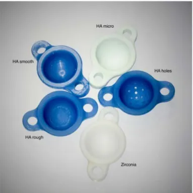
60 hollow domes were manufactured.
They were shaped as half spheres with an inner diameter of 6 mm and an outer diameter of 8 mm. Twelve of them were made of dense hydroxyapatite with a rough inner surface and a mean Sa of 1,1 μm (HA rough). Twelve were manufactured with the same properties as HA rough but had a smooth inner surface (HA smooth) with a mean Sa of 0,3 μm. Twelve were made of microporous hydroxyapatite with a rough inner surface with a mean Sa of 1,1 μm (HA μ). Twelve were manufactured with the same properties as HA rough including the rough inner surface but with macroscopic holes on top (HA holes). Twelve were made of Zirconia with similar properties as HA rough (Zirconia)
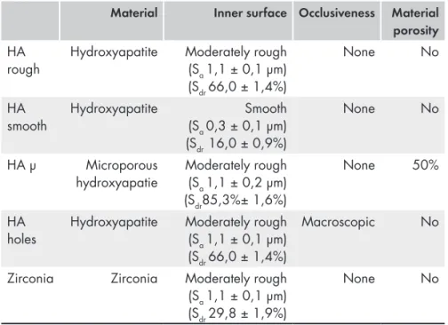
Table 1. The different space maintaining devices used in paper I - III
Material Inner surface Occlusiveness Material
porosity
HA
rough Hydroxyapatite Moderately rough (Sa 1,1 ± 0,1 µm) (Sdr 66,0 ± 1,4%)
None No
HA
smooth Hydroxyapatite (Sa 0,3 ± 0,1 µm)Smooth
(Sdr 16,0 ± 0,9%)
None No
HA µ Microporous
hydroxyapatie Moderately rough (Sa 1,1 ± 0,2 µm)
(Sdr85,3%± 1,6%)
None 50%
HA
holes Hydroxyapatite Moderately rough (Sa 1,1 ± 0,1 µm)
(Sdr 66,0 ± 1,4%)
Macroscopic No
Zirconia Zirconia Moderately rough (Sa 1,1 ± 0,1 µm)
(Sdr 29,8 ± 1,9%)
None No
The hydroxyapatite components were produced from a hydroxyapatite powder (Plasma Biotal Limited, Derbyshire, United Kingdom), which was further processed with water and a dispersant (polycyclic acid) to prepare a ceramic suspension by ball milling. The suspension was sprayed into liquid nitrogen (freeze granulation) and the frozen granules were freeze-dried. Ceramic green bodies of hydroxyapatite and Zirconia (TZ3YSBE, Tosoh Corporation, Tokyo, Japan) were prepared by isostatic compaction of the hydroxyapatite and zirconia granules at a pressure of 300MPa.
Based on the CAD model of the components, a five axis CNC-machine (Dechel-Maho, DMU 60, Deckel Maho, Pfronten, Germany) was used to form the green body into the desired macroscopic shape. To ensure that all components, despite different characteristics had the same sample size even when different sintering temperature were used during the fabrication process, the sintering shrinkage was measured at different sintering temperatures and different values were used to compensate for the enlargement of the CAD model depending on the fabrication process.
The smooth surface of the dense hydroxyapatite was obtained by sintering the machined components at 1250°C for 2 hours. The
dense and rough surface was obtained by presintering the machined material at 900°C, blasting with 110μm alumina particles at 1 bar followed by sintering at 1250°C for 2 hours. The rough micro porous surface was produced by presintering the machined material at 900°C for 2 hours, blasted with 110μm alumina particles at 1 bar and an additional heat treatment at 900°C. The surface on Zirconia was produced by presintering at 900°C. The inner surface was blasted in the presintered state with 110μm alumina particles at 1 bar, and sintered at 1450°C for 2 hours. Some Alumina particles could be detected on the blasted HA surfaces (HA rough, HA holes, HA μ). An image analysis from SEM was made and it was estimated that 0,2% of the μ HA surface was covered with Alumina particles and 0,5% of the Ha rough- and HA holes surfaces were covered with Alumina particles. No Alumina particles could be seen on the Zirconia surface.
Surface topography
The topography of the inner surfaces of the spacemaintaining devices was characterized using an interferometer (MicroXam; ADE Phase Shift Technology, Inc., Tucson, AZ). Three devices per group were randomly selected for analysis. Each device was measured at 5 positions (one at the most inner top and four at the inner radius areas). The parametric calculation was performed after form errors and waviness were removed with a 50 μm X 50 μm Gaussian filter. The following 3-D parameters were selected: Sa (μm) = the arithmetic average height deviation from a mean plane and Sdr (%) = the developed surface ratio.
Animals and anaesthesia and surgery
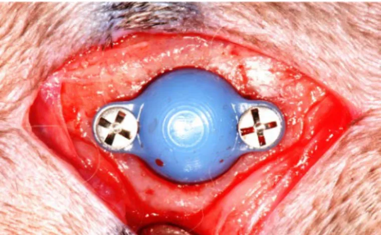
The study was approved by the Malmö/Lund regional animal ethics committee (approval no. M 314-10). Thirty lop-eared rabbits of mixed sex with a mean body weight of 4,15 kg were used in study I-III. Two implants were inserted on each rabbit’s calvaria according to a randomized scheme. Before surgery, the rabbits’ skulls were shaved and disinfected with chlorhexidine (5mg/ml, Pharmacia AB, Stockholm, Sweden). The animals were anaesthetized by intramuscular injection of a mixture of 0.15 ml kg-1 medetomidine (1 mg ml-1 Dormitor; Orion Pharma, Sollentuna, Sweden) and 0.35 ml kg-1 ketamine hydrochloride (50 mg ml-1 Ketalar; Pfizer AB,
Sollentuna, Sweden). Lidocaine hydrochloride (Xylocaine 10mg/ ml; AstraZeneca AB, Södertälje, Sweden) was administered as local anaesthetic at each insertion site at a dose of 0,5 ml. Sterile conditions were maintained during surgical procedure. An incision through the skin and periosteum was made along the midsagittal line on top of the skull measuring approximately 2 cm. The periosteum was carefully resected from the bone. A trephine bur was used to make one slit in the cortical bone on each side of the midline to a depth of 0,5mm. The area was constantly irrigated with saline solution. Two space maintaining devices were fitted to the bone slits and fixated with 2 titanium screws each (De Puy Synthes 04.503.203 Ø1.5 mm 3mm self drilling, De Puy Synthes, Zuchvil, Switzerland). The periosteum was then closed in position using Vicryl 4-0 continuous stitching before the skin was closed in the same fashion. Postoperatively, buprenorphine hydrochloride (0.5 ml Temgesic; Reckitt Benckiser, Slough, UK) was administered as an analgesic for 3 days. No antibiotics were used.
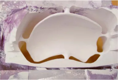
Figure 5. One ceramic space maintaining device attached to the skull
of a rabbit
Preparation of samples
At twelve weeks postoperatively, the rabbits were euthanised with an overdose (60 mg ml 1) of sodium pentobarbital (Apoteksbolaget AB, Stockholm, Sweden). Samples of the space maintaining devices including the attached underlying bone were retrieved and placed in 4% formaldehyde for 24 h, after which they were placed in 70% ethanol. All samples were processed for undecalcified ground
sectioning according to the technique described by Donath (Donath, 1985) In brief, after a series of dehydrations and infiltrations in resin, the samples were embedded in light-curing resin (Technovit 7200 VLC; Heraeus Kulzer Wehrheim, Germany). At this stage, 48 of the domes were analyzed with micro CT. The embedded specimens were divided into two identical blocks. Thereafter, one central cut and ground section from one of the two blocks was prepared from each sample by using Exakt sawing and grinding equipment. The sections were ground to a final thickness of approximately 25 μm and stained with toluidine blue. It was extremely difficult to make the sample any thinner because the ceramic peeled off if ground further. The second block was prepared for scanning electron microscopy (SEM). The blocks were ground and polished with a fine-grain abrasive paper (SiC-Paper, grit 4000, Struers A/S, Denmark) that contained particles of 9 μm, 3 μm and 1 μm. They were then coated with a thin conductive carbon layer with a thickness of approximately 1000Å by vacuum evaporation (AGAR SEM Carbon Coater, Germany). These specimens were examined with SEM (JEOL JXA-8600; JEOL, Tokyo, Japan). The interface between the ceramic material and newly formed bone was evaluated.
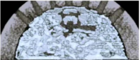
Three-dimensional trabecular bone analysis study 1
A micro CT equipped with a micro focus X-ray tube (focus size 868 mm, MCT-100MF; Hitachi Medical Corporation, Tokyo, Japan) produced a 3D image of each spacemaintaining device from 201 image slices. The tube voltage, tube current, magnification and voxel size were 70 kV, 100 μA, x 6, and 21.0 x 21.0 x 21.0 μm, respectively. Trabecular structure analysis software (TRI/3D BON; Ratoc System Engineering Co. Ltd, Tokyo, Japan) calculated the 3D trabecular structure parameters from the image, information from 110 slices from the very top of the space maintaining device and down. From binary images, the trabecular structure parameters were measured, including tissue volume (TV), bone volume (BV), bone surface per bone volume (BS/ BV), and trabecular thickness (Tb-Th). The star volume of marrow space (V*m.space) and the star volume of trabeculae (V*tr) were measured by star volume analysis. The trabecular connectivity was measured using node-strut analysis described by Garrahan et al. (Garrahan et al., 1990). Nodes (Nd;
connective point of three or more trabeculae), termini (Tm; terminal of trabeculae) and cortex (Ct; connective point of trabeculae and cortical bone) was identified by node-strut analysis, and the parameter of the number (n) of Nd per tissue volume (nNd/TV), the number of Tm per tissue volume (nTm/TV), the number of Ct per tissue volume (nCt/TV), and the total strut length per tissue volume (TSL/TV) was obtained by node-strut analysis.
Histological analyses study 2 and 3
Histological evaluations were performed using light microscopy (Eclipse ME600; Nikon, Japan). The material surface on the inside of the domes was photographed using 40 x magnifications. The pictures were stitched together using Adobe Photoshop Elements (Adobe Systems, San Jose, USA) creating a panoramic image. The bone to material contact was measured in percentage using the free software ImageJ (National Institutes of Health, Bethesda, Maryland, USA). First the total surface was measured from where the newly formed bone began and ended. Secondly the amount of bone in contact with the material surface was measured and calculated and this figure was divided from the total surface giving the percentage of bone in contact with the surface.
The bone volume was measured as percentage of new bone formed related to the total volume using the same method as described above with the magnification of the objective x10. No stitching of images was needed. Only the volume of newly formed bone was measured.
Samples with more than 15 measuring points were measured twice as a control of the measuring technique. If the measures differed more than 1% the samples were measured a third time and an average of the measurements were accepted as the true result. This had to be done with approximately one third (18) of the samples.
Statistics
Differences in the mean values between the control group (n = 12) and the three experimental groups (n = 12/group) were calculated for each parameter to obtain the median percentages. The means for each parameter were compared using Mann-Whitney U test. Values of p>0.05 were considered statistically significant. IBM SPSS Statistics was used for all analyses.
Paper IV
Three patients, two female and one male, volunteered to be included and were consented for surgery. They all had inadequate bone volumes for optimal installation of implants and required bone augmentation prior to implant surgery.
Patient 1 had posterior maxillary bone deficiency in the transverse direction.
Patient 2 had anterior maxillary bone deficiency in the vertical and transverse direction.
Patient 3 had posterior mandibular bone deficiency in the vertical direction.
All patients were examined preoperatively with one panoramic image and a cone-beam computed tomography, volume 60x60, 360 degrees, (3D Accuitomo 170, J Morita, Tokyo, Japan) through the area of interest.
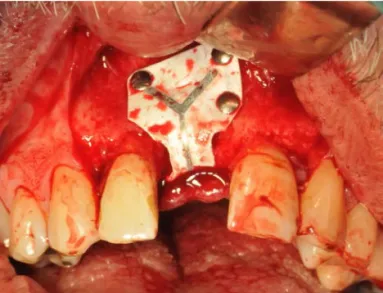
The CT images of each patient were used to build a plastic model of the jaw. On this model a wax was moulded to represent the bone that was supposed to be regenerated. This was covered with a sheet of wax that was formed to represent a membrane or barrier. Based on this wax-model, a five axis CNC-machine (Dechel-Maho, DMU 60, Deckel Maho, Pfronten, Germany) was used to mill a cold isostatically pressed and presintered green body of Zirconia (TZ3YSBE, Tosoh Corporation, Tokyo, Japan) to the desired macroscopic shape.
The size of the machined membrane was enlarged in order to compensate for the sintering shrinkage performed at 1450°C. The inner surface was blasted in the presintered state with 110μm alumina particles at 1 bar, corresponding to the manufacturing method of the space maintaining devices used in study II. The outer surface was characterized with the same interferometer method used in studies I-III. The inner surfaces were impossible to measure as the interferometer collided with the flanks of the material. After manufacturing the devices were cleaned in ultrasonic bath of 70% alcohol for 15 min followed by a brief heating to 1200 °C. They were again cleaned in an ultrasonic bath for 3x15min using 90% alcohol. Thereafter they were put in an autoclave (Getinge Quadro Avanti, Getinge, Sweden) and sterilized using a standard program (134 °C/3min).
Surgery
The intraoral area was initially cleaned with Chlorhexidine 1mg/ml (Hexident®, Meda AB, Solna, Sweden). Local anaesthetic, 20 mg/ ml Lidocaine + 12,5 μg/ml epinephrine (Xylocain Dental Adrenalin, Dentsply Limited, Surrey, Great Britain) was administered. A mucoperiosal flap of full thickness was raised using crestal incision and vertical releasing incisions on the buccal aspect of the surgical site. The cortical plate of the defect was perforated with a small roundbur to provoke bleeding. Fixation of the individualized ceramic membranes where done with Synthes® (De Puy Synthes, Johnson & Johnson, New Brunswick, New Jersey, USA) fracture screws. No adaptation was needed and membranes were stable using only 1 screw. Membranes were cleaned with 3% hydrogen peroxide prior to soft tissue closure. In patient 3, bone chips were harvested from the mandibular ramus on the left side with a Safescraper Twist (Safescraper®, META, Reggio Emilia, Italy) and placed between the underlying bone and the ceramic barrier. The membrane on the right side was placed without any bone chips.
After fixation of the ceramic barriers, the mucoperiosteal flaps were coronally repositioned with significant apical undermining. Split-thickness periosteal release incisions were completed, to aid primary tension-free closure using Vicryl ® 4-0 (Ethicon Inc., Johnson & Johnson, New Brunswick, New Jersey, USA). All patients received antibiotic (phenoxymethylpenicillin) 1g three times per day for 1 week postoperatively.
After a mean of 7 months of healing, the augmented sites were reopened using the similar surgical technique. The ceramic barriers and fixations screws were removed. After that installation of dental implants followed. Before the implants were installed the patient were again examined with one panoramic image and a cone-beam computed tomography, volume 60x60, 360 degrees, (3D Accuitomo 170, J Morita) over the area of interest. Samples of the newly grown bone and covering soft tissues were taken and sent for histological preparation and analysis.
Measuring of gained bone volume and soft tissue
topographic
Data from pre- and postoperative cone-beam-tomography investi-gations were analysed in 3D software (SimPlant Pro, Materialise Dental, Belgium). The amount of gained bone volume in 3 dimensions postoperatively was compared to the same area from the preoperative investigation by an overlay function.
The intra oral soft-tissue profile of the alveolar defect in patient 2 was documented using a three-dimensional metering equipment
Figure 7. The individualized ceramic
membrane attached to mandible on patient 3
(PRIMOS® optical 3D (Primos 5.7 and Primos Body 6.6), GFMess-technik Germany). The measurements were performed at two different occasions: 1) preoperatively and 2) after removal of the membrane. The difference was measured in a 90 degree cut from the alveolar crest.
Sample preparation
Samples of the regenerated bone were retrieved using a trephine bur and placed in 4% formaldehyde for 24 h, after which they were placed in 70% ethanol. All samples were processed for undecalcified ground sectioning according to the same technique used in paper I-III. The sections were ground to a final thickness of approximately 40 μm and stained with toluidine blue. Histological evaluations were performed using a light microscope (Eclipse ME600; Nikon, Japan).
Samples of the soft tissue found between the ceramic membranes and the newly formed bone were retrieved and placed in 4% formaldehyde. The samples were processed for soft tissue sectioning. After processing, the tissues were embedded in paraffin and serial sections 3μm thick were prepared and stained with Hematoxylin-Eosin and van Gieson. Histological evaluations were performed using a light microscope (Leica DMD 108, Leica microsystems, Wetzlar, Germany).
RESULTS
Surface measurements
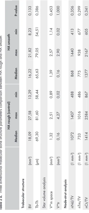
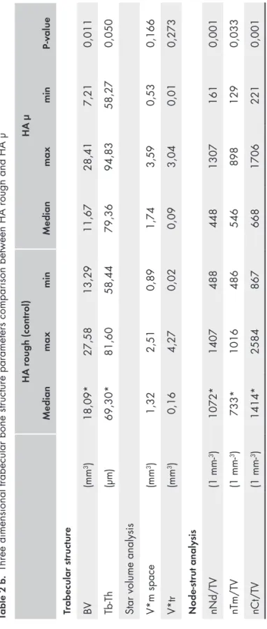
The interferometry measurements of the inner surface of the space maintainers used in paper I – III revealed statistically significant differences in roughness parameters between the ceramic space maintaining devices considered smooth (HA smooth) and the other four groups that were considered rough (HA rough, HA μ, HA holes, Zirconia). Only a small variation within the samples was found for the Sa values. Based on the average Sa value of 0.3 μm for HA smooth and Sa value of 1,1 μm for the other groups, the space maintainers can be categorized as smooth and moderately rough. The microporous hydroxyapatite surface had an Sdr of 85,3% while the Zirconia surface had an Sdr of 29,8%. For exact measured values see table 1.
Three dimensional results from micro-CT investigation
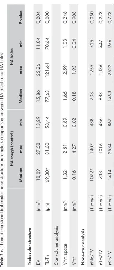
All hydroxyapatite space maintaining devices (HA rough, HA smooth, HA μ and HA holes) were possible to examine with micro-CT. The space maintaining devices made of Zirconia were too dense. The 3 dimensional trabecular bone microarchitecture on the 4 different hydroxyapatite ceramic space maintaining devices is described in table 2 a-c. The bone volume (BV) values for each hydroxyapatite space maintaining device are presented in diagram 1.
The differences in BV between HA rough and HA smooth were not significant. Nonetheless, HA rough produced a slightly larger median bone volume, BV of 18,09 mm3 compared to 16,22 mm3 for
HA smooth (p=0,133). The median number of trabecular connective points was also higher for HA rough with 1072 (max 1407/min 488) compared to 868 (max 1440/ min 413) for HA smooth (p=0,356).