THESIS
ALTERNATIVE SPLICING AND ITS REGULATORY MECHANISMS IN
PHOTOSYNTHETIC EUKARYOTES
Submitted by
Alicia Link
Department of Biology
In partial fulfillment of the requirements
For the Degree of Master of Science
Colorado State University
Fort Collins, Colorado
Fall 2011
Master’s Committee:
Advisor: A. S. N. Reddy
Stephen Stack
3
Copyright by Alicia Link 2011
All Rights Reserved
ABSTRACT
ALTERNATIVE SPLICING AND ITS REGULATORY MECHANISMS IN PHOTOSYNTHETIC EUKARYOTES
In recent years, alternative splicing (AS) of pre-mRNAs, which generates
multiple transcripts from a single gene, has emerged as an important process in
general proteome diversity and in regulatory gene expression in multicellular
eukaryotes. In Arabidopsis over 40% of intron-containing genes are alternatively
spliced. However, mechanisms by which AS is regulated in plants are not fully
understood, primarily due to the lack of an in vitro splicing system derived from
plants. Furthermore, the extent of AS in simple unicellular photosynthetic
eukaryotes from which plants have evolved is also not known. My research
addresses these two attributes of splicing in plants.
In Part 1 of my thesis, I have investigated an aspect of AS regulation in
plants. We have previously shown that an SR-related splicing regulator called
SR45 regulates AS of pre-mRNAs in Arabidopsis by altering splice site selection
(Ali et al. 2007). In this work using bimolecular fluorescent complements, I have
demonstrated that SR45 interacts with U2AF
35, an important spliceosomal
protein involved in 3’ splice site selection in plant cells. This interaction takes
place in the nucleus, specifically in the subnuclear domains called speckles,
iii
which are known to contain splicing regulators and other proteins involved in
transcription. My work has shown that SR45 interacts with both paralogs of
U2AF
35and I mapped the domains in SR45 that are involved in its interaction
with U2AF
35. In addition, my studies have revealed interaction of the paralogs as
hetero- and homodimers. Interestingly, U2AF
35was found to interact with
U1-70K, a key protein involved in 5’ splice site selection. Based on this work and
previous work in our laboratory, a model is proposed that explains the role of
SR45 in splice site selection.
In the second part of my work I studied the extent of alternative splicing
(AS) in the unicellular green alga Chlamydomonas, that shares a common
ancestor with land plants. In collaboration with Dr. Asa Ben Hur’s lab, we have
performed a comprehensive analysis of AS in Chlamydomonas reinhardtii using
both computational and experimental methods. Our results show that AS is
common in Chlamydomonas, but its extent is less than what is observed in land
plants. However, the relative frequency of different splicing events in
Chlamydomonas is very similar to higher plants. We have found that a large
number of genes undergo alternative splicing, and together with the simplicity of
the system and the use of available molecular and genetic tools. This organism
is an experimental system to investigate the mechanisms involved in alternative
splicing. To further validate predicted splice variants, we performed extensive
analysis of AS for two genes, which not only confirmed predictions but also
revealed novel splice variants, suggesting that the extent of AS is higher than we
predicted.
AS can also play a role in the regulation of gene expression through
processes such as regulated unproductive splicing and translation (RUST) that
involves nonsense-mediated decay (NMD), a mechanism of mRNA surveillance
that degrades transcripts containing premature termination codons (PTCs). The
basic mechanism of NMD relies upon many factors, but there are three critical
proteins, termed the UP-frameshift (UPF) proteins due to their ability to
up-regulate suppression of nonsense transcripts. UPF1, UPF2, and UPF3 appear to
be conserved across animals and plants. Our analysis of AS has found that in
Chlamydomonas, many splice variants have a premature termination codon
(PTC). However, to date, the mechanism of NMD has not been investigated in
Chlamydomonas. Analysis of the Chlamydomonas genome sequence shows
that UPF1, 2, and 3 proteins are present, and we have shown that they share
some sequence similarity with both plants and humans, indicating that the
process of NMD may be present in this organism. To address the role of UPFs
in NMD in Chlamydomonas, we have utilized the artificial miRNA approach. I
have generated stably transformed Chlamydomonas cell lines that are
expressing amiRNA for UPF1 and UPF3 that will be useful in analyzing NMD of
selected genes as well as all PTC-containing transcripts globally.
v
ACKNOWLEDGEMENTS
Completing this thesis has been one of the most difficult things in my life I’ve ever done, but also one that I am most proud of. First of all, I’d like to thank Dr. Reddy for his outstanding role as my advisor and seemingly perpetual happiness throughout my education in graduate school at CSU. He took me under his wing when continuing school seemed futile, and gave me the chance I needed to
accomplish my dream. For that, I am forever indebted to him. Second of all I’d like to thank the Reddy lab for their help and guidance. In particular, I’d like to thank Dr. Irene Day and Julie Thomas, whose support and amazing fortitude carried me throughout my degree. I would also like to thank Dr. Salah Abdel-Ghany, who showed up at the end of my term, but was eager to help and truly meets the definition of an “upright man.” I’d also like to thank Dr. Asa Ben Hur and his
students for their collaboration on the Chlamydomonas AS project. I’d like to thank my committee not only for being there for me and giving me support, but for being the inspiring people they are. I chose them because I looked up to them. I’d also like to thank Dr. June Medford for her support during the tough times.
Most importantly, I’m sending thanks to Dr. Paul Kugrens, who passed on during my time at CSU, but left an everlasting impression on me and will always be in my heart. His passion for science was contagious, and if I hadn’t met him, I never would have wondered what “pond scum” was. Science really hasn’t been the same for me without him, but I believe he provided for me the tools for success in life. I’d
also like to thank his family for their continuing support and for being a great part of my life. I know I wouldn’t have succeeded without them.
Next, I thank my parents. I couldn’t have asked for a better family, and I wake up each day grateful to have them in my life. Without my mother, I never would have had the perseverance to finish, and without my father, I wouldn’t have the confidence and “guts” to succeed. I’d also like to thank my brother for his support. My grandparents also have been extremely supportive of my education, and when I grow to be their age, I hope to have lived as rich of a life as they have.
I never would have made it without my fiancé, Paul. He has been my lighthouse in the middle of the storm, my constant, and my true partner. He continues to inspire me everyday with his innovative thinking, joyful presence, and determination. Meeting him has changed my life, and I know if he is with me, I can face anything and succeed. I’d also like to thank my best friend Stephanie Long for her friendship and early morning calls. She kept me sane through this experience and has through many others. She is a wonderful friend and I’m lucky to have her. Last but not least, I’d like to thank Tallulah the Cat and Charlie the Dog. Even though they can’t talk I know they love me.
I read one of Dr. Kugrens’ student’s thesis once and was greatly moved by the quote in their acknowledgements section, so I shall end mine in the same way with this quote by Antony van Leeuwenhoek: “This was for me, among all the marvels that I have discovered in nature, the most marvelous of all…no more pleasant sight has come before my eyes than these many thousands of living creatures, seen all alive in a drop of water.”
vii
TABLE OF CONTENTS
Chapter 1: In vivo interaction of a splicing regulator (SR45) with U2 auxiliary
factors (U2AF
35)
Introduction………1
Materials and Methods……...………...………...9
Results………..………...12
Discussion………..………...19
Chapter 2: Alternative splicing in Chlamydomonas reinhardtii
Introduction………..……….……..24
Materials and Methods………..29
Results………...32
Discussion……….…………..38
Chapter 3: Analysis of nonsense-mediated decay in Chlamydomonas reinhardtii
using artificial miRNA constructs
Introduction………..………...47
Materials and Methods………..…………53
CHAPTER 1: IN VIVO INTERACTION OF A SPLICING REGULATOR (SR45) WITH U2 AUXILLARY FACTORS (U2AF35)
(The results presented here are from a manuscript titled “Interactions of SR45 with U2AF35 and U1-70K: Insight into the spliceosome assembly on 5’ and 3’ splice sites by Day, Golovkin, Link, Ali, and Reddy” which has been submitted for publication)
Introduction
Precursor mRNA (pre-mRNA) in eukaryotes contains coding sequences known as exons, and intervening noncoding sequences known as introns. Pre-mRNA splicing is essential for the expression of most genes in eukaryotic organisms. This process involves the removal of introns and the covalent joining of exons in pre-mRNA. Four conserved core elements, which include 5’ and 3’ splice sites, a branch point, and a polypyrimidine tract upstream of the 3’ splice site that are necessary for splicing.
Multicellular eukaryotes are known to have less conserved sequences around the splice sites. As a result, additional regulatory sequences adjacent to the splice sites, which are called enhancers or repressors, are needed for correct and efficient definition of splice sites (Ali et al. 2007; Ast 2004; Reddy 2007).
Pre-mRNA splicing takes place in a large complex known as the spliceosome, which is made up of 5 small nuclear ribonucleoparticles (U1,2,4,5,6 SnRNPs) and many non-snRNP factors(Reddy 2007; Reddy 2001; Sharp 1994; Zhou et al. 2002). A fully assembled spliceosome contains around 300 different proteins and is one of the most complex cellular components investigated so far (Rappsilber et al. 2002; Zhou et al. 2002). The first step in spliceosomal assembly is the formation of the ATP-independent
2
early (E) complex, and this is where the 5’ splice site is recognized and subsequently bound by U1 snRNP. The branch site and 3’ splice site are then recognized by U2 snRNP and U2 auxiliary factors (U2AFs). Following this step, the A complex (the association of U2 with the branch site/3’ splice site region) is altered in an ATP-dependent fashion in order to stabilize binding to this region (Figure 1.1). Then, the U4/U6/U5 tri-snRNP joins the assembly of the spliceosome to form the B-complex. This complex then undergoes a series of conformational rearrangements, one of them involving a tri-snRNP associated protein kinase (SRPK2), which phosphorylates the RS domain of Prp28, an RNA helicase. This step is required for the stable association of Prp28 with the tri-snRNP as well as for tri-snRNP association with assembling
spliceosomes during B complex formation (Mathew et al. 2008). After the dissociation of U1 snRNP, the U4/U6 base-paired interaction is unwound by Brr2, which is a
U5-assocated helicase (Wahl et al. 2009; Will 2006). Then, U1 and U4 leave the
spliceosome, and U6 replaces U1 at the 5’ splice site (Figure 1.1). This then marks the progress of the B complex moving towards being the catalytically active B* complex, which performs the first step of splicing. However before the B* complex is formed however, the Bact complex is the transitional state between the B and B* complex. The Bact complex is void of U1, U4/U6 snRNPs, but contains the NineTeen Complex
proteins (NTC) (Fabrizio et al. 2009). The Bact complex is then transitioned to B* following ATP hydrolysis by RNA helicase Prp2, which dissociates and gives the final B* complex. A protein called Prp16 transitions the B* complex to the C complex, which contains U5, U6 and U2 and is capable of completing the second step of splicing (Valadkhan and Jaladat 2010) (Figure 1.1).
In contrast to constitutive splicing, a process called regulated alternative splicing (AS) produces multiple transcripts that encode different proteins from the same gene,
4
which then aids in post-transcriptional gene regulation and generates proteomic diversity. Both of these processes are essential for the function of many genes in eukaryotes (Ast 2004; Graveley 2001; Maniatis and Tasic 2002; Reddy 2007; Reddy 2001). Recent human genome-wide studies have suggested that 95% of pre-mRNAs from intron-containing genes are alternatively spliced (Pan et al. 2008; Wang et al. 2008). In addition to this, genome-wide studies in Arabidopsis have indicated that over 40% of intron-containing genes undergo AS (Filichkin et al. 2010 ).
The process of AS lengthens or shortens exons by altering the position of one of their splice sites, which results in both alternative 3’ and 5’ splice sites. AS also involves exon skipping or intron retention, where important regulatory events can be controlled by the failure to recognize an exon or excise an intron. It has been shown that abiotic/biotic stresses effect the pattern of splicing, showing that AS is regulated and the pattern of splicing is adjusted according to what kind and level of stress a plant experiences (Iida et al. 2004; Palusa et al. 2007). It has also been shown that AS has a critical role in flowering time, where temperature changes regulate this action, and it has been shown that varying AS transcripts are part of making this process proceed smoothly
(Balasubramanian et al. 2006). In addition, wound-response is affected by patterns of AS, and a separate study linked AS to disease resistance in plants (Bove et al. 2008; Dinesh-Kumar and Baker 2000). Thus, it seems that AS in plants is a posttranscriptional regulatory mechanism, which in turn eventually effects gene expression and
subsequently plant form and function (Reddy 2007).
In metazoans, the key players for the recruitment of U1 to the 5’ splice site and U2 to the 3’ splice site are members of what is known as the serine/arginine-rich (SR) protein family (Nilsen 2003; Reddy 2001). SR proteins are currently characterized in the following ways: they must contain one or two N-Terminal RNA recognition motifs
(RRMs) followed by a downstream serine/arginine rich region known as the RS domain of at least 50 amino acids and a minimum of 20% RS or SR dipeptides (Barta et al. 2010). These proteins aid in identifying the 5’ splice site by interacting with U1 and pre-mRNA concurrently (Graveley 2000; Reddy 2004). In addition to this, SR proteins are thought to regulate the selection of weak alternative splice sites. SR proteins bind to these less conserved sequences and therefore promote the recruitment of U1 to the correct 5’ splice site (Eperon et al. 1993; Graveley 2000; Jamison et al. 1995; Kohtz et al. 1994; Reddy 2004; Zahler and Roth 1995). All of these functions combined lend the SR protein family the reputation of being a major player for increasing transcriptome complexity as well as proteomic diversity (Graveley 2000). Cell-free extracts have been utilized to study splicing in animals, and it has been found that there are RNA-RNA, RNA-protein, and protein-protein interactions involved in the splicing process.
Serine/arginine (SR) proteins are responsible for many different roles in mRNA splicing (Barta et al. 2008; Graveley 2000; Lorkovic et al. 2008). About 18 SR proteins have been identified, while only 12 are present in humans (Barta et al. 2010). SR proteins have been found to be localized in interchromatin granule clusters in the nucleus as well as in the nucleoplasm (Ali et al. 2008a; Ali and Reddy 2008a; Fang et al. 2004; Lorkovic et al. 2008).
Plant introns differ from animal introns in their size, nucleotide composition, branch point sequence and polypyrimidine tract (Reddy 2001). Because of this, plant intron-containing transcripts are usually either not processed correctly or not processed at all in mammalian splicing extracts (McCullough et al. 1991). There is no plant-derived cell-free splicing extract available for plant splicing factors analysis, and because of this, in vivo methods have to be used to investigate the interaction network of proteins
6
SR45 is an SR-like protein with two RS domains, one being N-terminal and the other C-terminal. A second SR-like protein, known as SR45a, has been identified and shares the same domains as SR45, but only shows a 26% identity to SR45 with most of this being in the RS domains (Ali et al. 2007; Tanabe et al. 2007). SR45 has been shown to be an essential splicing factor in complementation assays, and has orthologs in other flowering plants, but none in algae. SR45 interacts with U1-70K, one of the U1 snRNP specific proteins, and their association in nuclear speckles was shown using bimolecular fluorescence complementation (BiFC) (Ali et al. 2007; Ali et al. 2008a). BiFC is a technique where two halves of a fluorescent protein are separately fused to two putative interaction proteins. When the two proteins interact, the two halves of the protein come together resulting in reconstitution of fluorescent protein. The fluorescent signal can then be detected through confocal microscopy (Hu et al. 2002). SR45 is alternatively spliced and produces two splice forms. In the SR45 mutant, sr45-1, the splicing patterns of many other SR genes are affected, suggesting its role in AS (Ali et al. 2007). The mutant phenotype indicates that SR45 has a role in multiple plant-specific developmental processes, including plant size, flowering time, and organ morphology. In addition to this, sr45-1 plants show delayed flowering, altered leaf morphology, and flowers with abnormal petal and stamen numbers, (Ali et al. 2007). One splice form of SR45 rescues the flower phenotype while the other splice form rescues the root phenotype, as shown in a gene complementation study (Mount and Zhang 2009).
To further understand the role of SR45 in splicing, we used it in a yeast two hybrid system, which resulted in isolation of U2AF35, a spliceosomal protein. It is part of the U2AF complex, which is involved in spliceosomal assembly. The large subunit of the U2AF complex is U2AF65, which binds to the polypyimidine (Py) tract. U2AF35 is the small subunit of the U2AF complex, which is involved in the recognition of the 3’ splice
site. This recognition occurs through the contact of the AG dinucleotide at the 3’ splice site. For introns with weak Py tracts, the U2AF35 interaction with the 3’ splice site is critical for U2AF (Merendino et al. 1999; Wu et al. 1999; Zorio and Blumenthal 1999). In Arabidopsis, two paralogs of U2AF35 have been characterized, known as U2AF35a and U2AF35b. I investigated in vivo interactions of U2AF35 with SR45 using BiFC. Here, I have shown that SR45 interacts with both paralogs and mapped the domains in SR45 that are involved in its interaction with U2AF35. In addition, my studies have shown interaction of the paralogs as hetero- and homodimers. A protein alignment of the paralogs of U2AF35 present in Arabidopsis, humans, and rice was performed which showed that there is a C-terminal domain that is unique to plants (Figure 1.2). We have tested a truncated version of this domain to investigate its role in U2AF35 interaction with SR45. My studies have also shown that U2AF35 and U1-70K interact. Since SR45 interacts with both U1-70K and U2AF35, it is likely that it has a role in 3’ to 5’ splice site selection and could possibly bridge the 5’ and 3’ components of the spliceosome.
! +!
Figure 1.2. U2AF35 proteins from Arabidopsis, Rice, and Human are aligned using MegaAlign. Identical amino acids are shown in reverse contrast lettering. Red boxes indicate that the C-terminal region is highly conserved in plants, but does not share the same identity with human U2AFs.
10 20 30 40 MA E H L A S I F G T E K D R V N C P F Y F K I G A C R H G D R C S R L H NRP 40 AtU2AF35a.seq MA E H L A S I F G T E K D R V N C P F Y F K I G A C R H G D R C S R L H NRP 40 AtU2AF35b.seq MA E H L A S I F G T E K D R V N C P F Y F K I G A C R H G D R C S R L H NKP 40 OsU2AF35a.seq MA E H L A S I F G T E K D R V N C P F Y F K I G A C R H G D R C S R L H NRP 40 OsUAF35b.seq MA EYL A S I F G T E K DKV N CSF Y F K I G A C R H G D R C S R L H NKP 40 HsU2AF35a.seq MA EYL A S I F G T E K DKV N CSF Y F K I G A C R H G D R C S R L H NKP 40 HsU2AF35b.seq 50 60 70 80 TI S P TLL L S N MY Q R P D MI T P G V D A Q G Q PLD PRKI Q E H F E D 80 AtU2AF35a.seq TI S P TLL L S N MY Q R P D MI T P G V DPQ G Q PLD PSKI QDH F E D 80 AtU2AF35b.seq S VS P TLL L S N MYLR P D MI T P GI D A Q GNPI D PEKI QA DF E D 80 OsU2AF35a.seq TVS P TI VLAN MY Q R P D MI T P G V D A Q G Q PI D PEKMQ E H F E D 80 OsUAF35b.seq TFSQTI ALLNI YR NPQ N S S Q S ADG L R C A V S D V E MQ E HY D E 80 HsU2AF35a.seq TFSQTI LI QNI YR NPQ N S A Q T ADG S H C A V S D V E MQ E HY D E 80 HsU2AF35b.seq 90 100 110 120 FFE DLF E E LG - KFG EI E S L NI C D N L A D H MI G N V Y V Q FKE E 119 AtU2AF35a.seq FYE D I F E E LN - KFG E V E S L N V C D N L A D H MI G N V Y VLFKE E 119 AtU2AF35b.seq FYE D I F E E LS - KYG EI E S LHV C D NFA D H MI G N V Y V Q F R E E 119 OsU2AF35a.seq FYE D I YE E LS - KFG E V ETL N V C D N L A D H MI G N V Y V Q F R E E 119 OsUAF35b.seq FFEE VFTEME EKYG E V EE MN V C D N LGD HL VG N V Y VKF RRE 120 HsU2AF35a.seq FFEE VFTEME EKYG E V EE MN V C D N LGD HL VG N V Y VKF RRE 120 HsU2AF35b.seq 130 140 150 160 DQ A A A A L Q A L Q G R F Y S G R P I I ADF S P V T D F R E A T C R Q Y E E 159 AtU2AF35a.seq D HA A A A L Q A L Q G R F Y S G R P I I ADF S P V T D F R E A T C R Q Y E E 159 AtU2AF35b.seq DQ A ARA L Q A LTG RYY S G R P I I VE F S P VSD F R E A T C R Q Y E E 159 OsU2AF35a.seq EQ AVA AH NA L Q G R F Y S G R P I I VEYS P V T D F R E A T C R QFE E 159 OsUAF35b.seq E DAE KAV I DLN NRWF NGQP I HA ELS P V T D F R E ACC R Q Y EM 160 HsU2AF35a.seq E DAE KAV I DLN NRWF NGQP I HA ELS P V T D F R E ACC R Q Y EM 160 HsU2AF35b.seq 170 180 190 200 NNC N R G G Y C N F MH V KL VS R E L R R K LFG RYR R SYRR GS R S R 199 AtU2AF35a.seq N S C N R G G Y C N F MH V KQI S R E L R R K LFG RYR R SYRR GS R S R 199 AtU2AF35b.seq N S C N R G G Y C N F MH V KEI GRDL RK RLFGH L HR SRRS HSH GR 199 OsU2AF35a.seq N S C N R G G Y C N F MH V KQI GR E L R R K LYG- - GR SRR- - SH GR 195 OsUAF35b.seq G ECTR G GFC N F MHLKPI S R E L R RELYG RRRK K HR- - S R S R 198 HsU2AF35a.seq G ECTR G GFC N F MHLKPI S R E L R RELYG RRRK K HR- - S R S R 198 HsU2AF35b.seq 210 220 230 240 S R SR S I S P RN - KRD N DRR D P S H R E F S HRD R D R E F Y R HGS G 238 AtU2AF35a.seq S R S- - I S P RR - KRE H SR- E R ER G- - D VRD R D R - - - - HGN G 229 AtU2AF35b.seq S R S- - PS PY H YR RD Y DRR S S SRS - - - - RD H D D - - - 225 OsU2AF35a.seq S R S- - PS P RH RRG NRDRD D F RRE R D G YRGGG D G Y R G GGG G 233 OsUAF35b.seq S RE - - - R RSRSR- - - DR G- - - - RGGG G - - - G- - 216 HsU2AF35a.seq S RE - - - R RSRSR- - - DR G- - - - RGGG G - - - G- - 216 HsU2AF35b.seq 250 260 270 280 K R - - - SSERS E - - - RQ ERDGS RG R RQ ASP 261 AtU2AF35a.seq K R - - - SSDRS E - - - RHD RDG G GRR RH GSP 252 AtU2AF35b.seq - - - Y YR G G- - - S HDY Y RG GSR R- - SS 243 OsU2AF35a.seq G G G D G Y R G G DSYR G GG G G G R R G G G SRYD RY D DG G R RR H G S 273 OsUAF35b.seq - - - G G- - - G G G GGR- - E R 226 HsU2AF35a.seq - - - G G- - - G G G GGR- - E R 226 HsU2AF35b.seq 290 300 310 KRG GSP G G GR E G S E E R R A R I E Q WN R E R EE K E E G G A 296 AtU2AF35a.seq KRSR SP R N VR E G S E E R R A R I E Q WN R E RD E - - - - G V 283 AtU2AF35b.seq ERHR SS Y D - S DG S E E R R AQI E Q WN R E R EA - - - A Q V 274 OsU2AF35a.seq P PR RARS P VR ESS E E R R AKI E Q WN R E R E 301 OsUAF35b.seq DR R R S R D R- - - E RS GRF 240 HsU2AF35a.seq DR R R S R D R- - - E RS GRF 240 HsU2AF35b.seq 290 300 310 G G N V D -P V R R -K E -G E K V E A -V R E G S E E R R A R I E Q WNWN R E R E E R E G S E E R R A R I E Q WNWN R E R D E S D G S E E R R A Q I E Q WNWN R E R E A R E S S E E R R A K I E Q WNWN R E R E - - - E R S G R F - - - E R S G R F !
MATERIALS AND METHODS
Constructs
Full-length and truncated mutants of SR45 were amplified with forward and reverse primers containing SalI and XmaI sites, respectively and cloned into pSPYNE-35S/pUC-SPYNE and pSPYCE-35S/pUCSPYCE (Ali and Reddy 2008b). Primers listed in Day et al 2011 (in preparation) were used to amplify (U2AF35a, c and Ctrb using DNA as a template. PCR was done using Takara Ex Taq (Fisher Scientific) according to manufacturer’s specifications (two-step). Fragments were digested with SalI/KpnI and ligated into pSPYNE-35S/pUC-SPYNE and pSPYCE-35S/pUCSPYCE vectors digested with the same enzymes. Full length U1-70K/YFPN was constructed previously (Ali et al.
2008b). Plasmid DNA was prepared using a Qiagen Midi-Prep kit or a Maxi-Prep kit.
DNA concentration was quantified using a spectrophotometer.Transient Expression of BiFC constructs in protoplasts
A) Protoplast isolation
Protoplasts were prepared from healthy leaves from 4-week-old WT Arabidopsis thaliana ecotype Columbia. Leaves were sliced into thin strips and were immersed in enzyme solution (0.4 M mannitol, 20 mM KCl, 20 mM MES pH 5.7) containing 2.0% cellulase and 0.2% macroenzyme. After vacuum infiltration the mixture was shaken in a 500 ml flask at low speed, RT for 3-4 hours and filtered through 75 µm mesh. The protoplasts were harvested by centrifugation (200xg for 2 minutes), resuspended in W5 medium (154 mM NaCl, 25 mM CaCl2, 5 mM KCl and 2 mM MES pH 5.7) and placed on
ice for 30 minutes. Protoplasts were pelleted again and resuspended in MMG (0.4 M mannitol, 15 mM MgCl2, 4 mM Mes pH 5.7) medium at a concentration of 2x10⁶/ml.
10
B) Transfection
Plasmid DNA was added at a concentration of 1 mg/ml and an equal amount of 40% PEG was added, the tubes were inverted several times to mix, followed by
incubation at RT for 30 minutes. The protoplasts were washed twice with two volumes of W5 and resuspended in 1 ml of W5 they were dispensed into a 6-well plate and kept in the dark in a 22°C incubator for 16 hours to allow for expression. A 50 µl aliquot was transferred to a glass bottom petri dish with a 30 to 70 mm cover slip and observed immediately under the microscope.
C) Confocal Microscopy
Transfected protoplasts were examined using a Zeiss LSM 510 Meta laser scanning confocal microscope. All samples were viewed using the YFP channel (Ali et al. 2008a). Protoplasts were first viewed at 40x to see transformation efficiency, and then single protoplasts were viewed and photographed using the 63x, N.A. 1.4 oil immersion apochromat objective. The YFP filter was set up with excitation at 514nm, 458/514 dichroic, and emission 560-615 BP filter.
Figure 1.3. BiFC assay using SR45 and U2AF35. Confocal images of Arabidopsis protoplasts expressing full-length SR45-YFPc paired with U2AF35a-YPFn, full length U2AF35b-YFPn, or U2AF35Ctrb-YFPN as indicated on the right. The images in the right-most panels show a zoomed in view of the nuclear region shown in the images under the YFP column. Bars = 10 µm.
12
RESULTS
In vivo interaction of SR45 with both U2AF35 paralogs
Previously, SR45 has been shown to co-localize with U1-70K in speckles in the nucleus (Ali et al. 2008a). In addition, both paralogs of U2AF35 have also been detected in nuclear speckles (Wang and Brendel 2006b). Recently, we have isolated U2AF35b as an interacting partner of SR45 using yeast two-hybrid screens. The aim of my work is to determine where in the cell they interact and which domains of SR45 are responsible for this interaction. To determine the location of the interaction between SR45 and U2AF35, we utilized BiFC as an in vivo proof of association. BiFC is based upon the
reconstitution of two split halves of yellow fluorescent protein (YPF), which upon reconstitution, results in fluorescence. Each half of YFP is fused to two putative
interacting proteins, and if those two proteins interact, the split halves come together and YFP can be visualized (Walter et al. 2004). Fusions to the N-terminal region of YFP (YFPn) were constructed by cloning U2AF35a, U2AF35b (full length), and U2AF35Ctrb, which lacks the C-terminal region) into a BiFC vector.
In previous studies, SR45 had been cloned in a similar way as a fusion to the C-terminal region of YFP (SR45/YFPc) into a BiFC vector. Arabidopsis protoplasts were than transformed with both constructs and examined for fluorescence using confocal microscopy. Figure 1.3 shows fluorescence in protoplasts transformed with SR45/YFPc and U2AF35a/YPFn or U2AF35Ctrb/YFPn. YFP fluorescence is largely seen in nuclear speckles with some fluorescence visualized in the nucleoplasm, which is similar to the localization of SR45 (Ali et al. 2008a). These results support the interaction of SR45 with both U2AF35a and U2AF35b. Since plant U2AF35 has a plant-specific C-terminal region (Figure 1.3), we tested if this region is necessary for U2AF interaction and
localization using a truncated version of U2AF35b. Interestingly, this construct showed YFP reconstitution but it is mostly in the nucleoplasm as seen in Figure 1.3, supporting the importance of the C-terminal domain for the localization of U2AFs in speckles.
Protein Alignment of U2AF35 paralogs
A protein alignment was performed for U2AF35 paralogs using MegaAlign for Arabidopsis, Rice, and Human. The two paralogs of Arabidopsis share 84% similarity, and they are about 65% similar to rice U2AFs. However, AtU2AF35s share only 26% similarity with human U2AFs and plant U2AFs have a short conserved C-terminal domain that is absent in humans.
U2AF35 proteins form hetero- and homodimers
U2AF is a heterodimer consisting of U2AF35, the smaller subunit, and U2AF65, the larger subunit. Förster resonance energy transfer (FRET) has been recently shown that the smaller subunit, U2AF35 in animals interacts with itself (Chusainow et al. 2005). BiFC was utilized to test if plant U2AF35 subunits interact with each other, as well as with its paralogs. U2AF35 proteins (a,b, and Ctrb) cloned into BiFC vectors as YFP
N-terminal and YFP C-N-terminal fusions were used for BiFC studies. Protoplasts were transformed as before with U2AF35a/YFPc, U2AF35b/YFPc, or U2AF35Ctrb/YPFc together with U2AF35a/YFPn, U2AF35b/YFPn, or U2AF35Ctrb/YFPn. Figure 1.4 shows protoplasts transformed with each set of U2AF35 proteins mentioned above. Each shows
fluorescence indicating that U2AF35 proteins can form both homo-and heterodimers, and that the C-terminal domain, which is not present in the truncated b form (Ctrb), is not essential for this interaction. It also appears that the localization of the dimer pairs is different amongst the protoplast transformations. U2AF35b dimers appear in speckles but with more fluorescence in the nucleoplasm than when dimerized with U2AF35a.
! %'!
Figure 1.4. Confocal images show Arabidopsis protoplasts expressing (Top Pictures) U2AF35a-YFPn paired with full-length U2AF35a-YFPc or U2AF35b-YFPc or U2AF35Ctrb-YFPc, (Middle pictures) full-length U2AF35b-YFPn paired with U2AF35b -YFPc or U2AF35Ctrb-YFPc, or (Bottom pictures) U2AF35Ctrb-YFPn paired with U2AF35Ctrb-YFPc. Images in the right most panels show a zoomed in view of the nuclear region shown in the images under the YFP column. Bars = 10 µm.
The U2AF35b and the U2AF35Ctrb dimer showed even more diffuse fluorescence in the nucleus.
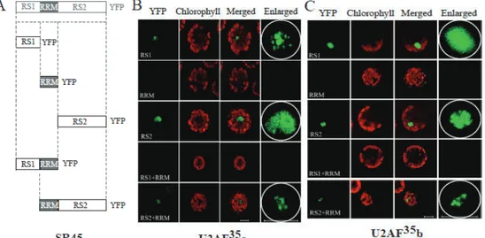
RS1 and RS2 of SR45 associate with U2AF35s independently.
SR45 is comprised of an N-terminal RS domain (RS1), a central RRM domain, and a C-terminal RS domain (RS2). BiFC was utilized to identify the domains of SR45 that interacts with U2AF35 proteins. In order to do this, a series of SR45 deletion mutants were introduced in a BiFC vector as fusions to YFPc, and then used in BiFC assays with the U2AF35 proteins (Figure 1.5). Constructs included were: RS1/YFPc, RRM/YFPc, RS2/YFPc, RS1/RRM/YFPc, and RRM/RS2/YFPc (Ali et al. 2008a). The SR45/YFPc deletion constructs were tested with U2AF35a/YFPn, U2AF35b/YFPn, and U2AF35Ctrb/YFPn. Protoplasts transformed with each variant of U2AF35/YFPn and those that contained either RS1 or RS2 showed fluorescence, which indicated an interaction of these two domains in SR45 with U2AFs (Figure 1.5B-D). Protoplasts transformed with the RRM/YFPc domain and each variant of U2AF35YFPn exhibited no fluorescence, indicating RRM is not involved in SR45 interactions with U2AFs. While the
RS2+RRM/YFPc domain showed some diminished fluorescence with each
U2AF35/YFPn protein (Figure 1.5B-D), it is interesting to note that the RS1+RRM/YFPn domain exhibited fluorescence only when paired with U2AF35Ctrb (Figure 1.5B,C,D).
In the case of U2AF35a/RS2, fluorescence was much more diffuse throughout the nucleus when compared to the full-length SR45 (Figure 1.2B), although small speckles were present (Figure 1.5B). Protoplasts were also transformed with U2AF35b+RS1, and the fluorescence was very diffuse throughout the nucleus, with very fine speckles. In contrast to this, U2AF35b+RS2 shows fluorescence predominantly in the speckles (Figure 1.5C). U2AF35Ctrb/RS1 was similar to U2AF35b, showing a more diffuse pattern with RS1,
! %)!
Figure 1.5. Interaction of SR45 domains with U2AF35 proteins. A. Schematic diagrams of the SR45 domains used in BiFC. B-D. Confocal images of
Arabidopsis protoplasts expressing U2AF35a-YFPn (B), full-length U2AF35b-YFPn (C), or U2AF35Ctrb-YFPN (D) paired with the deletion mutant indicated on each panel. The images in the right most panels show a zoomed-in view of the nuclear region shown in the images under the YFP column. Bars = 10 µm.
but in contrast, the RS2, RS1+RRM, and RS2+RRM constructs exhibited speckles which were much more visible with some still remaining in the nucleoplasm (Figure 1.5D). These results combined suggest that U2AF35 can interact with the RS1 and RS2 domains independently, but other domains of the protein alter the strength of that interaction.
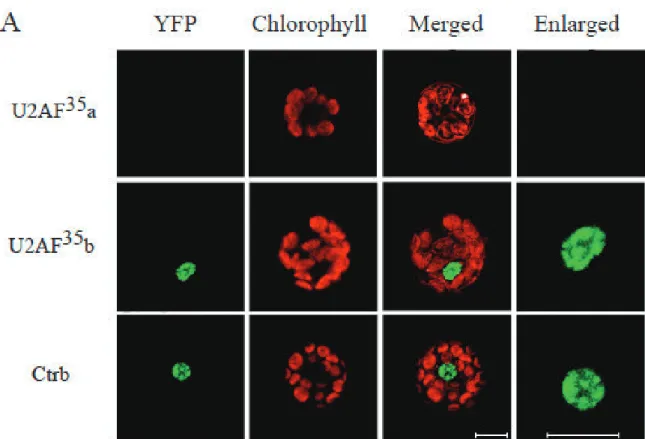
U2AF35 interacts with U1-70K
U1-70K and SR45 both interact with U2AF35 and have the same localization pattern in the nucleus. Because of this, we investigated whether U1-70K and U2AF35 interact utilizing BiFC. The full-length and truncated versions of U2AF35b showed
interactions with U1-70K, but U2AF35a did not. Each of the U2AF35b types that interacted with U1-70K showed fluorescence localized in the nuclear speckles, with some diffusion of fluorescence into the nucleoplasm (Figure 1.6).
! %+!
Figure 1.6. Interaction of U1-70K and U2AF35. A. Confocal images of Arabidopsis protoplasts expressing U1-70K-YFPn paired with U2AF35a-YFPc, full-length
U2AF35b-YFPc, or U2AF35Ctrb-YFPc as indicated on the left. Images in the right most panels show a zoomed-in view of the nuclear region shown in the images under the YFP column. Bars = 10 µm.
DISCUSSION
Eighteen SR proteins have been identified in Arabidopsis to date (Barta et al. 2010), whereas there are only 12 SRs in humans. In general, plants have many more SRs as compared to animals. Recent studies support evidence that there could be some functional importance to this variance as some aspects of pre-mRNA splicing in plants could vary from animals (Lazar and Goodman 2000; Lopato et al. 2002; Lorkovic et al. 2008; Reddy 2004; Tanabe et al. 2007). Previous studies have shown that SR45 binds to U1-70K, and hence, it could be involved in the 5’ site recognition in splicing. In this study, we identified an interaction between SR45 and U2AF35, which points to a connection between the 5’ and 3’ binding site proteins. This interaction is supported by in vivo studies using BiFC. We have seen through this study that SR45 interacts with U2AF35a and U2AF35b. More evidence to support this interaction comes from the interaction observed through BiFC utilizing mutant phenotypes of SR45 and U2AF35. It is known that SR45 knockout plants have altered development and delayed flowering in long- and short-day photoperiods, along with an abnormal number of floral organs (Ali et al. 2007). Similarly, plants with a T-DNA insertion in U2AF35a or U2AF35b RNAi also are late flowering under similar conditions and have abnormal flowering morphology (Wang and Brendel 2006b). In addition to this, the expression of FLC (Flowering Locus C) has been found to be much higher in both U2AF35b and SR45 mutants (Ali et al. 2007; Wang and Brendel 2006b). Another study has shown that the SR protein SR45a interacts with U2AF35b but not with U2AF35a in yeast two-hybrid assays (Tanabe et al. 2007). Both SR45a and SR45 have two RS domains (N- and C-terminal to the RRM domain), but share only 26% similarity.
In animals U2AF, a heterodimer consisting of U2AF35 and U2AF65, is involved in 3’ splice site recognition (Mollet et al. 2006; Wu et al. 1999). We report here that in
20
Arabidopsis both paralogs of U2AF35 form homo- and heterodimers in vivo. Human U2AF35 has also previously shown dimerization utilizing FRET analysis (Chusainow et al. 2005). These results suggest that two components of U2AF35 may pair with U2AF65 that make up the U2AF complex. BiFC analyses showed that this plant-specific domain is not essential for either interaction with SR45 or dimerization. Nevertheless, there were differences in the pattern of localization to speckles and nucleoplasm for proteins lacking this domain. The human U2AF35 has three splice forms and there are two U2AF35 proteins in humans but none of these have the C-terminal domain found in plant U2AF35 (Mollet et al. 2006), suggesting a plant specific function associated with this C-terminal domain.
BiFC analysis of the SR45 domains and U2AF35 interactions showed that both the RS1 and RS2 domains of SR45 interact independently with both U2AF35 paralogs. There were some differences in the localization of the U2AF35/SR45 interactions with different domains of SR45 and/or different paralogs of U2AF35, where the distribution between the speckles and nucleoplasm was altered. Previous studies have shown a similar result when the same domains of SR45 were utilized to study U1-70K
interactions with SR45 (Ali et al. 2008a). The fact that there was no interaction observed with any RRM construct with SR45/U1-70K, but there was observed fluorescence with U2AF35a, b, and Ctrb along with RS2+RRM of SR45 and RS1+RRM with U2AF35Ctrb suggests that interactions between SR45 with either U1-70K or U2AF35 are regulated differently. However, in both cases it seems as though all three domains are necessary for the specificity found with the full-length SR45. In the case of SR45a, the RS1 domain by itself did not interact with U2AF35b, which suggests that SR45a and SR45 interact with U2AF35 differently (Tanabe et al. 2007).
BiFC also has suggested that both U2AF35a and U2AF35b interact with U1-70K. Previous studies of SR proteins in animal systems have shown SR proteins interact with both U2AF35 and U1-70K, and may function as bridging factors between the 5’ and 3’ splice site factors (Wu and Maniatis 1993). However, no evidence of human U1-70K and U2AF35 interaction has been suggested recently and a FRET analysis was negative for interaction (Ellis et al. 2008). In contrast to this, our studies with BiFC indicate that both Arabidopsis U2AF35b and U1-70K associate. Arabidopsis contains three genes that encode for the large U2AF subunit (U2AF65) (Wang and Brendel 2006b), along with two paralogs of U2AF35 that may interact in very specific ways to modulate both splicing and alternative splicing. As we have shown here, SR45 interacts with both U2AF35a and U2AF35b, but in contrast, SR45a only interacts with U2AF35b. The possibility that the five similar human U2AF35 proteins (of these three are isoforms) and the single U2AF65 subunit may form heterodimers with different functional activities has previously been suggested (Kielkopf et al. 2004).
Based on our results we proposed a model (Figure 1.7) illustrating the roles of SR45, U1-70K, and U2AFs in splice site selection (Day et al in preparation). Both U2AF35 and U2AF65 have RRM-like motifs, which are a novel class of protein recognition motifs called UHMs (U2AF homology motif), that bind RNA weakly and need accessory proteins in order to assist proper binding (Kielkopf et al. 2004). Experiments with U2AF35, SR proteins, and enhancer sequences have revealed that U2AF35 mediates interactions between U2AF65 and proteins that are bound to enhancers (Zhou et al. 2002). In addition to this, several human SR proteins have been found to interact with both U1-70K and U2AF35 as a bridge between the 5’ and 3’ splice sites. This interaction has been confirmed for 2 of the SR proteins (SRSF1/ASF and SRSE2) utilizing FRET studies in vivo (Ellis et al. 2008; Wu and Maniatis 1993). We have shown using BiFC, as
! &&!
Figure 1.7. Model of SR45 roles in splicing. The RRM domain of SR45 binds to RNA
and may do so at either exonic (ESR) or intronic (ISR) splicing regulators. The RS domains then interact with splicing factors U1-70K and U2AF35 to recruit them to the 5’ and 3’ splice site, respectively. Interaction of both proteins with SR45 may bridge the 5’ and 3’ splice sites. White boxes indicate exons and horizontal line between and on either side of each exon indicate introns. Consensus sequences at 5’ and 3’ splice sites in plants are shown. Colored boxes in exons and intron represent ESRs and an ISR, respectively. SR45 may also interact with ESR/ISR through other SR proteins such as SR33, which is known to interact with SR45 (Golovkin and Reddy 1999). (From Day et al. submitted)
well as yeast two-hybrid assays and immunoprecipitation studies (Day et al in
preparation), that SR45 interacts in vivo and in vitro with both U1-70K and U2AF35, and subsequently may bridge these two sites. It could be that SR45 binds to specific ESRs and/or ISRs while other SR and SR-like proteins may bind to others, providing specificity to the splicing of genes. Methods such as RNA-ChIP (Chromatin immunoprecipitation) and CLIP (Crosslinking and Immunoprecipitation) are powerful assays (Niranjanakumari et al. 2002; Ule et al. 2005) that may provide some insight for in vivo RNA targets of RNA-binding proteins towards the identification of RNA sequences recognized by SR45.
24
CHAPTER 2: ALTERNATIVE SPLICING IN CHLAMYDOMONAS
REINHARDTII
(The results presented here were published in “Genome-wide Analysis of Alternative Splicing in Chlamydomonas” BMC Genomics, (Labradorf et al, 2010))
Introduction
The coding regions called exons in eukaryotic genes are disrupted by intervening non-coding sequences called introns. The process of pre-mRNA splicing which removes introns and covalently joins exons is both efficient and precise, and this is an important step for gene expression. Pre-mRNA splicing, whether constitutive or alternative, is carried out by macromolecular machinery known as the spliceosome, which consists of U1, U2, U4/U6, and U5 small ribonucleoprotein particles (snRNPs), and many other non-snRNP protein factors (Wahl et al. 2009). Years of research have established the accepted pathway for this stepwise process that allows for the spliceosome to become fully assembled. The assembly begins with the binding of U1 snRNP to the 5’ splice site, followed by the binding of U2 snRNP to the branchpoint at the 3’ splice site. Following this, the U4/6:U5 tri-snRNPs are joined to form the spliceosome (Matlin and Moore 2007; Smith et al. 2008). Aside from constitutive splicing, another process known as alternative splicing (AS) has been found to take place in many higher eukaryotic transcripts. This process has the ability to generate multiple transcripts from the same gene, thus potentially increasing the proteomic diversity and also introducing new ways in which gene expression may be regulated. In addition to this, the importance of AS is
further supported by recent evidence linking it to important biological pathways in development and disease (Cooper et al. 2009; Orengo and Cooper 2007). Effects AS has on biological pathways may be due to the fact that AS effects protein production and stability. Protein isoforms generated by a splice variant may either lose or gain a
function, have altered subcellular localization, and/or posttranslational modifications (Black 2003; Reddy 2007). In addition to this, AS regulates gene expression through processes such as regulated unproductive splicing and translation (RUST) and mRNA recruitment (Brenner et al. 2007a; Brenner et al. 2007b). Alternative splicing also plays a role in the evolution of organisms (Blencowe et al. 2007).
Recently, complete genome sequences of many multicellular eukaryotic organisms have become available, along with large sets of full-length cDNAs and expressed sequence tags (ESTs), which have permitted a comprehensive analysis of AS. Additionally, new techniques such as splicing-sensitive microarrays and next generation sequencing tools have provided the opportunity for a global analysis of AS (Blencowe et al. 2007; Blencowe et al. 2008; Johnson et al. 2003). These analyses have shown that pre-mRNAs in humans undergo AS in ~95% of multi-exon genes (Brenner et al. 2007a), whereas genome-wide studies in Arabidopsis have recently indicated that over 40% of intron-containing genes undergo AS (Filichkin et al. 2010 ). These levels may be an underestimate due to the low level of ESTs available in plants compared to animals, and because some AS events are currently not represented or may be under-represented in EST collections since they occur only in specific cells, tissues, growth conditions, or developmental stages (Barbazuk et al. 2008; Hirose et al. 1993; Reddy 2007; Yoshimura et al. 2002).
Alternative splicing in gene families which encode for serine/arginine rich (SR) proteins has been shown to be quite extensive, giving a five-fold increase in
26
transcriptome complexity due to AS (Palusa et al. 2007). It has been found in
mammalian systems that exon skipping is the most dominant type of AS, and in contrast 55% of AS events in flowering plants is due to intron retention (Campbell et al. 2006; Kim et al. 2007; Reddy 2007; Wang and Brendel 2006a). These differences in frequencies of splicing variations between plants and animals may be due to the differences in gene architecture and a regulatory mechanism that controls splicing (Reddy 2007; Wang and Brendel 2006a).
So far AS has not been studied extensively in unicellular autotrophs. The model green alga Chlamydomonas is of particular interest because it may be similar to the unicellular ancestor of land plants. Recently, the Chlamydomonas genome has been sequenced and a large number of available ESTs provide an opportunity to investigate post-transcriptional events including AS on a global level (Liang et al. 2008; Merchant et al. 2007; Vallon and Dutcher 2008). Chlamydomonas is a unicellular green alga that contains multiple mitochondria, two anterior flagella for mating as well as motility, and a single chloroplast that contains the photosynthetic apparatus along with many other critical metabolic pathways. Chlamydomonas diverged from land plants about one billion years ago, but still retains some animal as well as plant characteristics (Merchant et al. 2007). Chlamydomonas, like land plants, is an autotroph. It is similar to animals because it is also heterotrophic and is mobile (Harris 2001).
Comparative genomic analysis has traced Chlamydomonas genes back to the plant-animal common ancestor. Many Chlamydomonas genes are derived from the plant-animal common ancestor, and are have homologs in plants. Genes that are shared by Chlamydomonas and animals are derived from the last plant-animal common ancestor, although many genes that were once shared with animals have been lost in
angiosperms, namely those that encode for the eukaryotic flagellum and associated basal bodies (Li et al. 2004).
Another attribute which makes Chlamydomonas a useful model organism is that unlike plants, which are sessile, it is able to inhabit and survive in a wide variety of environments and conditions, due to regulatory genes that allow for extensive metabolic flexibility (Grossman et al. 2007).
For the last five decades, Chlamydomonas has been used as a model organism for photosynthesis, flagella function and structure, and many other biological processes. In addition to this, recent studies have utilized Chlamydomonas to investigate biofuels production, hydrogen production, and making human protein therapeutics. Although these investigations are just beginning, they look promising (Ghasemi et al. 2010; Mayfield et al. 2010; Rupprecht 2009).
Chlamydomonas reinhartii has a 120Mb genome, of which about 93% has been fully sequenced (Merchant et al. 2007; Vallon and Dutcher 2008). There are about 16,709 protein-coding genes predicted in the most recent version (v4) of the
Chlamydomonas reinhardtii genome and of these about half have cDNA/EST support. These protein coding genes contain on average 8.3 exons per gene and are intron-rich when compared with both unicellular eukaryotes and land plants. Interestingly, the average Chlamydomonas intron is much longer than that of Arabidopsis (~373bp), and although Chlamydomonas is a protist, the average number and size of its introns are more similar to multicellular organisms. In addition to this, only 1.5% of introns are short (<100 bp) and it was found that the bimodal intron size distribution, which is typical of most eukaryotes, was not observed (Merchant et al. 2007). Intron length has been
28
positively correlated in the past to an increased number of splicing events, resulting in separate isoforms.
Utilizing Chlamydomonas to analyze AS will allow for a much-needed
comparison of AS between unicellular photosynthetic eukaryotes and their related more complex flowering plants. This analysis will also allow us to gain insight into to how much AS has evolved during the evolution of land plants. In this regard, we have used both computational and experimental methods for a comprehensive analysis of AS in Chlamydomonas reinhardtii. Our results show that AS is common in Chlamydomonas, but its extent is less than in land plants. However, the relative frequency of different splicing events in Chlamydomonas is very similar to higher plants. Detailed results from the computational analysis are available on our “Chlamydomonas AS” site
MATERIALS AND METHODS
Cultures and Strains
Wild-type strain cc1690 and wall-less strain cc503 were obtained from the Chlamydomonas Center culture collection at Duke University. These were then used to inoculate 75ml of autoclaved TAP media in Erylenmeyer flasks (Harris 2009) with cotton stoppers. The cells were maintained at 22°C on a shaking platform in a growth chamber on a 12:12 light/dark cycle. Cells were subcultured during log phase at a starting density of 1x10⁵ cells/mL (Harris 2009). In order to obtain a cell pellet for RNA isolation, a 2ml aliquot was collected during log phase in 2ml tubes and centrifuged at 0.2 g for 2
minutes. Supernatant was removed and the procedure repeated until a combined pellet of 4-6mls was obtained. The pellet was then frozen immediately in liquid N2 and stored
at -20°C until RNA isolation.
RNA Isolation
Total RNA was isolated using an RNeasy Plant Mini Kit (Qiagen,
http://www.qiagen.com/). Prior to RNA isolation, the cell pellet was thawed on ice and frozen in liquid N2. This procedure was repeated 2-3 times in order to lyse the cells and the total RNA was isolated according to the protocol provided by the kit manufacturer. RNA amount was quantified spectrophotometrically at 260 nm. The RNA sample was treated with DNase I according to the manufacturer’s instructions (Invitrogen). The quality of RNA was verified by running an aliquot on a 1% agarose gel.
cDNA Synthesis
DNase-treated RNA (1.5µg) was used to synthesize first-strand cDNA with an oligo (dT) primer in a 20ul reaction volume using SuperScriptII (Invitrogen). After DNase treatment, 1 µl of oligo (dT) primer was added and the sample was centrifuged at 10,000
30
rpm for 30 seconds. This was incubated first at 65°C for 10 minutes and then on ice for 5 minutes. A cocktail solution was prepared for each sample which consisted of 4 µl 5x buffer, 2 µl 100mM DTT, 1 µl 10mM dNTP, 1 µl RNase Out enzyme, and 1 µl
SuperScript II. This cocktail was added to the sample after the ice incubation, and then it was kept at 42°C for 1 hour. As a final step, the sample was kept at 65°C for 10 minutes before PCR. Samples of cDNA were stored at -20°C prior to PCR.
PCR of Ornithine Decarboxylase 1 (ODC1) and Asparagine Synthase (ASyn) transcripts
One-twentieth of the first-strand cDNA was used for PCR amplification in a reaction volume of 20 ul. The primers were designed using the Primer3 Input (http://frodo.wi.mit.edu/) software. The control primers for TUA1 were designed according to Bisova et al 2005. Touchdown PCR (TD-PCR) was performed using a temperature range of 50-60°C based upon the primer Tm (Korbie et al 2008). An extended hot-start method was utilized in which the PCR sample was allowed to incubate at 95°C for 1.5 hrs prior to PCR cycling. The following TD-PCR conditions were used: initial denaturation performed at 95°C for 3 minutes, followed by 10 cycles where denaturation was at 95°C for 30 seconds, and an annealing temperature is 60°C for 45 seconds. The annealing temperature was set to decrease 0.5°C every cycle until the 10 cycles are complete. Extension was done at 72°C for 3.5 minutes. The next 20 cycles have a denaturing temperature of 95°C for 30 seconds, an annealing temperature of 50°C for 45 seconds, and an elongation temperature of 72°C for 3.5 minutes. The final extension occurs at 72°C for 5 minutes. Amplified PCR products were resolved by electrophoresis in 1% agarose gels. All PCR reactions were performed using Takara EX
TaqTM polymerase. Bands were extracted using a razor blade and stored at -20°C until gel extraction.
TOPO Cloning and Sequencing
Gel extraction was performed prior to TOPO cloning using the GeneJETTM Gel Extraction kit (Fermentas). After DNA was extracted, the sample was dried using the Speed-Vac (Savant SC110) and then the pellet was resuspended in 4 µl of water. The DNA was cloned using the TOPO TA Cloning Kit (Invitrogen). To a sterile tube 2.0 µl of PCR product, 0.5 µl of a kit-provided salt solution, and 0.5 µl of TOPO vector were added. This was then incubated for 10 minutes at RT and put on ice. To 2 µl of this mixture, 12.5 µl of TOP10 cells was added and heat shocked for 30 seconds at 42°C. This was then put on ice and 250 µl of kit provided SOC media was added. The sample was then shaken horizontally for 37°C. After 1 hr, the cells were spread on pre-warmed LB plates supplemented with carbinomycin, IPTG, and X-Gal. The plates were
incubated overnight at 37°C. The next day white colonies were picked from the plates to inoculate a 5 mL overnight culture was shaken at 37°C. Plasmid isolation was
performed using the QIAprep Spin Miniprep kit (Qiagen). Digestion was then performed with 20 µl plasmid, 2.0 µl water, 0.5 µl EcoRI, and 2.5 ul buffer. The sample was
digested at 37°C for 8 hours. The digestion was resolved by electrophoresis in 1% agarose gels. Upon confirmation of an insert, the plasmids were then sequenced at the Colorado State Macromolecular Center.
32
RESULTS
Computational Analysis
Our collaborator Dr. Ben Hur and his graduate students in computer sciences developed a pipeline for the detection and visualization of AS in Chlamydomonas utilizing the BLAT system to produce EST-to-genome alignments (Kent 2002), paired with a modified version of the Sircah tool for AS detection software (Harrington and Bork 2008). EST data for this analysis utilized a recently constructed EST dataset containing 252,484 ESTs that were processed using cDNA termini to anchor transcripts to their correct positions in the genome (Liang et al. 2008). The alignment and AS detection pipeline utilized in this study generated 498 ESTs aligned to the genome, which showed 611 AS events. These AS events were then summarized into splice graphs (2009a; Heber et al. 2002).
Experimental Verification
To verify the predicted splicing events, we chose two genes corresponding to ornithine decarboxylase 1 (ODC1, gene ID:OVA_SAN_estEXT_fgenesh2_kg.C_340012) and asparagine synthase (ASyn, gene ID: estExt_fgenesh2_kg.C_280076) with splice graphs seen in Figures 2.1 and 2.2. We then performed reverse transcription PCR (RT-PCR) to detect the splice variants that were predicted computationally. When DNAse-treated RNA with primers corresponding to these genes was used in PCR, there was no amplification, suggesting no DNA contamination within the RNA. The RT-PCR analysis performed with primers corresponding to the first and last exons of ODC1 revealed six splice variants (Figure 2.3B). An RT-PCR analysis performed for ASyn produced two splice variants (Figure 2.3C). When compared with the computational analysis,
Figure 2.1. ODC splice graph. Splice graph with the relevant EST evidence for the ODC1 gene that exhibits intron retention and alternate 3’ splice site. This figure was generated by Sircah as part of our pipeline.
! "'!
Figure 2.2. Asyn splice graph. Shown is a splice graph with the relevant EST evidence for the Asyn gene, which exhibits alternative 3’ splice site. This figure was generated by Sircah as part of our pipeline.
E)
36
ASyn. To verify these results, we cloned each isoform that was amplified and
sequenced these products. The types of AS events we discovered from this sequencing along with their effect on the predicted proteins is shown in Figures 2.3D and 2.3E. The RT-PCR results we obtained in this study show that ODC1 produces more isoforms than predicted by EST alignments. This suggests that although there is a considerable number of ESTs available for analysis, there still are more to be discovered and currently all AS events in a gene cannot be predicted based on the current collection alone.
A sequence analysis was performed on all six isoforms found in ODC1 and these studies revealed that five out of the six isoforms observed are due to AS of the 4th intron, which happens to be the largest in the ODC1 gene. In these isoforms, the AS events observed included intron retention, and Alt5’ and Alt3’ events. Of the six isoforms observed, only one produced the functional full-length protein product of 542 amino acids, which contains all of the seven conserved signature motifs of ODC1. Each of the remaining five isoforms were predicted to produce three truncated forms of this full-length protein containing 152-172 amino acids due to in-frame translation termination codons. All of these truncated forms do not contain conserved regions found in ODC1 and thus they are unlikely to be functional proteins. However five of the six isoforms contain a premature termination transcript (PTC), a component that is involved in the Nonsense-Mediated Decay (NMD) mRNA transcript surveillance system. It is interesting to note that all five of these splice variants with PTCs are highly expressed, and in some cases, expressed at a much higher level than the functional transcript (Figure 2.3C, compare the lowest band to the rest of the bands). For the ASyn gene, two of the four splice variants also encode truncated proteins (Figure 2.3E). Of these truncated
Although there were three predicted splice variants for ASyn, we verified only one of these predicted isoforms (Isoform 1). We were able to detect a novel isoform, which was not detected with ESTs (Isoform 2).
38
DISCUSSION
Properties of Introns
The vast majority of genes in Chlamydomonas have introns (~88%). Interestingly, although Chlamydomonas is considered to be a very simple organism, the percentage of intron-containing genes is much higher when compared with plants and humans. This is noteworthy, given that Chlamydomonas contains both animal and plant characteristics, which elicits provocative evolutionary questions about gene architecture and evolution. Previous studies, which compare gene architecture in flowering plants and animals, have shown that there are many significant differences between the two groups (Filipowicz et al. 2000; Reddy 2001). It has been shown that genes of land plants are not only shorter than animal genes, but that land plant genes also contain fewer exons with shorter introns (Reddy 2007). There are also differences in gene architecture between Chlamydomonas and land plants. For example, the average number of introns in Chlamydomonas is more similar to humans, whereas the median size of exons (132 bases) and introns (232 nucleotides) is more similar to flowering plants. Plant introns are rich in T or T/A, and this compositional bias is necessary for the recognition of splice sites and the efficient splicing of pre-mRNAs (Filipowicz et al. 2000; Reddy 2001).
The Chlamydomonas genome has a GC content of 64%, which is much higher than that of multicellular organisms. Four signals within the introns of protein coding genes of metazoans are necessary for precise splicing of mRNAs. These include two consensus sequences at the 5’ and 3’ splice sites with conserved GT and AG
dinucleotides, a polypyrimidine tract at the 3’ end of the intron, and a branch point found 17-40 nucleotides upstream of the 3’ splice site (Black 2003). In land plants a branch point is not that obvious, and the 3’ end of plant introns is very rich in T nucleotides
(Reddy 2001). In contrast to this, in Chlamydomonas, the 3’ end of introns is enriched in C in place of a polypyrimidine tract.
Extent and types of alternative splicing
Our computational analysis has shown that 498 clusters resulted in 611 AS events. Each of these splicing events is summarized in a splice graph similar to Figures 2.1 and 2.2. A website with splice graphs of all alternatively spliced genes and additional
information is available at http://combi.cs.colostate.edu/as/chlamy (Reddy et al. 2010). Out of the clusters that showed AS, 484 were associated with the genes predicted in the 4.0 version of the Chlamydomonas genome (2009b). Each of the observed AS events were classified into groups: Intron Retention (IR), Alternative 5’ splice site (Alt5’), Alternative 3’ splice site (Alt3’), events where both 5’ and 3’ ends of an intron are
alternative spliced (AltB), and exon skipping (ES). Our studies revealed that the relative frequency of each of these various types of splicing events is very similar to those observed in other plant species, with IR making up ~50% of those events (Table 2.1) (2009b).
Splice site strength
Splice sites that participate in AS are usually weaker than constitutive splice sites, and our observations were consistent with this trend in other organisms (Zheng et al. 2005), and all differences were statistically significant (Table 2.2). Of these differences, the most significant was shown to be at the 3’ splice site of Alt3’ events. The most prevalent form in each AS event was identified as the one supported by the largest number of ESTs. In the case of Alt5’ and Alt3’ events, it was found that the splice sites for the non-prevalent forms were weaker than those observed in constitutive splicing (Table 2.3). Each of these differences also proved to be highly statistically significant.
! '-!
Table 2.1 The prevalence of different types of alternative splicing events.
This table shows the number and frequency of each type of alternative splicing. Percentage of the total number of events is shown in parenthesis. The statics for Arabidopsis and rice are from [11]!
Table 2.2. Splice site strength in Alternative and Constitutive Splicing.
Table 2.3. Splice site strength for prevalent and non-prevalent forms.
Table 2.4. The effect of splicing on predicted proteins.
42
Length and GC content of retained introns and skipped exons
We compared the length of retained introns and skipped exons with those that didn’t exhibit AS. Our computational analysis revealed that retained introns are much shorter than those that do not exhibit AS. We found that the median size of retained introns is 127 bp compared to a median size of 232 bp in excised introns (Reddy et al. 2010). It was found that this difference was much more pronounced in Chlamydomonas, where median sizes are 93bp, compared with Arabidopsis, where median sizes are 100-200bp (Ner-Gaon et al. 2007). In addition to this, skipped exons were found to be shorter than exons that are not alternatively spliced. Chlamydomonas alternatively spliced exons exhibited a median size of 84 bp compared to the median size of 132 bp in constitutively spliced exons. Interestingly, it is known that in land plants introns have high AT content, whereas exons are GC rich. It has been reported that a high
percentage of A/T or T is an important factor for efficient splicing of introns in flowering plants. The presence of proteins that bind to U-rich sequences has also been found in plants (Lorkovic et al. 2000a; Lorkovic et al. 2000b). In our computational studies, we found that in Chlamydomonas, retained introns have a GC content of 57%, and that excised introns have a GC content of 62%. Moreover, short in-frame introns have a much lower GC content of 56%. We observed a similar pattern for skipped exons, which have a low GC content of 63%, when compared with constitutive exons, which have a GC content of 66%. These differences are highly statistically significant (Reddy et al. 2010). Interestingly, the opposite pattern is observed in Arabidopsis, where retained introns have a higher GC content (Ner-Gaon et al. 2007).
Impact of AS on predicted proteins
Alternative splicing usually results in the generation of a premature termination codon (PTC) (Black 2003; Brenner et al. 2007a). mRNA transcripts that contain a PTC are subject to degradation through NMD pathway (Chang et al. 2007; Maquat 2004). Many studies support that AS of pre-mRNAs is coupled to mRNA degradation though
regulated unproductive splicing and translation (RUST) (Brenner et al. 2007a; Brenner et al. 2007b; Palusa and Reddy 2010). Our computational analysis has shown that out of the 498 clusters showing AS, 483 of these correspond to the annotated genes. 416 of these have published start codons and 77 have a single AS event including a stop codon within a full-length EST. When alternative splicing occurred in coding region, 76 out of 77 clusters’ non-prevalent splice form led to a shorter protein because of the presence of a PTC (Table 4). Comparatively in Arabidopsis, 50% of AS events that occur in the coding region have a PTC (Wang and Brendel 2006a). It has been shown in plants that transcripts with a PTC undergo NMD, and some of the machinery involved in the NMD pathway has been reported in plants (Davies et al. 2006; Kurihara et al. 2009; Palusa and Reddy 2010; Schoning et al. 2008). The Chlamydomonas predicted proteome also contains components of NMD such as UPF1 and UPF3, along with exon-junction complex proteins, which point towards NMD playing a role in the gene expression of Chlamydomonas.
Alternative splicing motifs
It is thought that other sequences may be involved in regulated splicing due to the presence of four loosely conserved signals. Protein factors such as SR proteins and hnRNPs have been shown to regulate splicing by binding to splicing regulatory elements either in exons or introns and to enhance or to prevent the utilization of a splice site in