Dosimetry audit on the
accuracy of
192
Ir brachytherapy
source strength determinations in Sweden
Åsa Carlsson Tedgren
SSI Rapport
2007:13
Rapport från Statens strålskyddsinstitut tillgänglig i sin helhet via www.ssi.se
Ultraviolet, solar and optical radiation
Ultraviolet radiation from the sun and solariums can result in both long-term and short-term effects. Other types of optical radiation, primarily from lasers, can also be hazardous. SSI provides guidance and information.
Solariums
The risk of tanning in a solarium are probably the same as tanning in natural sunlight. Therefore SSI’s regulations also provide advice for people tanning in solariums. Radon
The largest contribution to the total radiation dose to the Swedish population comes from indoor air. SSI works with risk assessments, measurement techniques and advises other authorities.
Health care
The second largest contribution to the total radiation dose to the Swedish population comes from health care. SSI is working to reduce the radiation dose to employees and patients through its regulations and its inspection activities.
Radiation in industry and research
According to the Radiation Protection Act, a licence is required to conduct activities involving ionising radiation. SSI promulgates regulations and checks compliance with these regulations, conducts inspections and investigations and can stop hazardous activities. Nuclear power
SSI requires that nuclear power plants should have adequate radiation protection for the generalpublic, employees and the environment. SSI also checks compliance with these requirements on a continuous basis.
Waste
SSI works to ensure that all radioactive waste is managed in a manner that is safe from the standpoint of radiation protection.
Mobile telephony
Mobile telephones and base stations emit electromagnetic fields. SSI is monitoring developments and research in mobile telephony and associated health risks. Transport
SSI is involved in work in Sweden and abroad to ensure the safe transportation of radioactive substances used in the health care sector, industrial radiation sources and spent nuclear fuel.
Environment
“A safe radiation environment” is one of the 15 environmental quality objectives that the Swedish parliament has decided must be met in order to achieve an ecologically sustainable development in society. SSI is responsible for ensuring that this objective is reached. Biofuel
Biofuel from trees, which contains, for example from the Chernobyl accident, is an issue where SSI is currently conducting research and formulating regulations.
Cosmic radiation
Airline flight crews can be exposed to high levels of cosmic radiation. SSI participates in joint international projects to identify the occupational exposure within this job category. Electromagnetic fields
SSI is working on the risks associated with electromagnetic fields and adopts countermea-sures when risks are identified.
Emergency preparedness
SSI maintains a round-the-clock emergency response organisation to protect people and the environment from the consequences of nuclear accidents and other radiation-related accidents.
SSI Education
SSI rapport: 2007:13 november 2007 ISSn 0282-4434
The conclusions and viewpoints presented in the report are those of the authors and do not necessarily coincide with those of the SSI.
Författarna svarar själva för innehållet i rapporten.
edItorS / redaktörer : Åsa Carlsson Tedgren
tItle / tItel: Dosimetry audit on the accuracy of 192Ir brachytherapy source
strength determinations in Sweden / Kartläggning av status för dosimetri inom brachyterapi vid svenska sjukhus.
department / avdelnIng: Department of Occupational & Medical Exposures /
Avdelningen personal- och patientstrålskydd.
Summary: The absorbed dose delivered to the patient in brachytherapy is directly proportional to the source strength in terms of the reference air-kerma rate (RAKR). Veri-fication of this quantity by the hospitals is widely recognized as an important part of a quality assurance program. An external audit was performed on behalf of the Secondary Standard Dosimetry Laboratory at the Swedish Radiation Protection Authority (SSI). The aim was to investigate how accurately the source-strength in 192Ir brachytherapy is determined at Swedish hospitals. The SSI reference well-type ion chamber and calibrated equipment were used to measure the RAKR of an 192Ir source in each of the 14 Swedish afterloading units. Comparisons with values determined by vendors and hospitals were made. Agreement in values of RAKR as determined by SSI, hospitals and vendors were in all cases within the ±3% uncertainty (at a coverage factor of k=2), typically guaran-teed by the vendors. The good agreement reflects the robustness and easy handling of well-type chambers designed for brachytherapy in use by all Swedish hospitals. The 192Ir calibration service planned at SSI will solve the hospitals current problem with recalibra-tion of equipment. SSI can also advise hospitals to follow the IAEA recommendarecalibra-tions for measurement techniques and maintenance of equipment.
It is worthwhile for the hospitals to establish their own ratio (or deviation) with the vendor and follow it as function of time. Such a mean-ratio embeds systematic differences of various origins and have a lower uncertainty than has the RAKR alone, making it useful for early detection of problems with equipment or routines. SSI could also define requirements for the agreement between source strengths as determined by hospitals and vendors and couple this to an action plan, dependent on level of disagreement, and some kind of reporting to SSI. SammanfattnIng: Vid strålbehandling med brachyterapi är den absorberade dosen till pa-tienten direkt proportionell mot den använda strålkällans styrka, mätt i enheten referensluftker-marat (RAKR). En viktig del i kvalitetssäkring av brachyterapi är därför att noggrant bestämma denna storhet för varje strålkälla som tas i bruk. Målet med detta arbete har varit att undersöka noggrannheten i bestämningen av RAKR för brachyterapi med 192Ir vid svenska sjukhus. Undersökningen har gjorts via externa kontroller. Kalibrerad utrustning från Riksmätplatsen vid Statens Strålskyddsinstitut (SSI) har använts för att bestämma RAKR för en strålkälla i var och en av de 14 efterladdningsenheter för 192Ir-terapi som finns i Sverige. Resultaten har jämförts med de värden som sjukhusen själva har mätt upp samt med de värden som källtill-verkarna angivit. Värden uppmätta med SSI:s utrustning överensstämde för alla enheter inom 3 % med de värden som uppmätts av sjukhusen och angivits av källtillverkarna (som typiskt garanterar en noggrannhet av 3 % vid k=2 eller 95 % konfidensintervall). Den goda överens-stämmelsen speglar hur stabila och robusta de håljonkammare som används på alla svenska sjukhus är. Den vid SSI planerade kalibreringsservicen för håljonkammare i 192Ir kommer att lösa det problem med förnyad kalibrering som sjukhusen i Sverige för närvarande har. SSI kan bistå sjukhusen med råd att följa procedurer för användande och konstanskontroll av utrust-ning som ges av IAEA.
Sjukhusen kan också uppmanas att etablera sin egen kvot (eller avvikelse) med respektive käll-tillverkares RAKR-värde. En sådan kvot speglar systematiska skillnader av olika slag och har därför en lägre osäkerhet än RAKR ensam. Avvikelser i den etablerade kvoten (differensen) kan användas för att tidigt upptäcka problem med utrustning och mätrutiner på sjukhuset eller hos tillverkaren. SSI kan även formulera krav på överensstämmelse i RAKR mellan sjukhus och källtillverkare och koppla dessa krav till en handlingsplan beroende på avvikelsens storlek samt med rapportering till SSI.
List of contents
1. Introduction ... 3
2. Theory ... 4
2.1 Source strength specification ... 4
2.2 Standards for 192Ir... 5
2.3 Recommendations on equipment and its maintenance ... 6
3. Methods... 6
3.1 Equipment and constancy control routines ... 6
3.2 Chamber correction factors ... 7
3.3 On-site measurements ... 8
3.4 Estimation of uncertainty ... 10
4. Results... 10
4.1 Inventory of 192Ir sources and overview of equipment and procedures at Swedish hospitals ... 10
4.2 Constancy of the SSI equipment ... 11
4.3 Estimation of uncertainty ... 14
4.4 Audit results and discussion... 14
5. Conclusions ... 18
6. Acknowledgement ... 19
7. References ... 19
1. Introduction
Brachytherapy is radiation therapy applied through the insertion of sealed, radioactive sources within the patient. It is applied as intracavitary, intraluminal or interstitial therapy meaning placement within a natural body cavity, a lumen or through surgical insertion, respectively.
Use of brachytherapy has increased in Sweden during last years. Its main advantage is ability to give a highly localised dose to the target while irradiating small volumes of normal tissues. It is applied as only treatment or as a boost to external beam therapy for gynaecological malignancies, cancer of prostate, lung, head- and neck, and for certain paediatric malignancies.
The most common isotope is 192Ir, having a half-life of 74 days and a mean photon energy of around 350 keV. In Sweden the 192Ir treatments are carried out using remotely con-trolled after-loading equipment (one exception occurs at the Sahlgrenska hospital in Gothenburg where around 10 implants a year are performed with manually inserted 192 Ir-wires of low dose rate). With remote afterloading, a single miniature source of high activ-ity is stored in a radiation-safe afterloader. During treatment the source is “stepped” re-motely through source-channels, pre-inserted in the patient.
In radiation therapy, the requirement on accuracy in the determination of absorbed doses is high due to steep dose-response curves in tumours and normal tissues ((Brahme, 1984)). The strength of brachytherapy sources is determined by the quantity “reference air kerma rate”, RAKR. The absorbed dose delivered to patients is proportional to the
RAKR of the source, through the dose calculation formalism used in brachytherapy
treat-ment planning systems. The half-life of 192Ir is 74 days and hospitals therefore renew their sources with intervals of around 3 months. Each new source comes with a certificate on which the source vendor states the source strength (usually with an accuracy of ±5% at a confidence limit of 99,7%). An independent check of the strength of each source by the end user (i.e. the hospitals) before taking it into use is widely recommended as an impor-tant part of a quality assurance program (IAEA (2002), Nath et al. (1997), Venselaar and Pèrez-Calatayud (2004)). Using appropriate equipment, this assures traceability to do-simetric standards and an independent check of the value stated by the vendor.
In Sweden, 192Ir-brachytherapy is performed at 7 hospitals using 14 afterloading units. The current project is performed on behalf of the Swedish Radiation Protection Authority (SSI) with the aim of performing a dosimetry audit determining the source strength for a source in each of these units. The audit is carried out by the author (trained at SSI’s Sec-ondary Standard Dosimetry Laboratory) in collaboration with the personnel at SSI and using their calibrated equipment. The source strengths, as determined with SSI’s equip-ment, are compared to values specified by vendors and measured by hospitals. Through the site visits, information about the status of the hospital’s equipment and its handling were also gathered.
2. Theory
2.1 Source strength specification
The European recommendation for specifying brachytherapy source strength is as the “reference air kerma rate”, RAKR (ICRU (1985), ICRU (1997)). The RAKR is the air-kerma rate to air in air at a reference distance of 1 meter, , corrected for attenuation and scattering. RAKR refers to the quantity as determined along the perpendicular bisector of the source (commonly shaped as a cylinder), see Figure 1.
ref
d
Reference air-kerma rate, RAKR
ref
1.00 m
d
=
Figure 1 Brachytherapy source strength is specified by the reference air-kerma rate,
RAKR which is the kerma rate in air to air, at 1 meter along the perpendicular bisector of
the source corrected for attenuation and scattering.
In North America the source strength is instead specified through the “air-kerma strength”,
S
K, (Rivard et al. (2004)). The air-kerma strength is the air-kerma rate in air at a distance from the source, corrected for scattering and attenuation and multiplied with that distance squared, . The two definitions are related through:d
2 d 2 ref / K RAKR=S d (1)and hence, since dref is unity (1 m), numerically equal.
The dose calculation formalism in use by most treatment planning systems applies a parameterization of the single-source dose distribution in water as recommended by the Task Group # 43 of the American Association of Physicists in Medicine (Rivard et al. (2004)). The dose rate in water to a point at distance r and at polar angle θ from the source is in this way obtained through:
( )
(
( )
) ( ) ( )
0 0 , , , K G r D r S g r F r G rθ
,θ
θ
θ
= Λ & (2)where
S
Kis the air-kerma strength of the actual source and the remaining parameters are predetermined (through experimentally verified Monte Carlo simulations) according to rigorous rules set up and supplied in the TG43-report. From Eq. (2) it is clear that the dose delivered to the patient is directly proportional to the air-kerma strength of the source,S
K, or equivalently to the RAKR according to Eq. (1).2.2 Standards for
192Ir
Until recently no primary standard for high dose rate (HDR) 192Ir was available. The mean energy of 192Ir (350 keV) falls in between those of existing standards at most cali-bration laboratories. Several laboratories offer calicali-brations of ion chambers for 192Ir based on various methods of interpolation between radiation qualities below and above the mean energy of 192Ir (Goetsch et al. (1991), Büermann et al. (1994), van Dijk et al. (2004), Douysset et al. (2005)). A primary standard, in form of a graphite ion chamber, has recently been developed at the National Physics Laboratory (NPL) in Great Britain (Sander and Nutbrown (2006)). NPL started to offer calibrations against their standard in 2005.
The SSI well-chamber, used throughout this audit is a “HDR 1000 Plus”, manufactured by Standard Imaging Inc. It is designed for use in brachytherapy and complies with IAEA recommendations on suitable equipment (IAEA (2002)). The chamber was bought in 2002 with a calibration factor from the Accredited Dosimetry Calibration Laboratory at the University of Wisconsin in USA (UWADCL). UWADCL is a SSDL and uses the interpolation method by Goetsch et al. (1991) as reference for 192Ir calibrations. The SSI chamber was later (in 2005) calibrated against the NPL primary standard. The two differ-ent 192Ir calibration factors for the SSI chamber are given in Table 1.
Table 1 Data for the two calibrations of the SSI chamber. The calibration factor from
UWADCL has been recalculated to be valid at the reference conditions of 20 oC and 101,325 kPa. The uncertainty in calibration factors from NPL has since 2005 been further reduced and is know ±0,8% at k=2 (Sander and Nutbrown (2006)).
Calibration laboratory
Calibration factor Uncertainty at k=2 Source type UWADCL (2002) K S N =129,4 [Gym2/C] ± 2,5 % VariSource HDR NPL (2005) RAKR
N
=128,4 [Gy/C] ± 1,3 % MicroSelectron-classic HDRThe source type used in the calibration is also given in Table 1. Being strict, a calibration is valid for that source type only. Since the RAKR and SK refer to the value along the
per-pendicular bisector of the source (see Figure 1), the transfer of the initial, free-in-air measurement to the close-to 4π-geometry of the well-chamber (performed by the calibra-tion laboratory) can yield differences due to source design. A study on this matter showed
that differences between various common HDR 192Ir sources are lower than 2% (Stump et
al. (2002)). Uncertainties of calibration factors based on interpolation techniques are
typi-cal higher than 2% and it was hence concluded that factors for HDR 192Ir obtained with one type of source can be used for other fabricates without further corrections. For 192Ir, the source anisotropy is mainly determined by the length of the active material and most HDR sources are of similar dimensions. Some of the weaker PDR sources (PDR stands for pulsed dose rate and is an alternative treatment technique) have a shorter active length which makes them irradiate more isotropic. Differences in well-chamber calibration fac-tor for these sources are probably higher than was seen by Stump et al. (2002) in compar-ing HDR-sources, however no study on this issue exists. The main advantage with a pri-mary standard over those based on interpolation techniques is the reduced uncertainty of the calibration factor (c.f. Table 1) which might make it possible to reveal differences due to source design more careful in the future.
2.3 Recommendations on equipment and its maintenance
The International Atomic Energy Agency (IAEA) has published a report that provides detailed advices on calibration, equipment, measurement techniques, constancy controls and recalibration intervals (IAEA, (2002)). Secondary Standard Dosimetry Laboratories (SSDLs) and hospitals are both recommended to use well-type ionisation chambers as detectors for brachytherapy source strength. The chambers should be designed specially for brachytherapy and be open to the atmosphere. Hospitals are recommended to recali-brate their chambers at a SSDL with 2 years interval and to perform regular constancy controls of their chambers using long-lived check-sources. SSDLs are recommended to recalibrate at a Primary Standard Dosimetry Laboratory (PSDL) with 2 years interval. This interval might be prolonged to 5 years if the SSDL can assure constancy of the chamber at energies both below and above that of the 192Ir mean energy. The main photon emission lines of 192Ir fall in between 50 keV and 700 keV and use of 241Am and 137Cs as check-sources) is suggested (mean energies 60 keV and 662 keV, respectively.
3. Methods
3.1 Equipment and constancy control routines
The equipment used in the current audit is listed in Table 2.
The constancy of the equipment was checked before and after each external site visit. The thermometer, pressure meter and electrometer were checked through comparison with calibrated, stationary equipment at SSI. The stability of the well-type chamber was veri-fied through irradiation in SSI’s 60Co therapy level beam. A routine for constancy control with a 137Cs check-source for internal irradiation was also developed. The 137Cs source-holder is, however, currently broken which is why no check with this system was made
Table 2 Equipment used by SSI.
Equipment Name and serial number
Well-type ion chamber Standard Imaging “HDR 1000+”, s/n A022896 Thimble ion chamber Farmer NE2571, s/n 2565
Electrometer Keithley 6517A, s/n 0945145 Thermometer Vaisala, s/n 55110001 Pressure meter Paulin , s/n 10025
during the audit period. To comply with IAEA recommendations for SSDLs, regarding constancy check routines at energies below and above the mean energy of 192Ir, a routine for checking the stability of the well-chamber in a 241Am external beam was developed. Finally, at each hospital-visit, at least one source was measured with a redundant system consisting of a Farmer chamber in a PMMA-phantom. The ratio between the well-chamber and this system was used as an on-site check of the reference well-well-chamber.
3.2 Chamber correction factors
The well-type chamber is open to the atmosphere and corrections for temperature and pressure were performed through multiplying the measured current by the correction fac-tor:
(
)
(
)
0 , 0 273,15 273,15 T P T P k T P + = + ⋅ (3)where T is the temperature in oC and P is the pressure in kPa, T0 and P0 are the reference
conditions ( 20 oC and 101,325 kPa).
The recombination correction factor, , was determined using the two-voltage technique, according to S
k
S 300 1501
4
3
3
V Vk
I
I
=
⎛
⎞
− ⎜
⎟
⎝
⎠
(4)where
I
300V is the ionization current at the chamber’s working potential 300 V andI
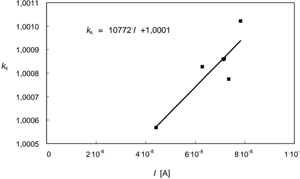
150V the current obtained at halved working potential i.e. 150 V. The ks as function ofioniza-tion current obtained in measuring a number of sources of varying strength is shown in Figure 2 together with a linear fit.
During the audit, the linear fit of Figure 2 was used to determine the recombination factor to correct currents when measuring HDR sources. For PDR sources, producing around ten times lower currents, no correction for recombination was applied.
ks = 10772.I +1,0001 1,0005 1,0006 1,0007 1,0008 1,0009 1,0010 1,0011 0 2.10-8 I [A] 4.10-8 6.10-8 8.10-8 1.10-7 ks
Figure 2 Recombination correction factors as determined with the two-voltage method at
different currents produced by HDR sources of varying strength and a linear fit to the data.
3.3 On-site measurements
The SSI on-site measurements comply with recommendations given by IAEA (IAEA (2002)). The equipment was always left in the treatment room on the day before meas-urements to reach thermal equilibrium with the surrounding. The air-temperature was determined within the well of the chamber. This was found important since the equipment come slowly into thermal equilibrium with the surrounding. A change in air-temperature of the room might not be fully reflected in the equipment until after several hours (see e.g. Figure 3.5 on page 48 in Venselaar and Pèrez-Calatayud (2004)). Storage within the room since the day before measurements does not ensure equivalence between the perature in the surrounding air and that in the well of the chamber since there can be tem-perature gradients within the room. Differences can exceed one degree when the place-ment during storage does not coincide exactly with that during measureplace-ments. We have once encountered a difference of several degrees between the chamber and the surround-ing air (not dursurround-ing this audit however). This occurred after a prostate treatment in which heated air was circulated beneath the quilt of the patient while the chamber was stored in a cupboard of the room.
The electrometer was turned on for at least one hour before measurements and the high voltage for at least 30 minutes. A system of low-scattering plastic boxes was used to get the centre of the chamber up at a distance of one meter above the floor. Care was taken to avoid walls and other equipment of the room as much as possible, to reduce scattering from these objects to contribute to the measured signal.
Figure 3 The SSI well-type ion chamber on top of the low-scattering plastic boxes used
to position the centre of the chamber at 1 meter above the floor.
Figure 4 The Farmer-in-Phantom system used as an on-site constancy control of the
well-type chamber. The plastic source-channel used to guide the source from the afterloading equipment to the irradiation position is also seen.
The chambers were pre-irradiated for a minimum of ten minutes before taking measure-ments. The point of maximum response was determined and three repeated source inser-tions were made. The electrometer was used in current-mode and therefore no account for source-transit time was needed. To assure the reproducibility in position it was checked that current-differences between the three insertions was less than 0,3% (IAEA (2002)). During each insertion the current was read on regular intervals at least ten times and lack of drift in the signal was assured. The time specified by date, hour and minute was used in decay corrections. Measurements were in all but one site done using a plastic source-channel to guide the source, as required on the NPL calibration-certificate. At one site problems with determining the length of the plastic channel arose and we had to use a
prostate needle of stainless steel (in use by this hospital). At a later time and another hos-pital, measurements to determine a correction factor, accounting for the difference be-tween use of a stainless steel needle and a plastic channel, were performed. This resulted in that a correction of 1,007 was applied to the signal determined using the stainless steel needle.
3.4 Estimation of uncertainty
The uncertainty in the determination of the ionization current and the reference air-kerma rate (RAKR) is determined following recommendations in the “Guide to the uncertainty in measurement” (GUM), ISO (1995)), for details see the Appendix.
4. Results
4.1 Inventory of
192Ir sources and overview of equipment
and procedures at Swedish hospitals
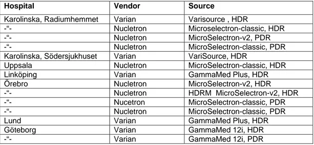
Brachytherapy with 192Ir is performed at 7 hospitals in Sweden, using in total 14 after-loading units, see Table 3.
Table 3 Inventory of the Swedish 192Ir after loading units. There are 9 nine HDR and 5 PDR units.
Hospital Vendor Source
Karolinska, Radiumhemmet Varian Varisource , HDR
-“- Nucletron Microselectron-classic, HDR -“- Nucletron MicroSelectron-v2, PDR
-“- Nucletron MicroSelectron-classic, PDR
Karolinska, Södersjukhuset Varian VariSource, HDR
Uppsala Nucletron MicroSelectron-classic, HDR
Linköping Varian GammaMed Plus, HDR
Örebro Nucletron MicroSelectron-v2, HDR
-“- Nucletron HDRM MicroSelectron-v2, HDR
-“- Nucetron MicroSelectron-classic, PDR
-“- Nucletron MicroSelectron-classic, PDR
Lund Varian GammaMed Plus, HDR
Göteborg Varian GammaMed 12i, HDR
-“- Varian GammaMed 12i, PDR
All the hospitals have well-type ionisation chambers that fulfil the IAEA recommenda-tions (IAEA (2002)). All except one have calibration factors traceable to UWADCL. One hospital relies instead on their own free-in-air measurements and interpolation technique (Drugge (1995)). This hospital is traceable to SSI through the interpolation qualities. The calibration factors in use are the ones obtained when purchasing the chambers, in some case as old as 7 years. No hospital has sent their chamber abroad for recalibration but await SSI to start offering this service.
All the Swedish hospitals measure the strength of each new 192Ir source. All, except one use the value determined by the vendor in the treatment planning system, i.e. the own
measurements are for verification purposes. Among the ones verifying the vendor value there is no strict tolerance level on the agreement although most hospitals tell they aim for and most often get an agreement within ±1%.
There are large differences in how the hospitals perform their measurements, both regard-ing the geometrical setup and in their handlregard-ing of the equipment. Most hospitals fulfil geometrical requirements on placing the chamber away from any walls and the floor while measuring. One hospital has their chamber permanently mounted on the wall in a corner of the treatment room. This placement is likely to increase the amount of scattered radiation reaching the chamber ((Podgorsak et al., 1992)). The hospital is aware of this but has not investigated the magnitude of the effect. Many calibration certificates require use of a plastic catheter for guiding the source to the chamber and most hospitals do so, however, some instead use a needle of stainless steel (of the type used for prostate treat-ments). Some hospitals leave the chamber to come into thermal equilibrium with the sur-rounding, allow time for stabilisation with high voltage supply on and pre-irradiate it before taking measurements. Others take measurements almost directly without precau-tions on stabilisation etc. Most hospitals determine the point of maximum response1 for the chamber for each new source and repeat source insertions to assure positional accu-racy (as recommended by e.g. the IAEA) whereas some trust earlier determinations of the point for maximum response and do not repeat source insertions.
Routines and methods for constancy control of the hospital’s chambers also vary. One hospital has a 137Cs check-source for internal irradiation, others irradiate their chambers in an external 60Co field, some do additional measurements with a redundant detector system whereas some do not check the constancy of their equipment at all. Among the hospitals with routines, some check the constancy of their equipment before each source strength determination whereas some only occasionally sometimes with intervals longer than one year.
Routines for controlling and calibrating other equipment (electrometers, thermometers, pressure meters) were not investigated.
4.2 Constancy of the SSI equipment
The stability of the SSI well-type chamber, as checked in the SSI external therapy level
60
Co beam, is shown in Figure 5.
Although the well-type chamber is permanently stored in the same room as the 60Co unit, the difference in temperature from the storage cupboard to the placement during irradia-tion is often 1 oC. Starting in 2006, the temperature is measured within the well of the chamber. The increased stability of the signal seen since 2006 is explained by the im-proved control of the temperature.
1
The point of maximum response of a well-type ionization chamber corresponds to the position of the source within the well that give raise to the highest signal. In most chambers there is a region of slowly varying sensitivity around this point which reduces positional uncertainty. The calibration of the chamber is usually valid for the point of maximum response.
0,997 0,998 0,999 1,000 1,001 1,002 1,003
Feb-05 May-05 Sep-05 Dec-05 Mar-06 Jul-06 Oct-06 Jan-07 Apr-07 Date
NK-60Co
/ N
K-60Co
Figure 5 Decay-corrected signal of the well-chamber normalised to its mean value
ob-tained by external irradiation in SSI’s therapy level 60Co beam.
NK-137Cs / N K-137Cs 0,997 0,998 0,999 1,000 1,001 1,002 1,003 Mar-06 Apr-06 Date
Figure 6 Decay-corrected signal of the well-chamber normalised to its mean value,
Figure 6 shows similar results using the 137Cs check source for internal irradiation of the chamber. The lack of results after April 2006 is due to a broken source holder that has not yet been repaired.
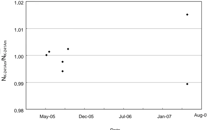
Figure 7 shows results of checking the constancy of the chamber in an external 241Am beam.The uncertainty of this routine is higher because the 241Am source is weak (thus producing a very low current in the chamber) and since the positioning is less precise. SSI normally uses the 241Am beam to calibrate instruments aimed for radiation protection purposes. 0,98 0,99 1,00 1,01 1,02
May-05 Dec-05 Jul-06 Jan-07
Date
N
K-241Am/N
K-241Am
Aug-07
Figure 7 Decay-corrected signal of the well-chamber normalised to its mean value,
ob-tained by external irradiation in SSI’s radiation protection-level 241Am beam.
Figure 8 shows the ratio of the currents, as measured by the well-type chamber to that of the Farmer-in-phantom, for different 192Ir sources. This ratio was determined at each site visited and used as an extra, on-site constancy control of the well-chamber. The standard deviation in the ratio is 1,2% which is comparatively high and depends on suboptimal properties of the phantom. The phantom could be improved by increasing the distance between the Farmer chamber and the source (from today’s 2 cm to around 5 cm, as used in most commercial phantoms), by drilling a straighter hole for the source-channel and by constructing inserts for source channels of varying diameter. Even though some proper-ties of the PMMA-phantom are not optimal it is valuable as an on-site check of the refer-ence equipment.
292 296 300 304 308 312
Dec-05 Mar-06 Jul-06 Oct-06 Jan-07 Apr-07
Date
current ratio: w
e
ll-ch
amber/Farmer-in-Phantom
Figure 8 The ratio of currents for 192Ir irradiation measured with the well-type chamber and the Farmer-in-Phantom system.
4.3 Estimation of uncertainty
The uncertainty in SSI’s determination of the ionization current (corrected for influence quantities), , is ± 0,2% which leads to a total uncertainty in reference air-kerma rate, RAKR, of ±0,7% (using the NPL calibration factor) at a coverage factor of k=1. This
cor-respond to uncertainties of ±0,4% and ±1,4% respectively at a coverage factor of k=2 (for details see the Appendix). The highest contribution to the total uncertainty in RAKR is contributed by the calibration factor. As determined here, the RAKR uncertainty disre-gards any contribution due to sources being of other design than the one used in the NPL calibration.
ion
I
4.4 Audit results and discussion
Figure 9a shows the RAKR as measured by SSI, vendors and hospitals. The PDR-sources are recognized by being around ten times weaker. (All results are decay-corrected to the time stated on the vendor certificate. The fifth PDR-source was initially delivered and used for HDR purposes and therefore appears here as being of HDR-strength).
Figure 9b repeats the results of Figure 9a in more detail and also shows the uncertainty limits; ±3% for vendors and ±1,4% for SSI at a coverage factor of k=2. (Most vendors state an uncertainty in RAKR of ±5% at a confidence limit of 99,7% which equals a cov-erage factor of k=3 following GUM ((ISO, 1995)) and hence translates to ±3% at k=2).
0 10 20 30 40 50 60 1 2 3 4 5 6 7 8 9 10 11 12 13 14 source number RAKR [mGy /h] SSI NK-NPL Vendor Hospital
Figure 9a Values of RAKR for the sources as determined by SSI (using the calibration
factor from NPL), vendors and hospitals. All source strengths refer to the date and time specified on the vendor’s certificate.
SSI (NRAKR-NPL) Vendor Hospital
42 46 50 54 1 2 3 4 5 6 7 8 9 10 source number RAKR [mGy /h] 4,2 4,3 4,4 4,5 4,6 4,7 11 12 13 14 RAKR [mGy /h]
Figure 9b Values of RAKR for the sources as measured by SSI (using the NPL
calib-ration factor), vendors and hospitals. Uncertainty limits from SSI and vendors are inserted.
Figure 10 presents ratios of RAKR with vendor values as determined by SSI and hospitals, numbering the sources as in Figures 9a and 9b.
Both SSI and hospitals are in good agreement with vendors, the ratio of RAKR-values in all cases within the guaranteed uncertainty (± 3% at k=2). The uncertainty in vendor val-ues has been reduced in the last years and the agreement seen in the Swedish audit is an improvement compared to what was seen in a similar investigations some 10 years ago (Venselaar et al., (1994)). This is probably thanks to the development and use of well-type chambers designed for brachytherapy both at hospitals, vendor laboratories and SSDLs. Well-type chambers are more robust and far easier to setup than is the earlier commonly used free-in-air jigs equipped with Farmer-type chambers.
0,97 0,98 0,99 1,00 1,01 1,02 1,03 1 2 3 4 5 6 7 8 9 10 11 12 13 14 source number RAKR -ratio with vend or ce rtificate SSI-NPL/Vendor Hospital/Vendor
Figure 10 Ratios of SSI- and hospital-determined RAKR to the same values on vendor
certificates
For source number 8, the hospital’s RAKR is 2,6% higher than the vendor’s value. Al-though not outside the uncertainty limits it is a tendency not verified by the SSI (see also the hospital-ratio with SSI in Figure 11). The hospital holding source number 8 uses their chamber mounted on the wall in a corner of the room during measurements, without any corrections. An increased value could be expected, due to photons back-scattered from the walls towards the chamber. Studies on room-scatter as function of distance to the floor and walls for brachytherapy well-type ion chambers were performed by Podgorsak
et al., (1992). They show that it is worse to be close to a wall than close to the floor and
say that even if corrections are applied, it is not advisable to place well-type chambers closer than 25 cm from a wall.
Embedded in ratios of RAKR are systematic differences due to traceability with different calibration laboratories. The hospital holding sources 1 and 2 uses their own interpolation technique for 192Ir calibrations (Drugge, (1995)) and is traceable to SSI through the
inter-polation quantities. The rest of the hospitals all have calibration factors from the UWADCL. Figure 11 shows values of RAKR from the UWADCL-traceable hospitals normalised to SSI-values obtained using either the SSI(NPL)- or the SSI(UWADCL)-calibration factors. Differences between hospitals and SSI using the SSI(UWADCL) cali-bration factor don’t exceed ±0,6%, excluding source number 8 (measured at the hospital with their well-chamber mounted on the wall). The hospital holding source number 8 uses their own value for verification purposes and enters that of the vendor in the treatment planning system. This means that if they stick to their geometry, the probable systematic error due to placement on the wall should be constant in time and embedded in their ratio with the source vendor.
0,97 1,00 1,03 3 4 5 6 7 8 9 10 11 12 13 14 source number RAKR -r atio Hospital/SSI(NPL) Hospital/SSI(UWADCL)
Figure 11 Comparing ratios of the hospitals RAKR determinations to those of SSI using
either the NPL or the UWADCL calibration factors for the SSI well-chamber.
Figure 12 shows results from a Swedish hospital on their difference with vendor values for different HDR-sources within the same afterloading unit (reproduced with permission from Marie Lundell at the Karolinska Hospital in Stockholm). Having observed an in-creased deviation, the hospital contacted the vendor which resulted in that a problem with the pressure meter at the vendor’s calibration laboratory was found and solved. Differ-ences have since then been stable and within ±1%. Figure 12 is an example of the useful-ness of visualising results as function of time.
-4 -2 0 2 4
jun-03 aug-04 sep-05 okt-06
Date
RAKR
difference with ve
ndor [%]
Figure 12 The difference in estimated RAKR at a Swedish hospital relative to the vendor
certificate for various HDR-sources within the same unit.
The establishment of a mean ratio (or mean difference) between the own hospital and the vendor for each afterloading unit is recommended. Such a ratio embeds the differences due to varying traceability as well as those due to source-type and setup. Provided that recommendations in equipment handling are followed (IAEA (2002)) and that measure-ments are performed in the same way each time (i.e. according to a written routine), the uncertainty in an established ratio has potential of being lower than that in RAKR. Most vendor certificates state an uncertainty in RAKR of ±3% at k=2. Assuming an uncertainty in the calibration factor of ±2,5% (the typical uncertainty at k=2, using the most common interpolation technique, see e.g. Table 1), this indicates an uncertainty of ±1% in the ioni-zation current (or charge) measurements at the vendor’s laboratory (including all correc-tions for influence quantities etc. in these measurements). SSI estimates their uncertainty in current measurements to ±0,4%, see the Appendix. Hospitals that follow recommenda-tions on use of equipment can probably do as good as the vendors (who probably includes some safety margin) which would give an uncertainty in ±1,4% for an established ratio. Variations in this ratio could be an early indicator on problems in some part of the sys-tem; either with the equipment or the measurement technique at the hospital or the vendor laboratory.
5. Conclusions
The accuracy in determining source strength for 192Ir brachytherapy is high at Swedish hospitals. All hospitals verify the strength of each new 192Ir source, using well-type chambers designed for brachytherapy. Values on source-strength measured by SSI and the hospitals all fell within the uncertainty limits guaranteed by the vendors.
The awaited start of a calibration service for 192Ir in Sweden will help solve the current problem of several hospitals having calibration factors that are too old according to inter-national recommendations. Routines for verifying the constancy of the equipment and in setting it up for measurements varies and some hospitals could improve their routines. With the awaited start of a calibration service for 192Ir in Sweden, the hospitals will most
likely have to transport their chambers, a procedure that necessitates systems for con-stancy control of the equipment before and after transport. Hospitals without external
60
Co beams can be recommended to purchase a 137Cs check-source for this purpose.
Hospitals can be recommended to establish their own ratio (or deviation) with the vendor and follow it as function of time. Such a mean-ratio embeds systematic differences of various origins and have a lower uncertainty than has the RAKR alone, making it useful for early detection of problems with equipment or routines.
There is room for SSI in advising hospitals regarding measurement techniques and main-tenance of equipment, for instance by advising them to follow the IAEA recommenda-tions. SSI could also define requirements for the agreement between source strengths as determined by hospitals and vendors. Such a recommendation could be coupled to an action plan, dependent on level of disagreement, and some kind of reporting to SSI.
6. Acknowledgement
Assistance from the physicists at the brachytherapy departments is greatly acknowledged. This project was funded by the Swedish Radiation Protection Authority (SSI project 1522.06).
7. References
Brahme A 1984 Dosimetric precision requirements in radiation therapy Acta Radiol. Oncol. 23 379-91
Büermann L, Kramer H-M, Schrader H and Selbacj H-J 1994 Activity determination of 192Ir solid sources by ionization chamber measurements using calculated corrections for self-absorption Nucl. Instr. and Meth. A 339 369-76
Douysset G, Gouriou J, Delaunay F, DeWerd L A, Stump K and Micka J 2005 Comparison of dosimetric standards of USA and France for HDR brachytherapy Phys. Med. Biol. 50 1961-78
Drugge N 1995 Determination of the Reference Air Kerma Rate for Clinical 192Ir Sources. Report from Department of Radiation Physics, Göteborg University, Göteborg, Sweden)
Goetsch S J, Attix F H, Pearson S W and Thomadsen B R 1991 Calibration of 192Ir high-dose-rate afterloading systems Med. Phys. 18 462-7
IAEA (International Atomic Energy Agency ) 2002 Calibration of photon and beta ray sources used in brachytherapy. (Vienna, Austria IAEA TECDOC 1274)
ICRU (International Commission on Radiation Units and Measurements) 1985 Dose and Volume Specification for Reporting Intracavitary Therapy in Gynecology. (Bethesda, MD: ICRU report no 38)
ICRU (International Commission on Radiation Units and Measurements)1997 Dose and Volume Specification for Reporting Interstitial Therapy. (Bethesda, MD: ICRU report no 58) ISO (International Organization for Standardization) 1995 Guide to the expression of uncertainty
Nath R, Anderson L L, Meli J A, Olch A J, Stitt J A and Williamson J F 1997 Code of practice for brachytherapy physics: Report of the AAPM Radiation Therapy Committee Task Group No. 56 Med. Phys. 24 1557-98
Podgorsak M B, DeWerd L A, Thomadsen B R and Paliwal B R 1992 Thermal and scatter effects on the radiation sensitivity of well chambers used for high dose rate Ir-192 calibrations
Med. Phys. 19 1311-4
Rivard M J, Coursey B M, DeWerd L A, Hanson W F, Huq M S, Ibbott G S, Mitch M G, Nath R and Williamson J F 2004 Update of AAPM Task Group No. 43 Report: A revised AAPM protocol for brachytherapy dose calculations Med. Phys. 31 633
Sander T and Nutbrown R F 2006 The NPL air kerma primary standard TH100C for high dose rate 192Ir brachytherapy sources. (Teddington, UK, NPL Report DQL-RD 004)
Stump K E, DeWerd L A, Micka J A and Anderson D R 2002 Calibration of new high dose rate
192
Ir sources Med. Phys. 29 1483-8
van Dijk E, Kolkman-Deurloo I-K K and G D P M 2004 Determination of the reference air kerma rate for 192Ir brachytherapy sources and the related uncertainty Med. Phys. 31 2826-33 Venselaar J L M, Brouwer W F M, van Straaten B H M and Aalbers A H L 1994 Intercomparison
of calibration procedures for Ir-192 HDR sources in The Netherlands and Belgium
Radio-ther. Oncol. 30 155-61
Venselaar J and Pèrez-Calatayud J eds 2004 A practical guide to quality control of brachytherapy equipment (ESTRO booklet no 8)
Appendix - Estimation of uncertainty
The ionization current in the well-chamber, corrected for influence quantities, , is de-rived as: ion
I
1/ 2 ln 2(
)
t Tion irr leak T P
I
I
I
k
k
e
⋅ −
=
−
⋅
⋅
⋅
(A1)where is the ionization current in the well-chamber during irradiation, is the leakage ionization current (obtained from the system in absence of irradiation), and
are the correction factors for ambient temperature and pressure and
irr
I
I
leak Tk
Pk
1/ 2 ln 2e
t T − is the cor-rection for decay with the half-life andt
the time since the vendor-issued calibra-tion.1/ 2
T
The experimental determination of the reference air-kerma rate, RAKR, of the sources is derived as:
RAKR ion
RAKR
=
N
⋅
I
(A2)where
N
RAKR is the calibration factor of the well-chamberThe uncertainty in and is derived as the combined uncertainty of the individ-ual input components in Eqs. A(1)-A(2), following recommendations in the GUM-report ((ISO, 1995) and assuming the inputs to be uncorrelated. Details on the uncertainty of the individual inputs are given below. The relative uncertainty in was determined to 0,2% and in to 0,7% at a coverage factor of 1, which corresponds to 0,4% and 1,4% respectively at a coverage factor of 2. This estimation of uncertainty is without account-ing for possible effects due to differences in sources design and hence strictly valid only for measurements of a MicroSelectron classic HDR source from Nucletron BV, as used in the NPL-calibration. The uncertainty for measuring sources of other design is not known in detail. For HDR -sources having an active length of the
ion
I
RAKR ionI
RAKR 192Ir material similar to that of the Microselectron Classic differences were less than the uncertainty in calibration factors and could therefore not be resolved, see Stump et al. (2002).
Uncertainties in the individual inputs
The uncertainties of the various factors of Eq. (A1) are derived below, at a coverage fac-tor of one.
Uncertainty in
N
RAKRThe uncertainty in the calibration factor is obtained with the calibration factor from the calibration certificate issued by the calibration laboratory. The uncertainty in the calibra-tion factor from NPL,
RAKR NPL
N
σ
− , is 1,3% at a coverage factor of 2 and hence 0,65% at a coverage factor of 1.Contribution Method for estimation
Uncertainty in NRAKR-NPL From calibration certificate
Combined 0,65%
Uncertainty in
I
irrContribution Method for estimation Type A 1s [%]
Type B 1s [%]
Positioning of source Standard deviation in signal from 3 re-peated insertions
0,005
Current reading Standard deviation of the mean in man-ual current readings from electrometer (The mean of 10-15 current readings are used as the signal value)
0,0025 (HDR)
or
0,01 (PDR)
Setup Standard deviation in repeated setups for one source
0,05
Maximum response curve
Measurements at maximum 5mm from point of maximum response of well-chamber
0,02
Calibrating e-m against “App 1”
Standard deviation in repeated calibra-tions of electrometer against “App 1” at SSI
0,05
Recombination The uncertainty in factor (derived with two-voltage method) assumed of similar magnitude as the correction.
0,1
Uncertainty in “App 1”
From Jan-Erik Grindborg, SSI 0,05
Stability Standard deviation in constancy checks 0,1
Combined; HDR 0,17 %
Combined; PDR 0,17 %
“App 1” is SSI’s calibrated reference electrometer. The standard deviation in current readings are somewhat higher for the weaker PDR-sources, however, the difference is not significant for the combined uncertainty which is dominated by the stability.
Uncertainty in
I
leakContribution Method for estimation Type A 1s [%]
Type B 1s [%]
Current reading Standard deviation in manual current readings
0,05
Calibrating e-m against “App 1”
Standard deviation in repeated calibra-tions against App 1 at SSI
0,05
Uncertainty in App 1 From Jan-Erik Grindborg, SSI 0,05
Stability Standard deviation in constancy checks 0,1
Combined 0,132 %
Uncertainty in
k
TThe signal is corrected for ambient temperature according to:
273,15
293,15
293,15
TT
k
=
+
where T is the temperature in degrees Celsius. Below is the uncertainty in the temperature
T derived.
Contribution Method for estimation Type A 1s [%]
Type B 1s [%]
Calibration against SSI-reference
Standard deviation in the correction from multiple comparisons of field and reference instruments, converted to uncertainty in T
0,25
Uncertainty in reading instrument
One decimal resolution yields uncer-tainty of 0,05 degrees
0,25
Uncertainty in reference instrument
From Jan-Erik Grindborg, SSI 0,005 0,006
Combined 0,35 % 0,35 0,006 An uncertainty in T of 0,35% amounts to an uncertainty in kT of 0,02%.
Uncertainty in
k
PThe signal is corrected for ambient pressure according to:
101,325
P
k
P
=
where P is the pressure in kPa. Below is the uncertainty in the pressure P derived.
Contribution Method for estimation Type A 1s [%]
Type B 1s [%]
Calibration against SSI-reference
Standard deviation in the correction from multiple comparisons of field and reference instruments, converted to uncertainty in P
0,12
Uncertainty in reading of instrument
From resolution of analog instrument 0,05
Uncertainty in ref in-strument
From Jan-Erik Grindborg, SSI 0,007 0,002
Combined 0,13%
An uncertainty in P of 0,13% amounts to an uncertainty in kP of 0,13%.
Uncertainty in 1/ 2 ln 2 t T
e
⋅ −The uncertainty in the decay correction arising from an uncertainty in the time of meas-urement between current measmeas-urement and that made at the vendor laboratory is consid-ered. Any uncertainty in the half time of decay is negligible.
Contribution Method for estimation Type A 1s [%]
Type B 1s [%]
Error in time of meas-urement specified by date:hour:minute
Errors in time of the experimentalists watch relative that of the vendor labora-tory: 5 minutes. Shortest period between the vendor calibration and measurement: 10 days
0,04
Combined
An uncertainty in t of 0,04 % amounts to an uncertainty in the decay correction of
1/ 2 ln 2 t T
e
⋅ − of 0,005%.2007:01 Statens ansvar för slutförvaring av använt kärnbränsle
SKI och SSI
2007:02 Strålmiljön i Sverige
Avdelningen för beredskap och miljöövervakning
Pål Andersson et.al. 310 SEK
2007:03 personalstrålskydd inom kärnkraft- industrin under 2005
Avdelningen för personal- och patientstrålskydd Stig Erixon, Karin Fritioff, Peter Hofvander, Ingemar Lund, Lars Malmqvist, Ingela Thimgren och Hanna Ölander Gür 70 SEK
2007:04 recent research on emf and Health risks. fourth annual report from SSI’s Independent expert group on electromagnetic fields, 2006
Avdelningen för beredskap och miljöövervakning 110 SEK
2007:05 doskatalogen för nukleärmedicin projekt; SSI p 1426.04
Avdelningen för personal- och patientstrålskydd Sigrid Leide-Svegborn, Sören Mattsson, Lennart Johansson, Per Fernlund och Bertil Nosslin 90 SEK
2007:06 personalstråldoser inom vård, forskning och icke kärnteknisk industri i Sverige under 1999-2005
Avdelningen för personal- och patientstrålskydd
Catarina Danestig Sjögren 100 SEK
2007:07 Inquiry into the radiological consequences of power uprates at light-water rectors worldwide
Avdelningen för personal- och patientstrålskydd Tea Bilic Zabric, Bojan Tomic, Klas Lundgren
and Mats Sjöberg 290 SEK
2007:08 Solvanor i Sverige 2006
Avdelningen för beredskap och miljöövervakning
Richard Bränström 100 SEK
2007:09 Säkerhets- och strålskyddsläget vid de svenska kärnkraftverken år 2006
SKI och SSI 100 SEK
2007:10 radiological consequences of radionuclide releases to sewage systems from hospitals in Sweden
Avdelningen för kärnteknik och avfall
Rodolfo Avila, Idalmis de la Cruz, Synnöve Sundell-Bergman
och Serena Hasselblad 430 SEK
2007:11 SSI:s granskning av SkB:s storregionala grundvattenmodellering för östra Småland (SkB rapport 06-64)
Avdelningen för kärnteknik och avfall
Björn Dverstorp 150 SEK
SSI-rapporter 2007
SSI reports 2007
2007:12 rapport från SSI:s vetenskapliga råd om ultraviolett strålning 2006Avdelningen för beredskap och miljöövervakning SSI:s vetenskapliga råd om ultraviolett strålning 110 SEK
2007:13 dosimetry audit on the accuracy of 192Ir brachytherapy source strength determinations in Sweden
Avdelningen för personal- och patientstrålskydd
Adress: Statens strålskyddsinstitut; S-171 16 Stockholm Besöksadress: Solna strandväg 96
Telefon: 08-729 71 00, Fax: 08-729 71 08 Address: Swedish Radiation Protection Authority SE-171 16 Stockholm; Sweden
Visiting address: Solna strandväg 96
Telephone: + 46 8-729 71 00, Fax: + 46 8-729 71 08
S
TATENS STRÅLSKYDDSINSTITUT, SSI, är en central tillsyns-myndighet som verkar för ett gott strålskydd för människan och miljön, nu och i framtiden.
SSI sätter gränser för stråldoser till allmänheten och för dem som arbetar med strålning, utfärdar föreskrifter och kontrollerar att de efterlevs. SSI håller beredskap dygnet runt mot olyckor med strålning. Myndigheten informerar, utbildar och utfärdar råd och rekom- mendationer samt stöder och utvärderar forskning. SSI bedriver även internationellt utvecklingssamarbete.
Myndigheten, som sorterar under Miljödepartementet, har 110 anställda och är belägen i Solna.
THESWEDISHRADIATIONPROTECTIONAUTHORITY (SSI) is a central
regulatory authority charged with promoting effective radiation protection for people and the environment today and in the future.
SSI sets limits on radiation doses to the public and to those that work with radiation. SSI has staff on standby round the clock to respond to radiation accidents. Other roles include information, education, issuing advice and recommendations, and funding and evaluating research.
SSI is also involved in international development cooperation. SSI, with 110 employees located at Solna near Stockholm, reports to the Ministry of Environment.