http://www.diva-portal.org
This is the published version of a paper published in FEBS Letters.
Citation for the original published paper (version of record):
Askerlund, P., Larsson, C., Widell, S. (1988)
Localization of donor and acceptor sites of NADH dehydrogenase activities using inside-out and
right-side-out plasma membrane vesicles from plants.
FEBS Letters, 239(1): 23-28
http://dx.doi.org/10.1016/0014-5793(88)80538-6
Access to the published version may require subscription.
N.B. When citing this work, cite the original published paper.
Open Archive article
Permanent link to this version:
Volume 239, number 1, 23-28 FEB 06392 October 1988
Localization of donor and acceptor sites of NADH dehydrogenase
activities using inside-out and right-side-out plasma membrane
vesicles from plants
Per Askerlund, Christer Larsson and Susanne Wide11
Department of Plant Physiology, University of Lund, PO Box 7007, S-220 07 Lund, Sweden
Received 2 August 1988
Inside-out and right-side-out plasma membrane vesicles from sugar beet (Beta &garis L.) leaves, prepared by aqueous two-phase partitioning, were used to localize donor and acceptor sites and to determine substrate affinities for plasma membrane-bound NADH dehydrogenase activities. NADH-ferricyanide and NADH-cytochrome c reductase activities were approx. 30% latent with inside-out vesicles and about 80% latent with right-side-out vesicles, indicating that both donor and acceptor sites for these activities are located on the cytoplasmic surface of the plasma membrane, and that
a possible transplasma membrane electron transport would constitute only a minor proportion of the total activity. Plasma membrane; Inside-out vesicle; Redox chain; Enzyme kinetics; NADH-ferricyanide reductase; NADH-cytochrome c reductase
1. INTRODUCTION
The redox activities associated with plant plasma membranes (PMs) are currently attracting much interest. Such activities have been observed with intact plants, cell cultures and purified PM preparations [l-3 1. These PM-associated activities have been suggested to participate in several physiological processes, such as ion transport and hormonal regulation of cell growth [2-51. With in- tact plants, tissues and cells, the PM redox system can reduce exogenous electron acceptors such as ferricyanide (FeCN) [3,6], which suggests the presence of a transplasma membrane electron- transport system. The natural electron acceptor is not known, however, in young roots of non- graminaceous plants for example, it may be Fe- chelates since they possess an Fe3+-reduction
Correspondence address: P. Askerlund, Department of Plant
Physiology, University of Lund, PO Box 7007, S-220 07 Lund, Sweden
Abbreviations: cyt., cytochrome(s); FeCN, ferricyanide; PM(s), plasma membrane(s); PpBQ, phenyl-p-benzoquinone
system which is induced by iron deficiency [7]. An auxin-stimulated NADH oxidase has been purified from soybean hypocotyl PMs [5], therefore 02 may be a natural electron acceptor in other tissues. The physiological reductant for the PM ox- idoreductases appears to be cytoplasmic NADH or NADPH [l-3,7], but oxidation of exogenous NAD(P)H by cells and tissues has been reported in several cases [6,8]. Isolated PM preparations of high purity have been reported to reduce artificial electron acceptors [9, lo], ascorbate free radical [ll] and to a lesser extent 02 [10,12,13] in the presence of NAD(P)H. NADH seems to be prefer- red to NADPH with PMs from some sources [lO,ll], however with PMs from different origins the activities are similar [9]. An NADH-FeCN reductase has been partially purified from PMs of maize roots [l 11.
In PM preparations consisting mainly of right- side-out (70-90% apoplastic side-out) [14] vesicles, NAD(P)H dehydrogenase activities show high latencies (large increase in activity on addition of Triton X-100) with impermeable electron accep- tors such as FeCN and cyt. c, as well as with duro- quinone [ 10,l 1 ,151. If it is assumed that the sole
Volume 239, number 1 FEBS LETTERS October 1988
effect of the detergent is to allow the added substrates to gain access to their active sites on the membrane surface, the reasons for the latency could be either that NAD(P)H or the acceptor has its active site on the cytoplasmic surface of the PM, or that both have their active sites on the cytoplasmic surface of the PM. A transplasma membrane electron-transport chain which could reduce Fe3+ at the apoplastic surface should have the donor and acceptor site(s) on the cytoplasmic and apoplastic surfaces, respectively. To obtain positive evidence for the orientation of donor and acceptor sites, it is necessary to have vesicles of op- posite orientation. Very recently, we succeeded in separating inside-out and right-side-out PM vesicles from leaves of sugar beet [16]. We have now used these PM vesicles to determine the orien- tation of the active sites for NADH and the elec- tron acceptors FeCN and cyt. c as well as the Km values for these substrates.
2. MATERIALS AND METHODS 2.1. Plant material
Light-grown 4-8-week-old sugar beet plants (Beta vulgaris L.), kindly supplied by Hilleshog AB, Sweden, were used. 2.2. Plasma membranes
PMs of inside-out and right-side-out orientation were prepared from sugar beet leaves as described in [16], except that dithiothreitol was omitted from the medium used during the final centrifugation as well as from the resuspension medium to decrease NADH-independent reduction of electron acceptors in the assays. Based on the latency of the Kc-stimulated, Mg*+-dependent ATPase, the inside-out fraction contained -90% sealed inside-out vesicles whereas the right-side-out frac- tion contained -90% sealed, right-side-out vesicles [16].
2.3. Enzyme assays
NADH-FeCN reductase activity was determined with an Aminco DW 2 spectrophotometer operated in the dual-beam mode by following the reduction of FeCN at 420 minus 500 nm at 25°C. The standard reaction mixture contained 25 mM Hepes-KOH (pH 7.3), or 50 mM Tris-acetate (pH 7.3), 0.33 M sucrose, 1 mM Ks[Fe(CN)e], 0.4 FM antimycin A, 1 mM KCN and 20-50 fig membrane protein in a volume of 1 .O ml. The reaction was initiated by the addition of 0.25 mM NADH. Cor- rection was always made for non-enzymatic reduction of FeCN. NADH-cyt. c reductase activity was measured under the same conditions as for NADH-FeCN reductase except that 4OpM cyt. c (final concentration) was substituted for Ks[Fe(CN).s]. The activity was recorded at 550 minus 600 nm. When K,,, values were determined the assay mixture also included 7.5 mM
24
MgCl2, and during measurement of K, for FeCN the concen- tration of NADH in the assay was increased to 0.5 mM.
NADH-PpBQ reductase was determined by following the ox- idation of NADH at 340 nm. A correction (11%) was made for the simultaneous decrease at 340 nm due to reduction of PpBQ, assuming an NADH/PpBQ stoichiometry of 1. The reaction mixture was the same as for NADH-FeCN reductase except that 0.2 mM PpBQ (final concentration), added in a volume of 5 ~1 dimethyl sulfoxide, was substituted for Kx[Fe(CN)b]. The reac- tion was started by the addition of membranes to allow subtrac- tion of non-enzymatic oxidation of NADH.
The values of the extinction coefficients used were 1, 19, 6.2 and 0.8 mM-‘.cm-’ for FeCN, cyt. c, NADH and PpBQ, respectively.
2.4. Protein
Protein was measured essentially as in [17], using bovine serum albumin as standard.
3. RESULTS
3.1. Latencies of NADH dehydrogenase activities The capacity of PM vesicles with opposite orien- tation to oxidize NADH in the presence of dif- ferent artificial electron acceptors was investigated. In the absence of Triton X-100 the ac- tivity was much higher with inside-out than right- side-out PM vesicles for all electron acceptors tested, whereas in the presence of Triton X-100 the activities were similar in both vesicle types (table 1). Thus, the latencies of NADH-dehydrogenase activities were low with inside-out vesicles and high with right-side-out vesicles. The considerable dif- ferences in latency between the two fractions were not due to different concentration optima for Triton X-100, since both showed maximal activity at the Triton concentrations used (fig.1). NADH- FeCN reductase activity reached a plateau above 0.015% Triton X-100, while that with cyt. c decreased at concentrations above -0.015% (fig.l), the exact optimum being dependent on the protein concentration (not shown). The Triton dependency with PpBQ was similar to that with FeCN (not shown). A greater latency was observed with PpBQ with inside-out PMs than with FeCN and cyt. c, however, with right-side-out PMs the latency was the same as with FeCN (table 1). No oxidation of NADH was observed in the absence of added electron acceptor, indicating that 0~ could not serve as an acceptor with these prepara- tions, at variance with previous reports for PM from other sources [12,13].
Volume 239, number 1 FEBS LETTERS October 1988
Table 1 ,. n
NADH dehydrogenase activities of inside-out (IO) and right- side-out (RO) plasma membrane vesicles with different electron
acceptors
Vesicle (unit) NADH-acceptor dehydrogenase activity FeCN Cyt. c PPBQ IO (spec. act.)” 655 + 186 200 f 58 198 f 56 IO + TX 100 (spec. act.) 936 k 192 263 f 53 326 + 40 Latency (o/o) 30* 13 25+ 6 40+14 RO (spec. act.) 177 + 47 43 + 9 40 f 22 RO + TX 100 P A A. NADH-FeCN B 0’ (spec. act.) 1090 + 224 202 + 63 270 + 30 Latency (%) 83+ 5 75 + 4 84 f 1 a nmol reduced acceptor .min-’ .(mg protein))’ (+ SD) in the
case of FeCN and cyt. c, and nmol oxidized NADH.min-’ .(mg protein)-’ + SD for PpBQ
Data represent means from measurements on the same 4 membrane preparations. Triton X-100 (TX 100) was used at 0.015% (w/v) in the case of cyt. c and at 0.025% (w/v) for FeCN and PpBQ. Latency is defined as:
(activity with TX 100) - (activity without TX 100) x loo% (activity without TX 100)
0. NADH-cyt. c l
OL I
0 0.01 0.02 0.03 Trlton X-100, % (w/v)
Fig. 1. Effect of Triton X-100 on NADH-FeCN (A) and NADH- cyt. c (B) reductase activities of inside-out (0) and right-side- out (A) plasma membrane vesicles. Protein concentration,
50pg.ml-‘.
3.2. Kinetic characterization of NADH-FeCN and
NADH-cyt. c reductase activities with
inside-out and right-side-out plasma
membrane vesicles
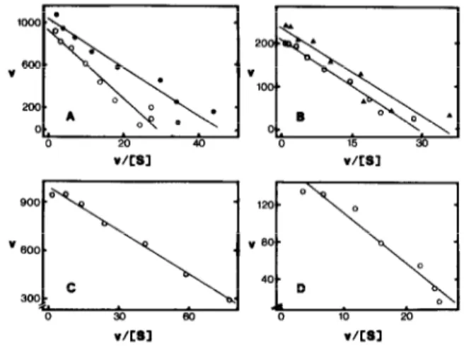
K,,, values for NADH were determined with inside-out and right-side-out PMs in the presence of Triton X-100, as well as with inside-out PMs in the absence of this detergent (fig.2A,B; table 2). We also attempted to measure &,(NADH) for FeCN reductase with right-side-out PMs in the absence of Triton X-100, however, the results were erratic due to the very low activities and are not presented here. No significant differences for K,,,(NADH) were observed between the two vesicle fractions with either FeCN, or cyt. c (table 2), nor was any evidence found in support of the existence of more than one component, i.e. straight lines were obtained with high regression coefficients (fig. lA,B). In contrast, there was a significant dif- ference in K,(NADH) values between NADH- FeCN and NADH-cyt. c reductase activities, the affinity for NADH of the latter activity being
-4-times higher (table 2).
The K,,, values for FeCN and cyt. c with inside- out vesicles could only be determined accurately in
V/CBl V/LB3
Fig.2. Eadie-Hofstee plots for determination of Km values. (A) K,,,(NADH) for NADH-FeCN reductase of inside-out PM vesicles in the absence (0, K,,, = 22pM, R = 0.96) and presence (0, K,,, = 32pM, R = 0.97) of 0.025% (w/v) Triton X-100. (B) K,,,(NADH) for NADH-cyt. c reductase of inside- out PM vesicles in the absence of Triton X-100 (0, K,,, =
7.1 PM, R = 0.99), and of right-side-out PM vesicles in the presence (A , K,,, = 6.8 gM, R = 0.94) of 0.015% Triton X-100. (C) K,(FeCN) for NADH-FeCN reductase of inside-out PM vesicles in the absence of Triton X-100 (K,,, = 9.0 ,uM, R = 1.0). (D) K,(cyt. c) for NADH-cyt. c reductase of inside-out PM vesicles in the absence of Triton X-100 (Km = 5.4pM, R =
0.98). v = nmol acceptor reduced.min-’ .(mg protein)-‘. S, substrate concentration (uM). R, regression coefficient obtained after linear regression analysis. 9 (A,B) or 7 (C,D)
Volume 239, number 1 FEBSLETTERS October 1988
Table 2
K,,, values for NADH, FeCN and cyt. c obtained with inside-out (10) and right-side-out (RO) plasma membrane vesicles
Vesicles Km W)
NADH-FeCN NADH-cyt . c
reductase reductase NADH FeCN NADH Cyt. c IO 30 + 8 8.0 + 1.4 7.6 + 0.7 4.1 f 1.3 (n = 3) (n = 2) (n = 2) (n = 2) IO + TX 100 25 t 3 NT 6.6 + 0.3 NT RO+TXlOO :1;“7 (n = 2) NT 8.0 + 1.4 NT (n = 3) (n = 2)
Concentrations of Triton X-100 (TX 100) as in table 1. NT, not tried. n, number of membrane preparations tested
the absence of detergent (table 2), since Triton X-100 released reducing compounds from the vesicles (originating from the homogenization medium) which interfered with measurements at very low concentrations of acceptor. No evidence for more than one component was obtained in determinations of K, values for FeCN and cyt. c (fig.2C,D). With right-side-out vesicles in the absence of Triton, activities were too low for the determination of K,,, values for either electron ac- ceptor.
During determination of K, values for NADH and FeCN it was noted that 7.5 mM Mg2+ in- creased the activities approx. 2-fold at low concen- trations of NADH and ferricyanide, respectively. This was probably due to screening of the net negative charge on the PM [18] by the cation, as has been reported for mitochondrial inner mem- branes [ 191. The effect of Mg2+ was not further in- vestigated, but 7.5 mM MgCl2 was always present in the assay medium during all measurements on the kinetics in order to maintain the surface poten- tial at a low value. This ensures that the K,,, values obtained are close to the ‘true’ values.
4. DISCUSSION
The membrane-impermeable electron donor NADH, and the impermeable electron acceptors FeCN and cyt. c, as well as the membrane- permeable acceptor PpBQ [20] were used to localize donor and acceptor sites of PM NADH
26
dehydrogenases using inside-out and right-side-out PM vesicles. The rationale was that impermeable substrates would only indicate the activities when both donor and acceptor sites are located on the same side of the membrane, whereas a permeable acceptor would also record transmembrane elec- tron transport provided that the donor site is on the exposed surface of the membrane vesicle. Ad- dition of detergent will rupture the vesicles, expose all sites and give 100% activity.
The latencies obtained with FeCN and cyt. c as acceptors clearly indicate that both the active sites for NADH-FeCN and NADH-cyt. c reductase are located on the cytoplasmic surface of the PM. Trans-membrane electron transport from NADH to these acceptors could at most constitute approx. 30% of the total activities, since the latencies with inside-out vesicles were only about 30% (table 1). However, this 30% increase in activity could also be due to contamination of inside-out vesicles by 30% right-side-out vesicles, although the latency of the K+-stimulated, Mg2+-dependent ATPase in- dicated only 10% contamination [16]. The hydrophobic acceptor PpBQ should be able to ac- cept electrons on both sides of the PM and NADH- PpBQ reductase should therefore only show NADH latency. The latency of this activity should thus provide a better measure of the degree of con- tamination by right-side-out vesicles. Surprisingly, PpBQ exhibited greatest latency (40%, table 1) with inside-out vesicles, which appears to exclude the possibility of transplasma membrane electron transport. However, an increase in activity on ad- dition of detergent may not only be due to ruptur- ing of vesicles with concomitant exposure of hidden sites, but may also be the result of stimula- tion of the activity by some other mechanism. This second possibility may explain the relatively high latency obtained with PpBQ and inside-out vesicles. Therefore, the possibility that trans- membrane electron transport makes a minor con- tribution to the total activity cannot be excluded. In agreement with our results, MorrC et al. [21] have observed reduction of FeCN by NADH only at the cytoplasmic surface in PM vesicles from soy- bean hypocotyls using a cytochemical method. Furthermore, Giannini and Briskin [22], working with a crude PM fraction from red beet storage tissue, obtained no evidence of transmembrane electron transport from NADH to FeCN.
Volume 239, number 1 FEBS LETTERS October 1988 However, results obtained with intact plant tissues
and cells [2,3,5,6], as well as with animal material [ 1,2] strongly suggest that transplasma membrane electron transport from NAD(P)H to FeCN does occur. Indeed, data obtained using animal material show that even if only 10% of the NADH-FeCN reductase activity measured with isolated PMs were transplasma membranous, it could still ac- count for the rates determined for intact cells [l]. The presence of one major NADH-FeCN reduc- tase with both donor and acceptor sites on the cytoplasmic surface of the PM was further sup- ported by the Km measurements (table 2, fig.aA,C). Eadie-Hofstee plots for determination
of the Km values for NADH and FeCN
demonstrated only one component, and the K,,, values for NADH were very similar irrespective of the vesicle fraction used and of the presence or absence of Triton X-100. Similarly, measurements of K,(NADH) and Km(cyt. c) for NADH-cyt. c reductase also indicated one major activity with donor and acceptor sites on the cytoplasmic sur- face of the PM. However, Km measurements yield- ed no information on the possible acceptor sites for FeCN and cyt. c on the apoplastic side of the PM, since it was not possible to measure the parameters for these acceptors in the presence of Triton (see section 3.2). The Km values for NADH of the FeCN and cyt. c reductases differed by a factor of 4, suggesting that the activities stem from different enzymes. Alternatively, K,(NADH) may be influenced by the redox state of the different electron carriers in a presumptive common chain from NADH to cyt. c via an acceptor site for FeCN [1,2].
It is possible that the NADH-FeCN and NADH- cyt. c reductase (and NADH-PpBQ reductase) ac- tivities of the PM may all be due to NADH-cyt. b5 reductase. Thus, the reduction of FeCN (and cyt. c) by this enzyme is not a transmembrane process. The presence of cyt. bs in PMs from sugar beet leaves is indicated by low-temperature spectra (Askerlund, P. et al., unpublished). Assayed as an- timycin A-insensitive NADH-cyt. c reductase ac- tivity, the NADH-cyt. bs reductase is widely used as a marker for the endoplasmic reticulum, however, in animal cells the activity is present in all endomembranes [l] and in plants it has been reported in glyoxysomes [23,24] and tonoplast [9]. In addition, both animal and plant PMs also
possess this activity [1,3,10,1 l] (table l), possibly because endomembranes undergo active exchange with the PM ([l] and references cited therein). Cyt. bs is required for reduction of cyt. c by NADH-cyt. bs reductase, but not in the case of FeCN which is reduced directly at the dehydrogenase [ 1,251. Triton X-100 was inhibitory at levels above ap- prox. 0.015% (w/v) with cyt. c but not with FeCN (fig.l), which may suggest that this detergent disrupts the connection between the dehydro- genase and cyt. bs in the PM.
In conclusion, our data show that donor and ac- ceptor sites for both NADH-FeCN and NADH- cyt. c reductase are located on the cytoplasmic sur- face of the PM whereas no evidence supportive of the donor or acceptor sites residing on the apoplastic surface was found. If a transplasma membrane redox chain does exist, it only con- stitutes a small proportion of total PM redox ac- tivity.
Acknowledgements: We wish to thank Mrs Adine Karlsson for
skilful technical assistance. We are grateful to Dr Ian M. Moller for valuable discussions. This work was supported by grants from the Swedish Natural Science Research Council (NFR) and the Carl Tesdorpf foundation.
REFERENCES
[l] Crane, F.L., Low, H. and Clark, M.G. (1985) in: The Enzymes of Biological Membranes (Martonosi, A.N. ed.) ~01.4, pp.465-510, Plenum, New York.
[2] Crane, F.L., Sun, I.L., Clark, M.G., Grebing, C. and Low, H. (1985) Biochim. Biophys. Acta 811, 233-264. [3] Msller, I.M. and Lin, W. (1986) Annu. Rev. Plant
Physiol. 37, 309334.
[4] Lilttge, U. and Clarkson, D.T. (1985) Prog. Bot. 47,
151 161 t71 181 PI [lOI 1111 WI 73-86.
Brightman, A.O., Barr, R., Crane, F.L. and Morre, D.J. (1988) Plant Physiol. 86, 1264-1269.
Rubinstein, B., Stern, A.I. and Stout, R.G. (1984) Plant Physiol. 76, 386-391.
Bienfait, H.F. (1985) J. Bioenerg. Biomembranes 17, 73-83.
Lin, W. (1982) Proc. Natl. Acad. Sci. USA 79, 3773-3776.
Barr, R., Sandelius, A.S., Crane, F.L. and Morrt, D.J. (1986) Biochim. Biophys. Acta 852, 254-261.
Buckhout, T.J. and Hrubec, T.C. (1986) Protoplasma 135, 144-154.
Luster, D.G. and Buckhout, T.J. (1988) Physiol. Plant. 73, 339-347.
Barr, R., Sandelius, A.S., Crane, F.L. and Morrt, D.J. (1985) Biochem. Biophys. Res. Commun. 131, 943-948.
Volume 239, number 1 FEBS LETTERS October 1988 [13] Meller, I.M. and BCrczi, A. (1985) FEBS Lett. 193,
180-184.
[17] Bearden, J.C., jr (1978) Biochim. Biophys. Acta 533, (141 Larsson, C., Kjellbom, P., Widell, S. and Lundborg, T.
525-529.
(1984) FEBS Lett. 171, 271-276.
[15] Btrczi, A., Larsson, C., Wide& S. and Moller, I.M. (1988) Plant Soil, in press.
[16] Larsson, C., Widell, S. and Sommarin, M. (1988) FEBS Lett. 229, 289-292.
[18] Moller, I.M., Lundborg, T. and Berczi, A. (1984) FEBS Lett. 167, 181-185.
[19] Edman, K., Ericson, I. and Meller, I.M. (1985) Biochem. J. 232, 471-477.
[20] Andersson, B. and Akerlund, H.-E. (1978) Biochim. Bio- phys. Acta 503, 462-472.
[21] Morre, D.J., Auderset, G., Penel, C. and Canut, H. (1987) Protoplasma 140, 133-140.
[22] Giannini, J.L. and Briskin, D.P. (1988) Arch. Biochem. Biophys. 260, 653-660.
[24] Luster, D.G. and Donaldson, R.P. (1987) Plant Physiol. 85, 796-800.
[23] Hicks, D.B. and Donaldson, R.P. (1982) Arch. Biochem. Biophys. 215, 280-288.
[25] Jollie, D.R., Sligar, S.G. and Schuler, M. (1987) Plant Physiol. 85, 457-462.