DISSERTATION
ALTERATION OF DIFFERENTIATION AND GROWTH OF NORMAL HUMAN EPIDERMAL KERATINOCYTES BY BENZO[A]PYRENE AND ARSENIC
Submitted by Damon Scott Perez
Department of Environmental and Radiological Health Sciences
In partial fulfillment of the requirements For the Degree of Doctor of Philosophy
Colorado State University Fort Collins, Colorado
COLORADO STATE UNIVERSITY
December 14th, 2004 WE HEREBY RECOMMEND THAT THE DISSERTATION PREPARED UNDER OUR SUPERVISION BY DAMON SCOTT PEREZ ENTITLED ALTERATION OF
DIFFERENTIATION AND GROWTH OF NORMAL HUMAN EPIDERMAL KERATINOCYTES BY BENZO[A]PYRENE AND ARSENIC
BE ACCEPTED AS FULFULLING IN PART REQUIREMENTS FOR THE DEGREE OF DOCTOR OF PHILOSOPHY
Committee on Graduate Work
__________________________________________ __________________________________________ __________________________________________ __________________________________________ Co-Advisor __________________________________________ Advisor __________________________________________ Department Head __________________________________________
ABSTRACT OF DISSERTATION
ALTERATION OF DIFFERENTIATION AND GROWTH OF NORMAL HUMAN EPIDERMAL KERATINOCYTES BY BENZO[A]PYRENE AND ARSENIC
Normal human epidermal keratinocytes (NHEK) were chosen as an in vitro model for mechanistic studies into how altered regulation of differentiation may play a role in the malignant transformation process in human cells. Initially, the cytotoxicity of four petroleum-derived hydrocarbons [benzo[a]pyrene (BaP), carbazole, dibenzothiophene, and isoquinoline] was investigated using the MTT assay; however, the research direction changed to focusing on examining the cellular effects, in NHEK, of BaP, the most toxic and carcinogenic polycyclic aromatic hydrocarbon among the four, and arsenic, another high priority skin carcinogen. This work demonstrates that BaP and arsenic inhibit terminal differentiation in NHEK. Arsenic also decreases proliferation in a manner suggestive of a G2 block. In contrast, BaP increases proliferation rates and induces rapid progression through the cell cycle, possibly by a shortened G2 phase. Differentiation is more sensitive to chemically-mediated perturbations than is proliferation, indicating that the former process may be the initial target at environmentally prevalent concentrations. To identify molecular alterations that are responsible for the observed chemical-specific effects, microarray analysis was carried out on NHEK treated with each carcinogen. From this analysis, BaP and arsenic altered 103 and 122 genes respectively. More sensitive real-time PCR revealed that BaP-treatment perturbed the expression of genes involved in cellular differentiation and growth. Altered genes include; α-integrin binding
protein-63, interleukin-1α, interleukin-1β, Ras guanyl releasing protein-1, retinoic acid- and interferon-inducible protein, and YY1-associated factor-2. Arsenic altered the expression of genes involved in cell cycle checkpoint regulation. These genes include; MAX binding protein, RAD50, retinoblastoma-1, retinoblastoma-binding protein-1, and transforming growth factor β-stimulated protein. Gene expression results suggest that BaP and arsenic target different steps in the pathways to growth and differentiation in this cell type and provide mechanistic clues as to how these chemicals favor transformation in target cells. Moreover, a quantitative biologically-based computer model of NHEK was developed providing an in silico experimental platform with which one can test chemical-mediated effects on cell cycle kinetics and differentiation. A clearer understanding of cellular growth and differentiation, both from a normal standpoint and from alterations induced by chemical exposure, will greatly aid the risk assessment process for
environmental contaminants.
Damon Scott Perez Environmental and Radiological Health Science Department Colorado State University Fort Collins, CO 80523 Spring 2005
ACKNOWLEGDEMENTS
I would like to thank my advisors Drs. Julie Campain and Raymond Yang for providing me the unique opportunity to work on a research project in which I gained invaluable scientific experience utilizing various experimental techniques and
technologies. I also thank my advisors and members of my committee (Drs. Michael Fox, Rajinder Ranu, and Marie Legare) for their support and guidance during my time at the Center for Environmental Toxicology and Technology at Colorado State University.
In addition, I wish to thank all the graduate students, post-doctoral fellows, and laboratory technicians I have worked with over the years. Specifically, I would like to thank Dr. Dong-Soon Bae for her guidance and instruction which aided me in my experimental research. I also cherish the close friendships I have developed with Drs. Ivan Dobrev, Ty Higuchi, and Ken Liao. If I didn’t attend graduate school at Colorado State University, I would have never met such wonderful, genuine people.
DEDICATION
I dedicate this work to my loving parents, Gilbert and Myra Perez, and to my brother and best friend, Darryl Perez. They have been my support, motivation, and joy during graduate school and throughout my entire life. The ones whose company I enjoy, laugh with, and love.
TABLE OF CONTENTS
CHAPTER 1: INTRODUCTION………1
1.1. Goals and Specific Aims………..1
1.2. Overview………..……2
CHAPTER 2: LITERATURE REVIEW - THE RELATIONSHIP BETWEEN ALTERED KERATINOCYTE DIFFERENTIATION AND CANCER………5
2.1. The Differentiation Process in the Epidermis………...…………...5
2.2. Regulation of Keratinocyte Differentiation……….8
2.2.1. Differentiation Markers……….………..8
2.2.2. The Switch Between Growth and Differentiation in Keratinocytes.. ………...………...9
2.2.3. Intermediate Pathways Involved in Keratinocyte Differentiation…. ………12
2.2.4. Transcription and Cell Cycle Regulation of Keratinocyte Differentiation………13
2.3. Altered Differentiation and Cancer in the Skin……….15
2.3.1. The Carcinogenesis Process………...15
2.3.1.1. Skin Cancer………..17
2.3.2. Initiation in the Skin………...17
2.3.4. Progression and Tumor Formation in the Skin………..19
2.3.5. The Altered Phenotype in Initiated Keratinocytes……….21
2.4. Chemicals of Interest……….24
2.4.1. Arsenic………...24
2.4.1.1. Arsenic-Induced Alterations in the Epidermis….……….25
2.4.2. Benzo[a]pyrene………..27
2.4.2.1. Benzo[a]pyrene-Induced Alterations in the Skin……….28
2.4.2.2. Benzo[a]pyrene-Mediated Perturbation in Genes Potentially Involved in Proliferation and Differentiation Regulation...30
2.5. Mathematical Models Describing Keratinocyte Differentiation…………31
2.5.1. Modeling the Cell Cycle………32
2.5.2. Modeling Keratinocyte Differentiation………..36
2.6. References………..38
CHAPTER 3: CYTOTOXIC EFFECTS OF BENZO[A]PYRENE, CARBAZOLE, DIBENZOTHIOPHENE, AND ISOQUINOLINE IN NORMAL HUMAN EPIDERMAL KERATINOCYTES………..71
3.1. Introduction………71
3.2. Materials and Methods………...74
3.2.1. Chemicals and Reagents………74
3.2.2. Cell Culture Techniques………75
3.2.2.1. Subculturing NHEK………..75
3.2.4. Statistical Analysis……….78
3.3. Results………78
3.3.1. The Ability of NHEK to Bioactivate Benzo[a]pyrene…………..78
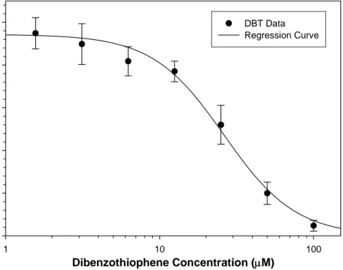
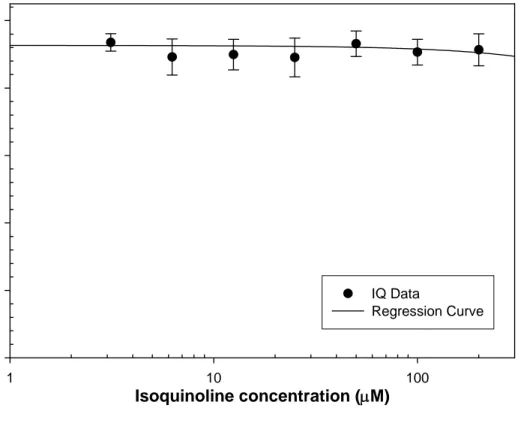
3.3.2. Cytotoxic Effects of Petroleum-Derived Hydrocarbons in NHEK... ………80
3.4. Discussion………..83
3.5. References………..88
CHAPTER 4: ARSENIC AND BENZO[A]PYRENE DIFFERENTIALLY ALTER THE CAPACITY OF DIFFERENTIATION AND GROWTH PROPERTIES OF PRIMARY HUMAN EPIDERMAL KERATINOCYTES………..99
4.1. Background………99
4.2. Abstract in the Original Publication………100
4.3. Introduction in the Original Publication………..101
4.4. Materials and Methods……….104
4.4.1. Chemicals and Reagents………..104
4.4.2. Cell Culture and Culture Reagents………..104
4.4.3. Cytotoxicity Assay………...104
4.4.4. Differentiation Assay………...105
4.4.5. Cell Division Rate Measurement Assay………..107
4.4.6. Flow Cytometric Analysis………...107
4.4.7. Statistical Analysis………...108
4.5. Results………..109
4.5.2. Arsenic and Benzo[a]pyrene Inhibit NNHEK Differentiation in
Vitro...110
4.5.3. Effects of Calcium on NHEK Differentiation In Vitro..………..111
4.5.4. Effects of Arsenic and Benzo[a]pyrene on Calcium-Induced Differentiation in NHEK………..113
4.5.5. Benzo[a]pyrene, Arsenic, and Calcium Alter Cell Cycle Distribution in NHEK………..114
4.5.6. Chemical Effects Seen on NHEK Proliferation Rate…………..115
4.6. Discussion………116
4.7. References………121
CHAPTER 5: GENE EXPRESSION CHANGES ASSOCIATED WITH ALTERED GROWTH AND DIFFERENTIATION IN BENZO[A]PYRENE OR ARSENIC EXPOSED NORMAL HUMAN EPIDERMAL KERATINOCYTES………...137
5.1. Abstract………137
5.2. Introduction………..138
5.3. Materials and Methods……….141
5.3.1. Chemicals and Reagents………..141
5.3.2. Cell Culture………..141
5.3.3. Treatment Regime and RNA Isolation………141
5.3.4. Microarray………142
5.3.5. Real-Time Reverse Transcription-Polymerase Chain Reaction…… ………...143
5.4. Results………..145 5.4.1. Characterization of Gene Expression Changes Associated with
Exposure to Benzo[a]pyrene or Arsenic in NHEK using
Microarray Technology………...145 5.4.1.1. Benzo[a]pyrene Altered Genes from Microarray Analysis.. ………..146 5.4.1.2. Arsenic Altered Genes from Microarray Analysis…….147 5.4.1.3. Common or Similar Genes Altered by both
Benzo[a]pyrene and Arsenic in NHEK………...148 5.4.1.4. Additional Information in Regards to Microarray
Experimentation………...149 5.4.2. Quantitative Analysis of Selected Chemically-Altered Genes by
Real-Time PCR………149 5.4.2.1. Benzo[a]pyrene Altered Genes Detected by Real-Time
PCR………..149 5.4.2.2. Arsenic Altered Genes Detected by Real-Time PCR….150 5.4.3. Comparing Real-Time PCR Data to Microarray Results………151
5.5. Discussion………152
5.6. References………159
CHAPTER 6: COMPUTATIONAL MODEL OF THE STEP-WISE PROCESSES OF TERMINAL GROWTH ARREST AND SQUAMOUS DIFFERENTIATION IN
KERATINOCYTES………193
6.2. Materials and Methods……….197
6.2.1. Cell Culture………..197
6.2.2. Cell Division Measurement Assay………...197
6.2.3. Flow Cytometric Analysis………...198
6.2.4. Examination of Cell Cycle Kinetics………198
6.2.5. Mathematical Models………...200
6.2.5.1. Two Stage Keratinocyte Differentiation Model: Model A... ………...…...200
6.2.5.2. Three Stage Keratinocyte Differentiation Model: Model B. ………..202
6.2.6. General Modeling Assumptions and Parameter Estimates …….204
6.2.6.1. Growth Fraction and Differentiation Rate Estimates in NHEK for Model A……….205
6.2.6.2. Estimating Parameters used in Model B……….206
6.2.7. Modeling Software………...208
6.3. Results………..208
6.3.1. Model A Simulations………...208
6.3.1.1. Chemically-Mediated Effects of the Cell Cycle Kinetics in NHEK………..209
6.3.1.2. Simulating Differentiation in Chemically-Exposed NHEK. ………..212
6.3.2. Model B Simulations………...212
6.5. References………220
6.6. Appendix A: ACSL Code for Model A – Chemically-Induced Alterations in NHEK Cell Cycle Kinetics and Differentiation………..224
6.7. Appendix B: ACSL Code for Model B – NHEK Cell Cycle Kinetics and Differentiation………..227
CHAPTER 7: CONCLUSIONS AND FUTURE DIRECTIONS………...267
7.1. Introduction………..267
7.2. Overview of Results, Conclusions, and Future Directions………..267
7.2.1. Cytotoxicity of Petroleum-Derived Hydrocarbons..………267
7.2.2. Chemically-Induced Effects on Differentiation and Proliferation in NHEK………..270
7.2.3. Gene Expression Changes Associated with Chemically-Altered Growth and Differentiation………..276
7.2.4. Mathematical Model of NHEK Cell Cycle Kinetics and Differentiation………..281
7.3. Final Comments………...284
Chapter 1
Introduction
1.1. Goals and Specific Aims
The overall objective of studies in our laboratory is development of predictive tools for detecting potentially carcinogenic chemicals and chemical mixtures. Toward this end, normal human epidermal keratinocytes (NHEK) have been chosen as an in vitro model for mechanistic studies into how altered regulation of differentiation may play a role in the transformation process in human cells. The epidermis is characterized by a highly regulated balance between epithelial cell growth and squamous differentiation. Although the exact mechanisms involved have not been completely elucidated, an imbalance between two such important and opposing processes has been strongly linked to the onset of carcinogenesis in this cell type. The studies described in this dissertation are aimed at developing a mechanistic understanding of how two diverse skin
carcinogens may alter the normal balance between growth and differentiation such that they favor malignant transformation. Through this work, I will also clarify whether signaling pathways involved in keratinocyte differentiation and proliferation are
disrupted by different carcinogens or whether only chemical-specific effects are evident. My hypothesis is that dermal carcinogens interfere with cellular differentiation and growth through dysregulation of controlling signal transduction pathways for
molecular biology information with computational modeling will improve our
understanding of keratinocyte growth and differentiation, and help predict chemically-induced cell kinetic changes due to carcinogen exposure. Developing a quantitative biologically-based computer model for the differentiation of human keratinocytes will provide an in silico experimental platform which may test this hypothesis much more efficiently. The effects of chemical exposures may then be simulated as perturbations of critical biological processes in an otherwise normal state.
My hypothesis will be tested in the following Specific Aims:
Aim 1. To characterize the time- and dose-dependent effects of two mechanistically diverse skin carcinogens, benzo[a]pyrene (BaP) and arsenic, on growth rate, cell cycle distribution, and squamous differentiation in human
keratinocytes.
Aim 2. To select chemically-altered genes which, based upon their known or putative function, are involved in the switch between growth and differentiation in keratinocytes.
Aim 3. To develop, and refine a biologically-based computer model for the process of squamous differentiation using the data in the literature and from Aims 1 and 2.
1.2. Overview
In the research presented herein, I will present how BaP and arsenic, through altered NHEK differentiation and proliferation, produce conditions that support cellular transformation. To better understand the process of cellular transformation, the Literature Review in Chapter 2 provides essential background information regarding the importance
of maintaining proper balance between cell growth and squamous differentiation in the skin. Also discussed is the linkage between phenotypic changes in keratinocytes and initiation of cell transformation that eventually lead to the onset of cancer. Relevant information concerning the chemicals of interest is presented along with their effects in skin as demonstrated in previously published work. To accomplish my goals and approach the Specific Aims listed above, these studies were organized in the following way: (1) Perform cytotoxicity studies, (2) examine chemically-induced phenotypic alterations (i.e. alterations in NHEK growth and differentiation), (3) Attempt to correlate gene expression alterations to the chemically-altered phenotype(s), and (4) construct a mathematical model to aid in understanding NHEK cell cycle kinetics and cellular differentiation perturbations induced by chemical exposure.
Cytotoxicity studies on four petroleum-derived hydrocarbons in NHEK (Chapter 3) were conducted to gather critical data for selecting reasonable concentrations to use in later studies examining chemical effects on NHEK differentiation, proliferation, and gene expression. Rather than continuing experimentation on each hydrocarbon, the research focus switched to specifically investigate the effects of the most cytotoxic hydrocarbon, BaP, and a commonly known skin carcinogen, arsenic, on NHEK differentiation and proliferation. Phenotypic changes produced due to chemical exposure were examined and is explained in detail throughout Chapter 4. More specifically, BaP- and arsenic-induced alterations of NHEK morphology, differentiation, cell cycle distribution, and potential doubling time were investigated. Subsequently, gene expression analyses under different chemical exposure scenarios were completed and documented in Chapter 5. In this chapter, the mechanisms and/or pathways that may have been involved in the
chemically-induced changes of growth and differentiation observed in Chapter 4 were examined. In the end, two mathematical models were constructed that illustrate keratinocyte behavior in terms of cell cycle kinetics and differentiation abilities (Chapter 6). The NHEK models used flow cytometry results displayed in Chapter 4 and other essential parameters from the literature as explained in detail throughout Chapter 6. Lastly, Chapter 7 explains the significance of this body of work and what conclusions are deduced from the resulting data. Also in this chapter, I will discuss future studies that could potentially branch off from many points within these experiments. Taken together, my results give a better understanding of how carcinogens modulate keratinocyte growth and squamous differentiation in NHEK and the molecular mechanism(s) behind their effects.
Chapter 2
Literature Review: The Relationship between Altered
Keratinocyte Differentiation and Cancer
2.1. The Differentiation Process in the Epidermis
All mammals are continually challenged by a variety of environmental hazards, and the first line of protection is provided by the epidermis. The mammalian epidermis is a highly organized tissue that forms an essential barrier between the organism and the surrounding environment. This protective function arises from finely regulated
proliferation and differentiation processes. The integumentary system consists of the skin, sweat glands, oil glands, hair, and nails. The skin itself is made up of two layers, which are the epidermis and the dermis. Some consider the layer just below the skin, the
hypodermis (subcutaneous layer or superficial fascia), as a third skin layer; however, this region is not technically part of the integumentary system. The studies presented within this dissertation utilized an in vitro keratinocyte model system which is representative of the outer most layer of the skin, the epidermis. Keratinocytes comprise approximately 95% of the cells in the epidermis (Eckert 1989; Green 1977; Green et al. 1982). Other major cell types which reside in the epidermis are melanocytes, Langerhans’ cells, and Merkel cells.
The epidermis has a thickness which varies from 0.075-0.15 mm over the body to 0.8 mm on the palms of the hands and 1.4 mm on the soles of the feet (Plewig 1970). The epidermis itself is divided into five layers, which are, from the inner to the outer most, the stratum basale or stratum germinativum (basal), stratum spinosum (spinous), stratum granulosum (granular), stratum lucidum (only found in thickened areas of the epidermis), and the stratum corneum (cornified) (Figure 2.1). Keratinocyte differentiation is a process whereby a relatively undifferentiated keratinocyte is converted into a fully mature
keratinocyte termed a corneocyte. Only the deepest or inner-most basal layer of the epidermis has the capacity for DNA synthesis and mitosis (Fuchs 1993). This layer provides a continuous supply of cells that repopulate the epidermis (Lavker et al. 1993; Yang et al. 1993). Cell regeneration is thought to be achieved by a hierarchy of
proliferative cells within the basal layer consisting of stem cells and transit-amplifying cells (Lajtha 1979).
In the early stages of epidermal differentiation, basal cells detach from the basement membrane and migrate to suprabasal layers, where they progress through an intricate series of stage-specific morphological and biochemical changes. The spinous layer, the second epidermal cell layer is characterized by dense desmosomes. Early markers of keratinocyte differentiation, including involucrin, and transglutaminase 1, are first expressed in this layer and becomes more prominent in the granular layer (Holbrook and Wolff 1987). The next layer, the stratum granulosum, is rich in granules. These granules contain products of keratinocyte differentiation that are used to assemble various terminal keratinocyte structures, including the corneocyte cornified envelope (Elias et al. 1988; Steven et al. 1990; Takahashi et al. 1992). The stratum lucidum is considered the
‘transition zone’ and is the layer that separates the dead from living epidermal layers. This is a region of systematic destruction of cellular organelles and nucleic acids that coincides with assembly of the keratin intermediate filament bundles and the cornified envelope. These cornified cells are the terminal product of keratinocyte differentiation. Corneocytes are dead, flattened polyhedron-shaped cells that occupy the stratum corneum and are uniquely adapted to provide a protective surface.
Corneocytes consist of a stabilized array of keratin filaments contained within a covalently cross-linked protein envelope (Matoltsy and Matoltsy 1966). Keratin
intermediate filament bundles course through their interior and connect at various points to the surrounding marginal band (i.e., the cornified envelope). The marginal band is a thick band that forms beneath the inner leaflet of the plasma membrane and is composed of a variety of proteins that are covalently connected by ε-(γ-glutamyl)lysine protein-protein crosslinks (Holbrook and Wolff 1987; Rice and Green 1977). These crosslinks are formed by the enzyme transglutaminase (Abernethy et al. 1977; Polakowska et al. 1991; Thacher and Rice 1985). Adjacent corneocytes are held together by modified desmosomes and by an interlocking system of ridges and grooves (Holbrook and Wolff 1987). The composite of billions of terminal keratinocytes (cornified envelopes) forms the protective layer on top of the epidermal surface; the dead cells that make up this layer eventually slough off into the environment. A typical cornified envelope is 12.0 µm in diameter and is chemically resistant to boiling detergents, chaotrophic salts, and reducing agents (Matoltsy and Matoltsy 1966; Sun and Green 1976). This chemical resistance is due to the presence of isodipeptide bonds, i.e., ε-(γ-glutamyl)lysine (Figure 2.2).
Turnover time for the corneocyte compartment has been determined to be 14 days in humans (Bergstresser and Taylor 1977). The average time required for a newly
differentiated basal cell to become a corneocyte is 31 days (Bergstresser and Taylor 1977). Thus, according to these results, the entire epidermal differentiation process takes an average of 45 days. A more recent study confirmed these results with a calculated epidermal turnover rate of 47 or 48 days (Iizuka 1994).
2.2. Regulation of Keratinocyte Differentiation
The transition from basal cell to corneocyte is a complex process that requires the simultaneous activation and inactivation of a wide variety of proteins and genes. In order for differentiation to produce a normal epidermal surface, these genes must be expressed at the correct time and location. A major goal of keratinocyte- and/or skin-related
research is to identify the mechanisms that regulate this complex process.
2.2.1. Differentiation Markers
The cytokeratins, cornified envelope precursor proteins, and transglutaminase are useful markers of gene expression in keratinocytes, both in vitro and in vivo (Fuchs 1990; Rice and Green 1977; Stoler et al. 1988; Yuspa 1994). The cytokeratins are a family of over thirty proteins that are assembled to form the intermediate filaments in epithelial cells (Steinert et al. 1985). Based on sequence homology and expression pattern, the keratins can be divided into acidic and neutral-basic families (Albers and Fuchs 1992; Steinert et al. 1985; Woodcock-Mitchell et al. 1982). Keratins are expressed in pairs to assure the presence of a neutral-basic and acidic partner to permit filament assembly (Woodcock-Mitchell et al. 1982). Specifically in the epidermis, cytokeratins 5 (K5) and
14 (K14) are expressed in the basal cells and form an intermediate filament network that is suitable for cells that are proliferating (Fuchs and Green 1980). In contrast,
cytokeratins 1 (K1) and 10 (K10) are expressed in suprabasal cells (Woodcock-Mitchell
et al. 1982). These keratins are involved in the formation of the intermediate filament
bundles that are present in differentiating keratinocytes (Aebi et al. 1983). The keratin bundles are stabilized by disulfide bonds and occupy the interior volume of the cytoplasm in terminally differentiated cells (Aebi et al. 1983). These bundles provide structural stability to the corneocyte. Profilaggrin, the precursor of filaggrin, is expressed in the suprabasal layers and is converted to filaggrin, which packages the keratin filaments into fibrils (Takahashi et al. 1992).
Involucrin and loricrin are envelope precursor proteins that are expressed in the epidermal suprabasal layers (Hohl et al. 1991; Mehrel et al. 1990; Rice and Green 1977; Takahashi et al. 1992; Thacher and Rice 1985). These proteins are crosslinked to form the cornified envelope by the enzyme transglutaminase which catalyzes the formation of inter-protein ε-(γ-glutamyl)lysine bonds. Thus the cornified envelope is a structure comprised of covalently crosslinked proteins that is assembled beneath the deteriorating plasma membrane during the final stages of keratinocyte differentiation.
2.2.2. The Switch between Growth and Differentiation in Keratinocytes
The onset of keratinocyte differentiation can be triggered by several distinct signals. These signals include activation of: (1) an extracellular Ca2+ receptor mechanism, (2) increase in cell-cell adhesions, and (3) detachment from the substratum (Dotto 1999).
involved during epidermal differentiation. Mouse and human basal keratinocytes with a high growth fraction can be cultured in medium with a calcium (Ca2+) concentration of 0.05 mM or less, whereas media with 0.1 mM Ca2+ has been shown to induce terminal differentiation (Boyce and Ham 1983; Hawley-Nelson et al. 1980; Hennings et al. 1980a; Hennings et al. 1980b; Shipley and Pittelkow 1987). These findings have facilitated the analysis of the regulation of keratinocyte growth and differentiation. In culture, Ca2+ -induced maturing keratinocytes cease proliferation, stratify, express the suprabasal
markers of differentiation, activate transglutaminase, cornify, and slough (desquamate) as a sheet of matured squames (Hennings et al. 1980b; Yuspa et al. 1989). As seen in vitro, a key regulator for keratinocyte maturation in vivo is a gradient of extracellular Ca2+ across the epidermis that is low in the basal compartment and increases in concentration within each outer epidermal layer (Elias et al. 1998; Malmquist et al. 1984; Menon et al. 1985). The binding of Ca2+ and/or other divalent or trivalent cations to transmembrane Ca2+ receptors initiates downstream signaling pathways which include phospholipase C (PLC) activation and increased intracellular Ca2+ (Filvaroff et al. 1994; McNeil et al. 1998; Xie and Bikle 1999). 1-α,25dihydroxy vitamin D3 (1,25(OH)2D) also promotes differentiation, by inducing involucrin and transglutaminase expression, via many of the same pathways as Ca2+, although in the absence of Ca2+ 1,25(OH)2D stimulated
differentiation is not as complete (Bikle and Pillai 1993; Bikle et al. 2003; Gibson et al. 1996; Hosomi et al. 1983; Johansen et al. 2000; Su et al. 1994). An opposing vitamin A gradient may also contribute to the regulation of keratinocyte gene expression and differentiation (Regnier and Darmon 1991).
Ca2+ has also been shown to trigger keratinocyte differentiation through a more indirect mechanism, such as by promoting close cell-cell adhesion. One of the earliest, and most important, changes associated with Ca2+-induced differentiation is the
establishment of close intercellular contacts, through adherens junctions and desmosome formation (Lewis et al. 1994; O'Keefe et al. 1987). Cadherin-mediated adherens
junctions may play a role in cell signaling events associated with growth and
differentiation. Moreover, transmembrane tyrosine kinase receptors, src kinase family members, cell-surface tyrosine phosphatases, and protein kinase C (PKC) family members have all been found to be associated with specific adherens junction
components and/or to be present at the adherens junction (Fagotto and Gumbiner 1996). The addition of Ca2+ to cultured keratinocytes induces stratification (i.e. sliding of cells over each other with a simultaneous loss of adhesion to the substrate) (Calautti et al. 1995). Induced detachment of keratinocytes from the matrix is associated with the
induction of biochemical markers of differentiation, as well as growth arrest (Di Cunto et
al. 1998; Li et al. 1996; Watt et al. 1988). Keratinocyte attachment to the underlying
extracellular matrix is mediated by integrin based structures, and these structures are thought to function as important signaling centers controlling the switch among
keratinocyte growth, differentiation, and apoptosis (Frisch and Ruoslahti 1997; Giancotti 1997). The down regulation of integrin expression parallels that of increasing Ca2+ concentrations in keratinocytes (Tennenbaum et al. 1996). Studies have demonstrated that attachment via the α4β6 integrin receptor (found only in basal layers) may serve as a positive determinant of keratinocyte proliferation, while the loss of α4β6 and α3β1 integrin receptor (involved in focal contacts) attachment may trigger signaling events contributing
to differentiation (Alt et al. 2001; Fuchs et al. 1997; Levy et al. 2000; Tennenbaum et al. 1996). These events may be controlled by Ras and MAPK-dependent pathways
(Mainiero et al. 1997).
2.2.3. Intermediate Pathways involved in Keratinocyte Differentiation
PLC- and PKC-dependent pathways, as well as induction of tyrosine
phosphorylation, play an important role in the switch between growth and differentiation. Intracellular PLC activation has been reported to occur within 30 seconds to one minute after Ca2+ treatment, in association with a very early increase in intracellular Ca2+
concentrations (Jaken and Yuspa 1988; Tang et al. 1988). Direct functional studies indicate that both PLC-γ activation and a rise in free intracellular Ca2+ are required for late aspects of keratinocyte differentiation (Li et al. 1995; Xie and Bikle 1999).
Phosphatidyl inositol bis phosphate (PIP2) breakdown results in inositol
1,4,5-triphosphate (IP3) production, which in turn triggers the increase in free intracellular Ca2+ by mobilization from intracellular stores. Diacylglycerol (DAG), the other product of PIP2 break-down, is also increased at very early stages following Ca2+ treatment (Jaken and Yuspa 1988). DAG is known to directly activate PKC. Among the 11 PKC isoforms, PKC-α, -δ, and -ε are activated upon Ca2+-induced differentiation (Alt et al. 2001;
Denning et al. 1995a; Denning et al. 1995b; Yang et al. 2003). Down regulation of these same isoforms are associated with decreased induction of late keratinocyte differentiation markers such as loricrin, filaggrin, and small proline rich protein (SPR-1) (Stanwell et al. 1996; Tesfagaigzi and Carlson 1999). Studies also show the possible involvement of other PKC isoforms (η, and ζ) in keratinocyte differentiation (Kashiwagi et al. 2002; Kashiwagi et al. 2000; Ohba et al. 1998; Osada et al. 1993; Osada et al. 1990).
The induction of tyrosine phosphorylation occurs as an early and specific event in keratinocyte differentiation in response to Ca2+ (Filvaroff et al. 1994; Filvaroff et al. 1990). Tyrosine phosphorylation of p62, and other similar proteins, causes the protein to associate with Ras-GTPase Activating Protein (Ras-GAP) in response to increased intracellular Ca2+, thus inhibiting the activation of Ras-GRP (Filvaroff et al. 1992). In keratinocytes, p62 tyrosine phosphorylation is induced specifically in response to increased extracellular Ca2+ (Filvaroff et al. 1992; Filvaroff et al. 1994). It has been suggested that tyrosine phosphorylation may serve as a negative feedback mechanism to inactivate PKC-δ during late stages of differentiation (Denning et al. 1996). The tyrosine kinase, Fyn, is activated a few hours after Ca2+-induced differentiation and may be responsible for several phosphorylating events (Calautti et al. 1995). Also, essential adherens junction components such as β-catenin, γ-catenin/plakoglobin, and p120-Cas (binds to E-cadherin) all become tyrosine phosphorylated by Fyn in cultured mouse keratinocytes within nine hours of Ca2+ treatment (Calautti et al. 1998). Interestingly, adherens junction formation has been recently reported to depend on a second signaling pathway involving the Rho and Rac GTPases, which control actin/cytoskeleton
organization and cell motility (Braga et al. 1997).
2.2.4. Transcription and Cell Cycle Regulation of Keratinocyte
Differentiation
There are distinct signaling pathways connected with transcription and cell-cycle control in keratinocyte differentiation. Evidence indicates that the two signaling pathways triggered by increased extracellular Ca2+ remain distinct even at the nuclear level. One of
tyrosine phosphorylation and, further downstream, to p21WAF1 promoter activation
(Missero et al. 1995; Prowse et al. 1997). Cyclin-dependent kinase inhibitor p21WAF1
generally plays a role in the inhibition of cell growth and its expression is typically increased in post-mitotic cells immediately adjacent to the proliferative compartment (Di Cunto et al. 1998). Guanine and cytosine binding factors (GC rich repeats) such as Sp3, as well as the transcriptional co-activator p300, appear to be involved (Ogryzko et al. 1996; Prowse et al. 1997). Tyrosine phosphorylation is a critical step in the activation of the signal transducters and activators of transcription (STAT) family of transcription factors, which regulate inducibility of the p21 promoter in response to epidermal growth factor (EGF) and interferon-γ (IFNγ) (Hauser et al. 1998; Look et al. 1995). The myc family has also been shown to be implicated in the control of keratinocyte growth and differentiation (Gandarillas et al. 2000; Gandarillas and Watt 1997; Hurlin et al. 1995; Pelengaris et al. 1999; Vastrik et al. 1995). Other key regulatory factors which could bind to the p21 promoter and with which Sp3 may interact include E2F family members (Hiyama et al. 1997). In addition, studies also indicate E2F-1 promotes the expression of keratinocyte proliferation-specific marker genes and suppresses differentiation-specific marker genes (Dicker et al. 2000; Paramio et al. 2000a).
The other pathway that is activated in response to Ca2+ leads to PKC and Fyn activation, AP-1 factor modulation, and differentiation marker expression (Eckert et al. 1997; Rossi et al. 1998; Rutberg et al. 1996; Rutberg et al. 1997; Whitmarsh and Davis 1996). The product of the Whn gene functions as a specific suppressor of this pathway (repressing involucrin and transglutaminase), while it does not affect the pathway leading to p21 promoter activation (Brissette et al. 1996; Jessen et al. 2000). Several other
transcription factors, such as those in the NF-κB/rel family (induced by PKC activation), as well as nuclear hormone receptors, are likely to play an important role in the balance between keratinocyte growth and differentiation (Hu et al. 2001; Li et al. 2000; Nehls et
al. 1996; Qin et al. 1999; Seitz et al. 1998). Unfortunately, the interconnections between
activation of these factors and the specific signaling pathways involved in keratinocyte differentiation remain to be established. Other signaling pathways and/or genes not mentioned here are also involved in the regulation of keratinocyte differentiation, however, this literature review focused on the most highly characterized pathways and signals which are thought to control this cellular process. Figure 2.3 depicts numerous factors and genes involved in regulating the switch between differentiation and
proliferation in keratinocytes.
2.3. Altered Differentiation and Cancer in the Skin
2.3.1. The Carcinogenesis Process
Cancer can be characterized as uncontrolled cell growth which eventually leads to tumor formation. Cancer may develop due to the disruption of the normally
well-controlled balance between cell division, death, and differentiation in living organisms. Ultimately cancer is a disease of abnormal gene expression which can occur through direct DNA insults (gene mutations, translocations, or amplifications) and abnormal gene transcription or translation. The process of carcinogenesis can be divided into three stages: initiation, promotion, and progression. Initiation is an irreversible stage involving a permanent heritable change in gene expression caused by direct DNA damage (somatic
frequent exposures over a substantial period of time. Within the promotion stage there are two operational stages, conversion and propagation. Conversion is a limiting step which allows cells to override local growth restraints or proliferative latency. Propagation is characterized by chronic mitogenic stimulation during which time initiated cell
populations expand. Finally, progression is an irreversible stage that is characterized by gross structural changes to chromosomes and genetic instability. Most chemicals work at distinct stages in this process and, therefore, must combine with other chemicals or other types of carcinogens to induce the development of malignant tumors. There are chemicals that, at high enough concentrations and/or prolonged exposures, can cause cancer by themselves, and are termed complete carcinogens. For example, BaP has been termed a complete carcinogen as shown in previous rodent skin research (Ashurst et al. 1983; Iskander et al. 2004; Jensen et al. 1993).
Somatic mutations leading to uncontrolled growth of cancer may be categorized as affecting two classes of genes: proto-oncogenes and tumor suppressor genes. Proto-oncogenes, such as ras and myc, encode proteins involved in the cascade of events leading to cellular proliferation including growth factors, growth factor receptors, and transcription factors. An activating mutation of a single copy of a proto-oncogene can lead to unrestricted cell growth. In contrast, tumor suppressor genes, such as p53 and
retinoblastoma (RB), are genes that normally control critical checkpoints in cell growth
and division. Typically, inactivation of both copies of tumor suppressor genes is required for unrestricted cell growth.
2.3.1.1. Skin Cancer
In general, there are two common types of nonmelanoma skin cancers: basal cell and squamous cell carcinomas. Basal cell carcinoma (BCC) is the most common form of skin cancer. It is a malignant neoplasm of basal cells of the epidermis. Ultraviolet (UV) irradiation has been a common culprit of many BCC cases. Less often, BCC may result from exposure to arsenic or certain industrial pollutants. Squamous cell carcinoma (SCC) is a malignant neoplasm of the keratinizing (transit-amplifying) cells within the epidermis and is the second most common skin cancer in humans. SCC usually occurs later in life than BCC. Although most cases of SCC are related to sun exposure, a smaller number develop in skin that has been injured or exposed to carcinogens. Subsequent sections will further discuss the events that drive the carcinogenic process in skin.
2.3.2. Initiation in the Skin
Initiating agents, such as the potent mutagen and carcinogen BaP, can potentially trigger the onset of carcinogenesis by causing mutations which can produce subtle changes in keratinocyte phenotype, unrecognizable in the context of the intact epidermis. Initiated keratinocytes in vitro display an altered response to signals for terminal
differentiation, a characteristic which provides a selective growth advantage under culture conditions favoring differentiation (Kilkenny et al. 1985; Kulesz-Martin et al. 1980; Yuspa and Morgan 1981). Exploitation of this difference has been particularly helpful in isolating keratinocytes of mouse and human origin in vitro (Hennings et al. 1987b; Kilkenny et al. 1985; Kulesz-Martin et al. 1983; Yuspa et al. 1986).
gene (c-rasHa) associated with papilloma formation (Balmain et al. 1984; Bizub et al.
1986; Harper et al. 1987). These genetic studies indicate that: (1) c-rasHa gene mutations
were frequently heterozygous in papillomas and could be detected in initiated skin prior to tumor formation (Nelson et al. 1992); (2) the initiating agent determined the existence, nature, and site of the c-rasHa mutation (Brown et al. 1990; Harper et al. 1986); and (3)
many human benign and malignant skin tumors, probably induced by UV light, also contain ras gene mutations (Ananthaswamy et al. 1989; Ananthaswamy et al. 1988; Corominas et al. 1989; Daya-Grosjean et al. 1993; Leon et al. 1988; Suarez et al. 1989).
2.3.3. Promotion in the Skin
Application of tumor promoters to initiated epidermis causes the selective clonal outgrowth of a percentage of the chemically-mutated cells to produce multiple benign squamous cell papillomas; each lesion represents an expanded clone of a single initiated cell (Boutwell 1974; Deamant and Iannaccone 1987; Iannaccone et al. 1987). In skin carcinogenesis, selective clonal expansion of the initiated population may result from differential sensitivity of initiated and normal keratinocytes to either cytotoxicity or growth stimulatory affects of exogenous tumor promoters (Finzi et al. 1987; Hartley et
al. 1987). Phorbol esters, such as 12-O-tetradeanoylphorbol-13-acetate (TPA), are both
potent exogenous tumor promoters in initiated cells that activate PKC and accelerates terminal differentiation in normal keratinocytes (Dlugosz and Yuspa 1993; Yuspa et al. 1980, 1982). In contrast to normal cells, initiated keratinocytes are resistant to terminal differentiation induced by PKC activators such as TPA (Hennings et al. 1987a; Hennings
et al. 1990a; Yuspa et al. 1983a). The differential response of normal and initiated cells
producing papillomas. Therefore, the process of tumor promotion by phorbol esters in
vivo recapitulates the clonal selection of initiated cells by differentiation-inducing agents
in keratinocyte culture (Hennings et al. 1992; Hennings et al. 1990a; Strickland et al. 1992).
In skin, squamous papillomas are characterized by a high rate of proliferation and by delayed expression of differentiation markers, properties which are analogous to the phenotype of individual initiated cells in vivo (Glick et al. 1993; Roop et al. 1988). The mechanisms of exogenous promotion is likely to be epigenetic in most cases since: (1) papillomas are diploid when they first emerge (Aldaz et al. 1988a; Aldaz et al. 1988b); (2) a single genetic change in normal keratinocytes is sufficient to produce a papilloma phenotype (Bailleul et al. 1990; Greenhalgh et al. 1993); and (3) most promoting agents are not mutagens (Yuspa and Dlugosz 1991). In the absence of exposures to exogenous tumor promoters, initiated skin rarely develops into tumors. Thus, exogenous promotion in general is a rate-limiting early event in carcinogenesis, but the actual tumor yield is determined by additional hereditary factors influencing both the initiation and promotion stages (DiGiovanni et al. 1991). The genes which determine hereditary susceptibility to initiators and promoters in mouse skin remain largely to be defined, and susceptibility appears to be multigenic (Bangrazi et al. 1990; Naito et al. 1988).
2.3.4. Progression and Tumor Formation in the Skin
Premalignant progression of a papilloma to a carcinoma in mouse skin is
generally a spontaneous process that is not enhanced by most exogenous tumor promoters (Hennings et al. 1983; Hennings et al. 1990b). Genetic studies indicate that nonrandom,
mouse papillomas; particularly prominent are trisomies of chromosomes 6 and 7 (Aldaz
et al. 1987; Aldaz et al. 1988a; Aldaz et al. 1989; Bianchi et al. 1990; Conti et al. 1986).
Premalignant progression and malignant conversion can be enhanced and accelerated by exposing animals bearing papillomas to mutagens (Hennings et al. 1983; Hennings et al. 1990b; O'Connell et al. 1986).
Malignant conversion of benign tumors is a relatively rare occurrence since less than 5% of squamous papillomas spontaneously convert to clinically detectable cancers (Hennings et al. 1983; Hennings et al. 1990b). Squamous papilloma cell lines in vitro can be converted to squamous carcinoma cells by introducing the following oncogenes:
c-rasHA (mut), v-rasHA, v-fos, c-myc, E1A, TGFα, p53 (mut), neu, and v-jun (Dotto et al.
1988; Finzi et al. 1988; Greenhalgh et al. 1989; Greenhalgh and Yuspa 1988; Harper et
al. 1986). In particular, changes in two cellular genes, c-rasHA and p53, have been
identified with malignant conversion in skin tumors. The mutated rasHA oncogene, which
is heterozygous in papillomas, is frequently homozygous in carcinomas (Bianchi et al. 1990; Quintanilla et al. 1986). An oncogenic rasHA gene can cause malignant conversion
of papilloma cells with a heterozygous c-rasHA gene mutation (Greenhalgh et al. 1989;
Harper et al. 1986). Mutations in the p53 tumor suppressor gene are rarely found in chemically induced papillomas, but are frequently detected in squamous carcinomas, particularly those induced by benzo[a]pyrene (Burns et al. 1991; Kress et al. 1992; Ruggeri et al. 1991; Ruggeri et al. 1993). In addition, topical administration of initiators and promoters to mice lacking an intact p53 gene results in rapid progression of
papillomas to the carcinoma stage (Kemp et al. 1993). Also, mutations in the p53 gene are frequently detected in human squamous and basal cell carcinomas of the skin (Brash
et al. 1991; Kanjilal et al. 1993; Rady et al. 1992; Ziegler et al. 1993). Overall, it seems
multiple mechanisms may influence this late stage of carcinogenesis.
2.3.5. The Altered Phenotype of Initiated Keratinocytes
A subpopulation of keratinocytes isolated from carcinogen-initiated skin or normal basal keratinocytes exposed to carcinogens in vitro resists the Ca2+-mediated induction of terminal differentiation and evolves as multiple foci which continue to grow in medium with >0.1 mM Ca2+. These foci show properties consistent with the initiated phenotype. Ca2+ resistant foci: (1) develop from keratinocytes initiated in vivo or in vitro (Kawamura et al. 1985; Kulesz-Martin et al. 1980; Yuspa and Morgan 1981); (2) are more frequent with exposure to strong initiators and higher initiator doses (Kilkenny et
al. 1985); (3) develop even if a delay of 10 weeks is interposed between initiation in vivo
and Ca2+ selection in vitro (Kawamura et al. 1985); and (4) develop into cell lines which produce papillomas or carcinomas when grafted into nude mice (Kulesz-Martin et al. 1983; Strickland et al. 1988).
Phenotypic alterations such as increased proliferation rates and blocks to
differentiation are commonly seen in initiated keratinocytes. The oncogenic activation of c-rasHA is thought to play a key role in facilitating the development of the initiated
phenotype in skin. When v-rasHA oncogene is introduced into cultured normal
keratinocytes, recipient cells form papillomas when grafted into nude mice, indicating that this single genetic change is sufficient to produce the initiated phenotype (Roop et al. 1986). However, other changes, in addition to ras mutations, are thought to be required to fully transform human keratinocytes (Kawamura et al. 1985). In vitro, mouse
keratinocytes expressing v-rasHA have a high proliferation rate and fail to terminally
differentiate in response to medium with >0.1 Ca2+ (Cheng et al. 1990; Roop et al. 1987; Yuspa et al. 1985; Yuspa et al. 1983b). This hyperproliferation induced by v-rasHA
transduction is caused by overexpression of and autocrine response to TGFα, a growth factor known to be elevated in papillomas (Glick et al. 1991; Imamoto et al. 1991; Lee et
al. 1992). When basal cells are cultured in 0.05 mM Ca2+ medium, v-rasHA keratinocytes
express a phenotype with a striking similarity to normal keratinocytes exposed to activators of PKC, such as phorbol esters and DAG (Dlugosz and Yuspa 1993; Roop et
al. 1987; Toftgard et al. 1985; Yuspa et al. 1983a). When v-rasHA keratinocytes are
exposed to medium with >0.1 mM Ca2+, they share similar features with phorbol ester-treated epidermal cells; Cytokeratins K1 and K10 are not induced, while loricrin and filaggrin are overexpressed (Dlugosz and Yuspa 1993). However, transglutaminase mRNA decreases in Ca2+-treated v-rasHA keratinocytes, and terminal differentiation is
blocked (Yuspa et al. 1985). An essential role for PKC activation in the v-rasHA
keratinocyte phenotype is supported by studies with the functional PKC inhibitor, bryostatin (Sako et al. 1987). This report suggests that qualitative and quantitative changes in keratinocyte PKC isoforms might be a consequence of v-rasHA oncogene
expression. Differential modification of isoforms, particularly activation of PKC-α and inhibition of PKC-δ, produce keratinocytes with enhanced proliferative capacity and reduced sensitivity to signals for terminal differentiation (Denning et al. 1993; Denning
et al. 1995b; Deucher et al. 2002; Dlugosz and Yuspa 1991).
In later stages of carcinogenesis, TGFβ, p53, fos, p21WAF1 and integrin alterations
inhibitors for cultured keratinocytes, and TGFβ-1 regulates basal cell proliferation in
vivo. The absence of TGFβ-1 in normal epidermis of TGFβ-1 knockout mice causes basal
cell hyperproliferation (Dahler et al. 2001; Glick et al. 1993). TGFβ isoforms are differentially expressed in increasingly malignant grades of keratinocytes (Gold et al. 2000). Tumor suppressor p53 has been shown to promote differentiation in keratinocytes (Paramio et al. 2000b). Several laboratories have reported that the introduction of a mutant p53 gene into neoplastic epithelial cells with endogenous wild-type p53 conveys resistance to the growth-inhibitory effects of TGFβ (Gerwin et al. 1992; Reiss and Sartorelli 1987). This suggests a link between mutations in p53, TGFβ, and premalignant progression. Squamous cancers produced by v-fos oncogene-mediated conversion are highly invasive and vascular, do not express K1 or K10, and have disseminated expression of α4β6 integrin throughout the tumor mass (Tennenbaum et al. 1996;
Tennenbaum et al. 1992). Changes in integrin distribution could have profound effects on the neoplastic phenotype since integrins are important mediators of cell and cell-extracellular matrix interactions (Hynes 1992; Owens and Watt 2001; Ruoslahti 1991). Other oncogenes which modify gene transcription, such as c-myc, EIA, or v-jun, do not cause phenotypic progression when transduced into papilloma cell suggesting the fos-related changes in genetic regulation are specific for malignant conversion. It is also interesting to note that p21WAF1 enhances papilloma formation but not malignant
conversion in mouse skin (Weinberg et al. 1999). In addition, p21WAF1 successfully
inhibits the growth of skin cancers, even those with alterations in p53, p21ras, RB gene
The initiated phenotype results from mutational events producing intrinsic changes in intracellular signaling pathways, particularly those related to terminal
differentiation. However, initiated cells remain responsive to extrinsic restraints imposed by surrounding normal cells and the microenvironment. Nevertheless, defects in the control of proliferation, differentiation, and apoptosis can lead to a disturbance of
epidermal homeostasis and have been implicated in cancer, as well as other skin diseases.
2.4. Chemicals of Interest
2.4.1. Arsenic
Arsenic is widely distributed, at low concentrations, in food, water, air and soil (Bettley and O'Shea 1975; Chappell et al. 1997; Knowles and Benson 1984). Arsenic combined with oxygen, chlorine, and sulfur is called inorganic arsenic, which represents the most common form of either arsenate or arsenite in the environment (Bettley and O'Shea 1975; Knowles and Benson 1984; Landolph 1994). Studies indicate that arsenite is considerably more toxic and carcinogenic than arsenate (Tsuda et al. 1995). Inorganic arsenic species have been ranked highest in priority on the list of Top 20 Hazardous Substances by the ATSDR and USEPA (ATSDR 1999a, 2003a). Exposure to arsenic occurs through arsenic-rich geological soil, nonferrous smelters, pesticide manufacturing and use, microelectronics, burning of fossil fuels, or from consumption of contaminated drinking water (Basu et al. 2001; Jessen et al. 2001). Although arsenic is a known human carcinogen, it is only slightly mutagenic during chronic exposure or at very high
Thus, arsenic has been characterized as a promoting, progressing, and/or co-mutagenic agent (Lee et al. 1988; Maier et al. 2002; Rossman et al. 2001; Rudel et al. 1996).
Potential carcinogenic actions for arsenic include oxidative stress, genotoxic damage, inhibition of DNA repair, epigenetic effects such as altered methylation, and activation of certain signal transduction pathways leading to aberrant gene expression. Unlike many carcinogens, arsenic fails to behave as a mutagen and cause point mutations in standard assays (Rossman and Goncharova 1998), but has been shown to induce large deletion mutations instead (Hu et al. 1998). There are numerous reports showing that arsenic exposure induces chromosome aberrations (Rossman et al. 2001), aneuploidy and micronuclei formation (Gurr et al. 1993; Wang and Huang 1994), DNA-protein cross-linking and sister chromatid exchange in animal and human cells (Dong and Luo 1993). Both nucleotide-excision-repair (NER) and base-excision repair (BER) of DNA are inhibited by very low, non-cytotoxic concentrations of arsenic (Hartwig et al. 2002). Arsenic inhibits the completion of DNA excision repair, perhaps via effects on DNA ligase, which is especially sensitive to arsenite in cells (Li and Rossman 1989). The activation of NADH oxidase by arsenic may produce O2- radicals. The clastogenic activity of arsenic is likely mediated through O2- and its secondary radicals (Hei et al. 1998; Lynn et al. 2000). Gene amplification induced by arsenic may also play a role in its carcinogenic effects (Hughes 2002).
2.4.1.1. Arsenic-Induced Alterations in the Epidermis
Although arsenic is a potent human skin carcinogen, the exact mechanism of arsenic induced carcinogenesis is currently unclear. Arsenic accumulates in the skin and
acanthosis (an increase in the thickness of the stratum spinosum of the epidermis), pigmentation disorders, and keratinocyte tumors including basal cell carcinomas and squamous cell carcinomas (Chen et al. 1985; Shi et al. 2004). Due to
arsenic-contaminated drinking water in many countries, development of neoplastic skin lesions has become a health problem of global proportions (Chowdhury et al. 2000; Tseng 1977; Wong et al. 1998). Hence, ingested arsenic can be absorbed and accumulated in the skin (Lansdown 1995). Many cases of nonmelanoma skin cancer have been reported among people exposed to arsenic through dietary, occupational, or medicinal means (Germolec
et al. 1998; Saha 1996; Schwartz 1997; Tseng 1977). The toxicity of arsenic has been
related to its high reactivity with vicinal sulfhydryl groups on macromolecules such as glutathione (GSH) and cysteine (Scott et al. 1993). The reason that the skin is unusually sensitive to arsenic toxicity may be due to its richness in sulfhydryl-containing
molecules, such as keratin (Lindgren et al. 1982; Yamauchi and Yamamura 1983). Arsenic (arsenite in particular) has been shown to be a potent differentiation inhibitor in keratinocytes by decreasing cornified envelope formation or by altering differentiation-associated markers (Jessen et al. 2001; Kachinskas et al. 1994; Perez et al. 2003). These studies demonstrate that involucrin, transglutaminase, SPR-1, and filaggrin expression are suppressed by arsenic exposure in various keratinocyte cell lines. In addition, arsenic has been shown to have dose-dependent effects on cell proliferation and to affect expression of a variety of growth regulatory factors in keratinocytes (Germolec
et al. 1996; Monzon et al. 1996; Vega et al. 2001). In particular, non-cytotoxic to slightly
toxic concentrations of arsenic have been shown to increase cell proliferation (hormesis), while toxic concentrations inhibit cell growth (Chen et al. 2000; Liao et al. 2004; Styblo
et al. 2002; Vega et al. 2001). These phenotypic changes due to arsenic exposure may be
related to altered expression of genes which modulate signal transduction pathways known to regulate keratinocyte proliferation and differentiation. Arsenic has been reported to be a potent stimulator of proto-oncogenes c-fos and c-jun expression and AP-1 transactivational activity (Cavigelli et al. AP-1996; Liao et al. 2004). The requirement of AP-1 for tumor promotion has been demonstrated in various cell models (Angel and Karin 1991). The induction of AP-1 activity by arsenic appears to be mediated by activation of PKC and MAPK family members (Huang et al. 2001a). NFκB activation is thought to be associated with initiation and accelerated tumorigenesis (Gilmore 1997). Studies also observed the activation of NFκB following arsenic exposure in keratinocytes (Huang et al. 2001b; Liao et al. 2004). TGFα overexpression has been associated with the neoplastic transformation in the skin, and arsenic has been shown to induce TGFα in human skin (Simeonova and Luster 2000). There are conflicting reports on arsenic-induced effects on tumor suppressor p53. One report failed to demonstrate an effect of arsenic on p53-dependent transcription (Huang et al. 1999); however, other reports show arsenic decreasing p53 expression (Hamadeh et al. 1999), or inducing p53
phosphorylation and accumulation (Salazar et al. 1997; Yih and Lee 2000).
2.4.2. Benzo[a]pyrene
Benzo[a]pyrene (BaP) is a ubiquitous environmental pollutant and has been proven to be a potent mutagen and carcinogen in both humans and animals. The polycyclic aromatic hydrocarbon (PAH) is also among the Superfund Top 10 Priority Hazardous Substances and is a common constituent in petroleum products (ATSDR
overcooked foods, and air pollutants formed by incomplete pyrolysis of combustible organic material (Hanelt et al. 1997; Miller et al. 2000; Parkinson and Newbold 1980).
BaP must be bioactivated in order to induce its adverse effects in an organism or target tissue. In the cell, BaP is metabolized mostly by cytochromes P450 1A1 and 1B1, as well as epoxide hydrolase, yielding syn- and anti-BaP-7,8-diol-9,10-epoxides (BPDE) (Guengerich et al. 1996; Guengerich and Shimada 1998; Hartwig et al. 2003). BDPE can then react and bind to DNA at the 2-amino group of guanine (BPDE-N 2-dG) in the minor groove, as well as N7 of guanine in the major groove. The majority of BPDE forms stable adducts at the N2 positions of guanine, which is considered to be the critical event in carcinogenicity (Sage and Haseltine 1984). If left unrepaired, mutation can occur, typified by a G to T transversion in the DNA (Figure 2.4) (Tran et al. 2002). This transversion predominates in many ras and p53 mutations found in a variety of cancers, including skin cancer (Amstad and Cerutti 1995; Cherpillod and Amstad 1995;
Colapietro et al. 1993; Hattori et al. 1996). If DNA repair mechanisms are not
functioning properly to repair adduct-induced transversions produced by BPDE, these mutations are likely to be inherited by daughter cells.
2.4.2.1. Benzo[a]pyrene-Induced Alterations in the Skin
There is potential for either environmental or occupational dermal exposure to BaP. The most likely source of dermal contact with toxic BaP levels would be in workers in the petroleum industry.Bioactivated BaP is a known skin carcinogen, acting as a mutagen and an initiating agent during transformation (Ashurst et al. 1983; Mager et al. 1977). When applied dermally, BaP induces cytokinetic abnormalities and inflammation, followed by skin tumors (Albert et al. 1991; Albert et al. 1996). Many of the earliest
studies of BaP were carried out using the murine skin system (Lee and O'Neill 1971; Turusov et al. 1987). The chemical alters differentiation in multiple cell types (Edmondson and Mossman 1991; Reiners et al. 1991). This activity is likely due to mutagenesis of proteins in crucial signal transduction paths. Clearly, much work remains to be done to clarify the relationship between BaP exposure, abnormalities in
proliferation and differentiation, and development of neoplastic skin lesions.
Although there have been limited studies on BaP-mediated effects on keratinocyte proliferation and differentiation, the data which are available support BaP’s ability to alter these cellular processes. BaP has been proven to suppress keratinocyte
differentiation in cultured squamous carcinoma cells by inhibiting retinoid-induced transglutaminase and by inhibiting the expression of involucrin and transglutaminase in the absence of a differentiation inducing agent (Rice et al. 1988; Rubin and Rice 1988). In related studies, BaP exposure was also shown to alter the expression of differentiation-related cytokeratins in tracheal and bladder epithelial cells (Edmondson and Mossman 1991; Summerhayes et al. 1981). The fact that BaP is capable of altering the
differentiation program of committed cells may explain, in part, why exposures of the tracheal epithelium to BaP lead to squamous metaplasia and potentially squamous cell carcinoma (Benfield et al. 1981; Chopra and Joiakim 1991; Saffiotti et al. 1964; Yoshimoto et al. 1980). BaP is known to interfere with other cellular transduction pathways within the cell, namely those involving Ca2+ and epidermal growth factor (EGF) in lymphocytes. These cells exhibit glutathione (GSH) depletion and an elevation of intracellular Ca2+ upon BPDE treatment, possibly as a result of sulfhydryl damage produced by this BaP metabolite (Romero et al. 1997). As a result, alterations in Ca2+
homeostasis, cell activation, and proliferation in these cells can be compromised (Romero
et al. 1997). In support of this interpretation, cultures of epidermal keratinocytes exhibit
an increase in overall metabolism of BaP upon pre-incubation in high Ca2+ medium prior to BaP treatment (DiGiovanni et al. 1989). These results indicate that the expression and inducibility of enzymes involved in BaP metabolism can be regulated by Ca2+, a factor which is extremely important in keratinocyte differentiation (DiGiovanni et al. 1989).
2.4.2.2. Benzo[a]pyrene-Mediated Perturbation in Genes Potentially Involved in Proliferation and Differentiation Regulation
BaP also exerts profound effects on genes that have been shown to play crucial roles in keratinocyte proliferation, apoptosis, differentiation, and carcinogenesis. Evidence indicates that the p53 and ras genes are selective targets of metabolically activated BaP. More specifically, reports show a link between BaP exposure, G to T transversion mutation in p53, and skin carcinoma formation (Puisieux et al. 1991; Ruggeri et al. 1993). Activation of the c-rasHA proto-oncogene by BaP has been reported
previously (Marshall et al. 1984). Studies also demonstrate that BaP initiated papillomas exhibit a high incidence of specific ras mutations and that PKC levels constitutively decreased in these papillomas, indicating that activated ras gene is associated with and may contribute to the observed decrease in PKC levels (Colapietro et al. 1993). If this effect of ras and PKC were seen in BaP treated keratinocytes, inhibited differentiation and increased proliferation would be the most likely phenotypic alterations.
BaP-mediated interference in PKC-related signal transduction in vascular smooth muscle cells has also been reported (Ou and Ramos 1994; Ou et al. 1995). BaP-induced alterations in function of p53, Ras, and PKC have been shown to have an impact on cell growth and
carcinogenesis, however, whether these changes correlate with perturbed differentiation awaits clarification. In related studies done in hepatocytes, the time- and dose-dependent effects of BaP on c-fos, c-jun, c-myc, and c-rasHA expression were evaluated (Zhao and
Ramos 1998). Upstream from these and other genes, BaP can also mimic growth factor signaling pathways leading to alterations in cell growth and proliferation. This is best exemplified by BaP interfering with EGF signaling cascades which activate Ras-MAPK signaling pathways (Guyda et al. 1990; Ivanovic and Weinstein 1982). BaP has also demonstrated the ability to activate insulin-like growth factor (IGF-1) signaling pathways under insulin-deficient conditions in human mammary epithelial cells (Zhang et al. 1995). From all these studies, one can begin to acquire a sense of how BaP might alter various cellular processes; however, many more molecular studies are required to better understand and describe the mechanisms behind the alterations produced by BaP,
especially in the skin. Although a plethora of data exist on BaP, much work remains to be done to clarify the relationship between BaP exposure, abnormalities in cells proliferation and differentiation, alterations in cell signaling pathways, and the development of
neoplastic skin lesions.
2.5. Mathematical Models Describing Keratinocyte
Differentiation
In general, numerous mathematical cell-cycle models have been developed to describe the cell cycle kinetics and growth rates of various tumor and normal cell lines and tissues (Basse et al. 2003; Bertuzzi et al. 2002; Bertuzzi et al. 1997; Clairambault et
However, there are very few mathematical models that depict and incorporate skin differentiation. Modeling studies presented in this dissertation are largely based on the cell kinetic theories developed by Gordon G. Steel (Steel 1977). In subsequent sections, I review literature that was used to formulate and construct keratinocyte differentiation and cell cycle kinetics models presented in Chapter 6. The conceptual keratinocyte model (simple version) that was developed is depicted in Figure 2.5 and the schematic of the more complex version is shown in Figure 2.6.
2.5.1. Modeling the Cell Cycle
In order to mathematically describe the cell cycle one must understand the dynamics and kinetics that are involved. The cell cycle is separated into four main phases: G1 (Gap 1), S-phase (DNA synthesis), G2 (Gap 2), and M (Mitosis). After cell division occurs in mitosis, daughter cells re-enter the cycle at G1. It is at this point where cells, keratinocytes for example, have a ‘choice’. Cells can either: (1) continue through the cell cycle and enter into S-phase, (2) differentiate into a phenotypically new cell type, or (3) enter into a state of cell arrest in G0 called quiescence. This ‘decision’ depends on various endogenous and exogenous signals and growth factor availability. To consider all the factors which control the cell cycle in a model is a very difficult task because the cell cycle is a very complex system to describe mathematically. In order to better understand this process, it is sometimes required to first build a simplistic model (thus, following the Law of Parsimony) which describes the basic cell cycle and differentiation process in the cell type of interest. Cell cycle data are often obtained by various flow cytometric


![Figure 2.4. Benzo[a]pyrene: Metabolic activation, binding to DNA, and induction of G to T transversion](https://thumb-eu.123doks.com/thumbv2/5dokorg/5503549.143341/81.918.196.797.155.582/figure-benzo-pyrene-metabolic-activation-binding-induction-transversion.webp)


![Figure 3.1. Benzo[a]pyrene cytotoxicity with and without S9 in NHEK. BaP treatment ranged from 0.1625 to 10.0 µM for 24 hr in both S9 and without S9](https://thumb-eu.123doks.com/thumbv2/5dokorg/5503549.143341/107.918.232.774.172.611/figure-benzo-pyrene-cytotoxicity-nhek-bap-treatment-ranged.webp)

![Figure 3.5. Comparative lethal concentration analysis of benzo[a]pyrene, carbazole, and dibenzothiophene in NHEK](https://thumb-eu.123doks.com/thumbv2/5dokorg/5503549.143341/111.918.197.730.160.517/figure-comparative-lethal-concentration-analysis-pyrene-carbazole-dibenzothiophene.webp)
