PÄR JOHANSSON
ON HYDROXYAPATITE
MODIFIED PEEK IMPLANTS
FOR BONE APPLICATIONS
P ÄR JOHANSSON MALMÖ UNIVERSIT ON HYDR O XY AP A TITE MODIFIED PEEK IMPL ANT S FOR BONE APPLIC A TIONS DOCT OR AL DISSERT A TION IN ODONT OL OG Y
O N H Y D R O X Y A P A T I T E M O D I F I E D P E E K I M P L A N T S F O R B O N E A P P L I C A T I O N S
Malmö University
Faculty of Odontology Doctoral Dissertation 2017
© Pär Johansson 2017
ISBN 978-91-7104-801-1 (print) ISBN 978-91-7104-802-8 (pdf)
PÄR JOHANSSON
ON HYDROXYAPATITE
MODIFIED PEEK IMPLANTS
FOR BONE APPLICATIONS
Malmö University, 2017
Faculty of Odontology
Department of Prosthodontics
Publikationen finns även elektroniskt, se www.mah.se/muep
I dedicated this work to my best friend and partner in life, Kristin, for your unconditional love and support. I love you so much
This thesis is number 51 in a series of investigations on implants, hard tissues and the locomotor apparatus originating from the De-partment of Biomaterials, University of Gothenburg and the Depart-ment of Prosthodontics/Material Sciences, Malmö University, Swe-den.
1. Anders R Eriksson DDS, 1984. Heat-induced Bone Tissue
In-jury. An in vivo investigation of heat tolerance of bone tissue and temperature rise in the drilling of cortical bone.
Thesis defended 21.2.1984. External examiner: Docent K-G. Thorngren.
2. Magnus Jacobsson MD, 1985. On Bone Behaviour after
Irra-diation.
Thesis defended 29.4.1985. External examiner: Docent A. Nathanson.
3. Fredric Buch MD, 1985. On Electrical Stimulation of Bone
Tissue.
Thesis defended 28.5.1985. External examiner: Docent T. Ej-sing-Jörgensen.
4. Peter Kälebo MD, 1987. On Experimental Bone Regeneration
in Titanium Implants. A quantitative microradiographic and histologic investigation using the Bone Harvest Chamber. Thesis defended 1.10.1987. External examiner: Docent N. Egund.
5. Lars Carlsson MD, 1989. On the Development of a new
Con-cept for Orthopaedic Implant Fixation.
Thesis defended 2.12.1989. External examiner: Docent L-Å Broström.
6. Tord Röstlund MD, 1990. On the Development of a New
Ar-throplasty.
Thesis defended 19.1.1990. External examiner: Docent Å. Carlsson.
7. Carina Johansson Res Tech, 1991. On Tissue Reaction to
Metal Implants.
Thesis defended 12.4.1991. External examiner: Professor K. Nilner.
8. Lars Sennerby DDS, 1991. On the Bone Tissue Response to
Thesis defended 24.9.1991. External examiner: Dr J.E. Da-vies.
9. Per Morberg MD, 1991. On Bone Tissue Reactions to Acrylic
Cement.
Thesis defended 19.12.1991. External examiner: Docent K. Obrant.
10. Ulla Myhr PT, 1994. On factors of Importance for Sitting in Children with Cerebral Palsy.
Thesis defended 15.4.1994. External examiner: Docent K. Harms-Ringdahl.
11. Magnus Gottlander MD, 1994. On Hard Tissue Reactions to Hydroxyapatite- Coated Titanium Implants.
Thesis defended 25.11.1994. External examiner: Docent P. Aspenberg.
12. Edward Ebramzadeh MScEng, 1995. On Factors Affecting Long-Term Outcome of Total Hip Replacements.
Thesis defended 6.2.1995. External examiner: Docent L. Linder.
13. Patricia Campbell BA, 1995. On Aseptic Loosening in Total Hip Replacement: the Role of UHMWPE Wear Particles. Thesis defended 7.2.1995. External examiner: Professor D. Howie.
14. Ann Wennerberg, DDS, 1996. On Surface Roughness and Im-plant Incorporation.
Thesis defended 19.4.1996. External examiner: Professor P-O. Glantz.
15. Neil Meredith BDS MSc FDS RCSm, 1997. On the Clinical Measurement of Implant Stability Osseointegration.
Thesis defended 3.6.1997. External examiner: Professor J. Brunski.
16. Lars Rasmusson DDS, 1998. On Implant Integration in Mem-brane-Induced and Grafter Bone.
Thesis defended 4.12.1998. External examiner: Professor R. Haanaes.
17. Thay Q Lee MSc, 1999. On the Biomechanics of the Pa-tellfemoral Joint and Patellar Resurfacing in Total Knee Ar-throplasty.
Thesis defended 19.4.1999. External examiner: Docent G. Nemeth.
18. Anna Karin Lundgren DDS, 1999. On Factors Influencing Guided Regeneration and Augmentation of Intramembrane-ous Bone.
Thesis defended 7.5.1999. External examiner: Professor B. Klinge.
19. Carl-Johan Ivanoff DDS, 1999. On Surgical and Implant Re-lated Factors Influencing Integration and Function of Tita-nium Implants. Experimental and Clinical Aspects.
Thesis defended 12.5.1999. External examiner: Professor B. Rosenquist.
20. Bertil Friberg DDS MDS, 1999. On Bone Quality and Implant Stability Measurements.
Thesis defended 12.11.1999. External examiner: Docent P. Åstrand.
21. Åse Allansdotter Johansson MD, 1999. On Implant Integra-tion in Irradiated Bone. An Experimental Study of the Effects of Hyperbaric Oxygeneration and Delayed Implant Place-ment.
Thesis defended 8.12.1999. External examiner: Docent K. Ar-vidsson-Fyrberg.
22. Börje Svensson FFS, 2000. On Costochondral Grafts Replac-ing Mandibular Condyles in Juvenile Chronic Arthritis. A Clinical, Histologic and Experimental Study.
Thesis defended 22.5.2000. External examiner: Professor Ch. Lindqvist.
23. Warren Macdonald BEng, MPhil, 2000. On Component Inte-gration on Total Hip Arthroplasties: Pre-Clinical Evaluations. Thesis defended 1.9.2000. External examiner: Dr A.J.C. Lee. 24. Magne Røkkum MD, 2001. On Late Complications with HA
Coated Hip Arthroplasties.
Thesis defended 12.10.2001. External examiner: Professor P. Benum.
25. Carin Hallgren Höstner DDS, 2001. On the Bone Response to Different Implant Textures. A 3D analysis of roughness, wavelength and surface pattern of experimental implants.
Thesis defended 19.11.2001. External examiner: Professor S. Lundgren.
26. Young-Taeg Sul DDS, 2002. On the Bone Response to Oxi-dised Titanium Implants: The role of microporous structure and chemical composition of the surface oxide in enhanced osseointegration.
Thesis defended 7.6.2002. External examiner: Professor J.E. Ellingsen.
27. Victoria Franke Stenport DDS, 2002. On Growth Factors and Titanium Implant Integration in Bone.
Thesis defended 11.6.2002. External examiner: Associate Pro-fessor E. Solheim.
28. Mikael Sundfeldt MD, 2002. On the Aetiology of Aseptic Loosening in Joint Arthroplasties and Routes to Improved ce-mented Fixation.
Thesis defended 14.6.2002. External examiner: Professor N. Dahlén.
29. Christer Slotte CCS, 2003. On Surgical Techniques to In-crease Bone Density and Volume. Studies in Rat and Rabbit. Thesis defended 13.6.2003. External examiner: Professor C.H.F. Hämmerle.
30. Anna Arvidsson MSc, 2003. On Surface Mediated Interac-tions Related to Chemomechanical Caries Removal. Effects on surrounding tissues and materials.
Thesis defended 28.11.2003. External examiner: Professor P. Tengvall.
31. Pia Bolind DDS, 2004. On 606 retrieved oral and craniofacial implants. An analysis of consequently received human speci-mens.
Thesis defended 17.12.2004. External examiner: Professor A. Piattelli.
32. Patricia Miranda Burgos DDS, 2006. On the influence of mi-cro- and macroscopic surface modifications on bone integra-tion of titanium implants.
Thesis defended 1.9.2006. External examiner: Professor A. Pi-attelli.
33. Jonas P. Becktor DDS, 2006. On factors influencing the out-come of various techniques using endosseous implants for re-construction of the atrophic edentulous and partially dentate maxilla.
Thesis defended 17.11.2006. External examiner: Professor K.F. Moos.
34. Anna Göransson DDS, 2006. On Possibly Bioactive CP Tita-nium Surfaces.
Thesis defended 8.12.2006. External examiner: Professor B. Melsen.
35. Andreas Thor DDS, 2006. On platelet-rich plasma in recon-structive dental implant surgery.
Thesis defended 8.12.2006. External examiner: Professor E.M. Pinholt.
36. Luiz Meirelles DDS MSc, 2007. On Nano Size Structures for Enhanced Early Bone Formation.
Thesis defended 13.6.2007. External examiner: Professor Lyndon F. Cooper.
37. Pär-Olov Östman DDS, 2007. On various protocols for direct loading of implant-supported fixed prostheses.
Thesis defended 21.12.2007. External examiner: Professor B. Klinge.
38. Kerstin Fischer DDS, 2008. On immediate/early loading of implant supported prostheses in the maxilla.
Thesis defended 8.2.2008. External examiner: Professor K. Arvidsson Fyrberg.
39. Alf Eliasson 2008. On the role of number of fixtures, surgical technique and timing of loading.
Thesis defended 23.5.2008. External examiner: Professor K. Arvidsson Fyrberg.
40. Victoria Fröjd DDS, 2010. On Ca2+ incorporation and na-noporosity of titanium surfaces and the effect on implant per-formance.
Thesis defended 26.11.2010. External examiner: Professor J.E. Ellingsen.
41. Lory Melin Svanborg DDS, 2011. On the importance of na-nometer structures for implant incorporation in bone tissue.
Thesis defended 01.06.2011. External examiner: Associate professor C. Dahlin.
42. Byung-Soo Kang MSc, 2011. On the bone tissue response to surface chemistry modifications of titanium implants.
Thesis defended 30.09.2011. External examiner: Professor J. Pan.
43. Kostas Bougas DDS, 2012. On the influence of biochemical coating on implant bone incorporation.
Thesis defended 12.12.2012. External examiner: Professor T. Berglundh.
44. Arne Mordenfeld DDS, 2013. On tissue reaction to and ad-sorption of bone substitutes.
Thesis defended 29.5.2013. External examiner: Professor C. Dahlin.
45. Ramesh Chowdhary DDS, 2014. On efficacy of implant thread design for bone stimulation.
Thesis defended 21.05.2014. External examiner: Professor Flemming Isidor.
46. Anders Halldin MSc, 2015. On a biomechanical approach to analysis of stability and load bearing capacity of oral im-plants.
Thesis defended 28.05.2015. External examiner: Professor J. Brunski.
47. Francesca Cecchinato MSc, 2015. On magnesium-modified ti-tanium coatings and magnesium alloys for oral and orthopae-dic applications: in vitro investigation.
Thesis defended 20.11.2015. External examiner: Professor C. Stanford.
48. Jonas Anderud DDS, 2016. On guided bone regeneration us-ing ceramic membranes.
Thesis defended 27.05.2016. External examiner: Professor S. Lundgren
49. Silvia Galli DDS, 2016. On magnesium-containing implants for bone applications.
Thesis defended 08.12.2016. External examiner: Professor J.E. Ellingsen.
50. Bruno Chrcanovic DDS MSc, 2017. On Failure of Oral Im-plants.
Thesis defended 08.06.2017. External examiner: Associate Professor B. Friberg.
51. Pär Johansson DDS, 2017. On hydroxyapatite modified PEEK implants for bone applications.
Thesis to be defended 15.12.2017. External examiner: Profes-sor L. Rasmusson.
TABLE OF CONTENTS
THESIS AT A GLANCE ... 18
ABSTRACT ... 20
LIST OF PAPERS ... 24
Contribution by the respondent ... 25
ABBREVIATIONS, ACRONYMS AND SYMBOLS ... 26
PEEK AS A BIOMATERIAL ... 30
Definitions in biomaterial science ... 30
Basic principles of Polymers ... 31
Polymers in the Medical Field ... 33
Properties of PEEK Polymers ... 34
Processing and Thermal Transitions ... 35
Mechanical properties ... 37
Chemical and radiation stability ... 40
Clinical applications ... 41
Biocompatibility and bioactive modifications of PEEK ... 50
Definitions ... 50
Cytotoxicity and genotoxicity of bulk PEEK ... 50
Animal studies of bulk PEEK ... 51
Bioactive modifications of PEEK ... 52
AIMS ... 64
MATERIALS AND METHODS ... 65
Implant and Coating Preparation ... 65
Surface Characterization ... 67
Optical interferometry ... 67
Atomic force microscopy ... 68
Scanning electron microscopy ... 69
Chemical and Mechanical Characterization ... 71
X-ray photon spectroscopy ... 71
Coating adhesion in Sawbones® simulation model ... 71
Tensile testing ... 72
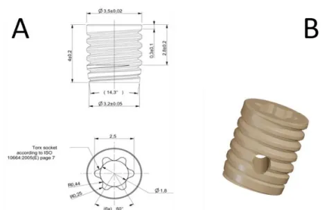
Experimental Animal Model ... 72
Animals and ethical approvals ... 72
Anesthesia ... 73
Surgical procedure and implantation ... 73
Experimental model ... 74
Post-operative care and euthanasia ... 75
Ex vivo analysis ... 75
Removal torque testing ... 75
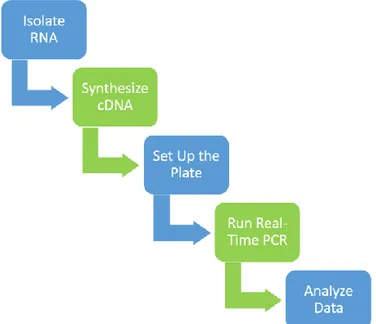
Gene expression analysis ... 76
Bone histomorphometry ... 79
X-ray computed microtomography ... 81
Statistics ... 83
SUMMARY OF RESULTS ... 85
Surface Characterization ... 85
Chemical and Mechanical Characterization ... 87
Ex vivo results ... 91
Removal torque results ... 92
Gene expression results ... 94
Histological results ... 96
Micro-CT results ... 105
DISCUSSION ... 109
Structural and chemical properties of the HA-coating ... 109
Biomechanical properties of HA coated implants ... 117
Bone formation around PEEK and HA-PEEK ... 119
Genetic mechanisms ... 127
CONCLUSION AND FUTURE PERSPECTIVES ... 131
POPULÄRVETENSKAPLIG SAMMANFATTNING ... 134
ACKNOWLEDGMENTS ... 136
REFERENCES ... 138
People are living longer today than ever before, and the number of older adults will increase exponentially over the next coming years. However, that does not necessarily mean that they are living health-ier. An aging population presents many opportunities but also chal-lenges in the public health care with a sharp increase in incidence of many diseases associated to the bone, teeth and joint systems. Or-thopedic and dental implants commonly used mostly consist of met-als, composites, polymers and ceramics.
Titanium has been widely used in the orthopedic and dental field to replace or support mineralized structures. Titanium possesses good mechanical, physical and biologic properties. However, tita-nium has in the clinic presented some critical drawbacks. The mod-ulus of elasticity is higher than that of bone which may cause stress shielding and bone resorption ultimately jeopardizing the implant retention. In addition, with rare cases of titanium allergy and patient desires for non-metallic treatments, alternatives to titanium are be-ing studied. Furthermore, the radiopacity of titanium causes inter-ference with the radiographic evaluation and the non-esthetic color may discolor the gingiva of thin biotypes.
Recently, advanced polymeric implantable materials have been in-troduced into the medical field. Polyetherether ketone (PEEK) is widely used in various orthopedic applications, such as spine im-plants, joint replacements and fracture fixations. In dentistry, PEEK can be manufactured as prosthetic components, fixed and removable bridges and dentures.
PEEK is a thermoplastic polymer with mechanical properties that are close to those of human bone, for example the elastic modulus. This will minimize the stress shielding upon loading and stress dis-tribution. Besides, PEEK holds excellent biocompatibility with no
toxic or mutagenic effects in vitro and in vivo.
However, unmodified PEEK is biologically inert with limited pro-tein and cell adhesion to the surface. Therefore, improving the bio-activity of PEEK is a significant challenge and a prerequisite to pos-sess all of its potentials as a biomaterial. There are currently 2 meth-ods to improve the bioactivity of PEEK; surface modification and composite preparation. Hydroxyapatite (HA) is the most widely used material due to its biocompatibility and osteoconductive po-tential in association with biomaterials. There are many developed and evaluated methods to deposit HA on metallic implants, but the only commercially used one is plasma spraying. However, the tech-nique has some severe disadvantages such as irregular coating thick-ness with deficient adherence to the implant surface. Clinical com-plications have been confirmed with inflammation caused by frag-ments of delaminated coating. To challenge these disadvantages, several alternative coating techniques have been introduced.
The aim of this thesis was to evaluate a novel surface modification of bioactive hydroxyapatite deposited by a spin-coating technique. The technique generates a crystalline, nanosized coating homoge-nously distributed over the implant surface. In particular, the thesis encompasses a comprehensive topographic analysis to evaluate the coating properties. In addition, the HA-PEEK implant will be
com-pared in vivo with an unmodified control with respect to its
oste-oconductive and bioactive properties.
In study I, the surface topography was characterized using interfer-ometry (IFM), scanning electron microscopy (SEM) and atomic force microscopy (AFM) of the same types of implants that were evaluated in the experimental animal models. The chemical charac-terization before and after the coating procedure was evaluated with X-ray photon spectroscopy (XPS). Mechanical testing of implants were completed in accordance with the tensile testing standard (ISO 527-2). Furthermore, the hydrophilic appearance after coating was
estimated by measuring the contact angle. The results from the ma-terial characterization reveal a hydrophilic coating with a minimally rough surface which preserved its mechanical properties after the coating procedure. The chemical analysis revealed that only Ca, P, C and O were present at the surface of HA-PEEK with a proportion as that of human bone.
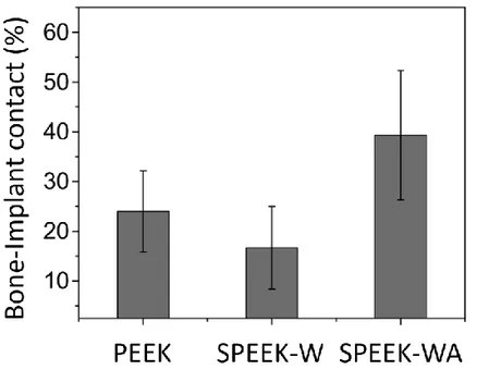
In study I and IV, PEEK implants were implanted in the rabbit tibia and femur and retrieved after 3, 12 and 20 weeks of submerged heal-ing and subjected to biomechanical analysis usheal-ing a removal torque device. The removal torque required to unscrew the implants was significantly higher for HA-PEEK after the 3 and 12 week retrieval time points. However, the absolute torque values were lower at 12 weeks compared to earlier time points, presumably caused by ab-sence of primary stability and dissolution of the HA-coating. After 20 weeks of healing the absolute values were even lower than for the implants recovered at 3 and 12 weeks. However, the retention was still significantly increased for the HA-PEEK implants.
In study II, III and IV, the osseointegration was histologically evalu-ated with respect to bone-implant contact (BIC) and bone area (BA) after the same healing time points as aforementioned. The perfor-mance of the HA-coating was most significant 3 weeks after implan-tation. However, after 12 and 20 weeks, the BIC turned out to be comparable between the groups. Furthermore, the implants im-planted in femur were designed with an apical perforation to evalu-ate the osteoconductivity in terms of area of bone. After 3 and 12 weeks the results revealed a significant improvement on the HA-PEEK. However, after an extended healing period of 20 weeks, the unmodified implants were presenting comparable results as the HA-PEEK group.
A more comprehensive description of the osseointegration can be achieved using computed micro-tomography. Three different diam-eters around the implant and inside the apical perforation were eval-uated with respect to bone density and trabecular properties. How-ever, there were no detectable differences between the PEEK and HA-PEEK. The outcome was limited to the resolution of X-rays. There was a minor contrast between resin and PEEK.
The implant surface, particularly the thread edges are subjected to mechanical forces during implant placement and removal. The sta-bility of the coating was evaluated in study II. The coating was well-preserved in deformities of the surface, but on the flat edges of the threads there were signs of deformation and absence of HA crystals. This thesis demonstrated that a nanosized HA-coating can be pro-cessed using a spin-coating technique with nanostructures and suffi-cient adhesion to the substrate.
Furthermore, the performance of the HA-coating was found to improve the surface osteoconductivity by increasing the level and speed of osseointegration. However, the effect was most prominent in the early stages of healing whereas the implants from the extended time points showed osteoconductive properties comparable to un-modified PEEK.
In addition, the ability of HA-coating to improve bone fusion in-side a perforation was the most clinical relevant outcome. Unmodi-fied PEEK implants are today widely used in spine surgery and the present surface modifications may improve the clinical outcomes where a rapid bone formation is essential.
This dissertation is based on the following papers, which will be re-ferred to in the main text by their Roman numerals. The papers are appended at the end of the thesis.
I. Biomechanical evaluation and surface characterization of a
nano-modified surface on PEEK implants: A study in the rab-bit tibia.
Johansson P, Jimbo R, Kjellin P, Currie F, Chrcanovic B, Wen-nerberg A.
International Journal of Nanomedicine 2013 Aug 14;9:
3903-11; doi: 10.2147/IJN.S60387.
II. Nanosized Hydroxyapatite Coating on PEEK Implants
En-hances Early Bone Formation: A Histological and Three-Di-mensional Investigation in Rabbit Bone.
Johansson P, Jimbo R, Kozai Y, Sakurai T, Kjellin P, Currie F, Wennerberg A.
Materials 2015 Jun 25;8(7):3815-3830; doi:
10.3390/ma8073815.
III: Polyether ether ketone implants achieve increased bone fusion when coated with nano-sized hydroxyapatite: a histomorpho-metric study in rabbit bone.
Johansson P, Jimbo R, Naito Y, Kjellin P, Currie F, Wenner-berg A.
International Journal of Nanomedicine 2016 Apr 6;11:
1435-42; doi: 10.2147/IJN.S100424
IV: Biomechanical, histological and computed X-ray tomographic analyses of hydroxyapatite coated PEEK implants in an ex-tended healing model in rabbit.
Johansson P, Barkarmo S, Hawthan M, Peruzzi N, Kjellin P, Wennerberg A.
Submitted to Journal of Biomedical Materials Research: Part A
Reprint permissions have been granted from:
Paper I and III: Dove Medical Press Limited, AUT171619 Paper II: MDPI, Open access license (CC, BY)
The respondent performed most of the work from planning, experi-mental work (with exception of the SEM imaging, EDX analysis, contact angle analysis and computed microtomography imaging) and analysis of the data. The respondent was also the main contrib-utor to writing the manuscripts.
Abbreviations
°C Degree Celsius
µm Micrometer
3D Three-dimensional
ACTB Beta-actin
AFM Atomic force microscopy
ALIF Anterior lumbar interbody fusion
ALP Alkaline phosphatase
ANAB Accelerated neutral atom beam
ASD Adjacent segment degeneration
BA Bone area
BGLAP Bone gamma-carboxyglutamate (GLA) protein
BIC Bone-implant contact
BMP2 Bone morphogenetic protein-2
BS/BV Bone surface per given bone volume
BV/TV Bone volume to total volume
CaCl2 Calcium chloride
CAD/CAM Computer aided design/computer aided
manufac-turing
CCD Charge coupled device
cDNA Complementary deoxyribonucleic acid
CFR-PEEK Carbon-fiber-reinforced polyetheretherketone
CoCr Cobalt chromium alloy
CoCrMo Cobalt chromium molybdenum
COL1A1 Collagen type I alpha 1
CS Calcium silicate
CT Computed tomography
DICOM Digital Imaging and Communications in Medicine
EDX Energy-dispersice X-ray spectroscopy
FDA Food and Drug Administration
FDP Fixed dental prostheses
FHA Fluorohydroxyapatite
GAPDH Glyceraldehyde-3-Phosphate Dehydrogenase
GPa Gigapascal
h Hours
H2SO4 Sulphuric acid
HA Hydroxyapatite
HA-PEEK Hydroxyapatite- polyetheretherketone
IFM Interferometer
IGF1 Insulin like growth factor 1
ISO International Organization for Standardization
kg Kilogram
kV Kilovolt
L-929 Mouse fibroblast cells
LDHA Lactate Dehydrogenase A
Ltd Limited liability company
MC3T3-E1 Mouse osteblastic cell line
MG-63 Human Osteosarcoma cell
micro-CT Micro-Computed Tomography
min Minute
mL Milliliter
mm Millimeter
MPa Megapascal
NaOH Sodium hydroxide
Ncm Newton centimeter
nm Nanometer
O2 Oxygen gas
PCR Polymerase chain reaction
PDMS Polydimethylsiloxane
PE Polyethylene
PEEK Polyetheretherketone
pH Potential of hydrogen
PLIF Posterior lumbar interbody fusion
PMMA Poly methyl methacrylate
PP Polypropylene
qPCR Quantitative polymerase chain reacton
Ra Arithmetic average of the rougnhess
RCT Randomized controlled trial
RFA Radiofrequency ablation
RNA Ribonucleic acid
RNase Ribonuclease
rpm Revolutions per minute
RTQ Removal torque
RUNX2 Runt-related transcription factor 2
s Second
SBF Simulated body fluid
SD Standard deviation
Sa Topographical parameter – mean height deviation
Sdr Topographical parameter – developed area ratio
Sds Topographical parameter – density of summits
SEM Scanning electron microscopy
SSP1 Secreted phosphoprotein
Tb.N Trabecular number
Tb.Th Trabecular thickness
TCP Tissue Culture Plate
TCPS Tissue culture polystyrene
TEM Transmission electron microscopy
Th.Sp Trabecular spacing
TiO2 Titanium dioxide
TJA Total joint arthroplasties
TLIF Transforaminal Lumbar Interbody Fusion
UHMWPE Ultra-high-molecular-weight polyethylene
USD United States Dollar
W Wolfram
w/w Weight by weight
VOI Volume of interest
XPS X-ray photoelectron spectroscopy
YSZ Yttria-stabilized Zirconia
Å Ångström
β-TCP Beta-tricalcium phosphate
μA Microampere
μCT Micro-computed tomography
μm Micrometer Element symbols Al Aluminum Ar Argon C Carbon Ca Calcium Cl Chlorine Co Cobalt Cr Chromium F Fluorine H Hydrogen He Helium Mg Magnesium Mo Molybdenum N Nitrogen Nb Niobium O Oxygen P Phosphorus S Sulfur Sr Strontium Ti Titanium V Vanadium Y Yttrium Zr Zirconium
The meaning of a ‘biomaterial’ has been debated and changed over the years but the European Society for Biomaterials derived the
to-day applied definition in medicine1-3. The latest definition is ‘a
ma-terial intended to interface with biological systems to evalu-ate, treat, augment or replace any tissue, organ or function of the body’4.
In biomaterial science a material is ‘a substance of which things are made’ and man-made materials are historically divided into metallic, ceramic and polymeric. All these materials have many subdivisions with plentiful applications in medicine2. Today biomaterials are used with good success for example in heart valve prostheses, hip replace-ments, dental implants, intraocular lenses and ventricular assist de-vices5.
Over the past decades remarkable technological improvements in quality, performance and versatility of instrumentation have been achieved using biomaterials when it comes to rehabilitate lost func-tions and aesthetics. Furthermore with high growth in the geriatric population and rising numbers of hip, knee and teeth replacements, reports have forecasted the biomaterial market to annually grow 13.2 % during 2016-20216,7. The global medical device market was in 2016 valued USD 339.5 Billion and projected to reach USD 435.8
‘Polymer’ originates from Greek and means ‘many parts’ and is a large molecule with many repeated subunits or monomer segments, usually composed of carbon, hydrogen and sometimes oxygen, chlo-rine and nitrogen9. The unique attribute of polymers is determined by the length and composition of the molecular chain10.
Table 1: Global medical device technology industry by region (in Billion US$) 2016-2020. Data obtained from Worldwide Medical Devices Forecast 2020 Report
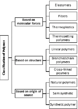
The classification system of polymers can be made based on the mo-lecular forces or the source of origin (Figure 1). Based on the source, polymers can be sub-divided in two groups; natural polymers and
synthetic polymers. Natural polymers are abundant in nature and
the ultimate natural polymers are the deoxyribonucleic acid (DNA) and ribonucleic acid (RNA) which are the origin of all life. Silk, wool and rubber are other natural polymers. The evolution of synthetic polymers started by the mid-1800s when rubber was accidentally mixed with sulfur which created a more stable rubber that did not deform or melt below 100 degrees Celsius (°C). A few decades later Goodyear was given the patent for vulcanized rubber, the origin of today’s car tires. Over the years countless variations of polymers
2016 2017 2018 2019 2020 North America 166.6 176.5 187.3 197.9 208.6 Asia-Pacific 68.7 72.6 77.6 82.9 88.6 Central/Eastern Europe 14.6 15.7 17 18.1 19.1 Middle East/Africa 10 10.8 11.5 12.5 13.2 Western Europe 79.5 85.1 92.6 101.4 106.2 Total 339.5 360.8 386.1 412.8 435.8
have been developed and grown to usage in larger numbers than aluminum, copper and steel industries combined11,12.
Figure 1. Schematic classification of polymers based on the molecu-lar forces, structure and origin of source.
The inter-molecular forces is another way to differentiate polymers. They can be divided into four types; Elastomers, fibers, thermoset-ting and thermoplastics (Figure 2). Elastomers have no or very few inter-molecule cross linkages giving an ability to stretch and regain
original shape after the force is removed. One example of elastomer
is natural rubber. Fibers hold the opposite feature of elastomers with strong hydrogen bonds or dipole-dipole interactions between the chains. In that way the molecules are closely arranged and the poly-mer possesses high tensile strength and minor elasticity. Nylon and silk are two common examples. A thermoplastic polymer have mon-omer chains that are networked with weak van der Waals forces and the material soften on heating and set on cooling. Polythene (as
‘plastic’) is a common thermoplastic polymer and can be manufac-tured into any shape and size. In contrast, when heat is applied to a thermosetting polymer the molecules cross-link and permanently set to become a solid component without the ability to re-modulate. Acrylic resin and epoxy are two common thermosetting poly-mers13,14.
Figure 2. Molecular structure of polymers. The arrangement of the chains determines the properties of the polymer. Branched chains as elastomers and networked thermosets once set have increased strength but less flexibility compared to thermoplastics.
Rapidly developing science of polymers in medicine was seen during the Second World War where e.g. polyesters and polyamides were introduced for sutures. Since that time considerable research with numerous newly introduced polymers has been conducted and turned the biomedical polymer field into a billion euro industry15. The pursuit of constantly improving polymers is motivated by the need for biomaterials in modern therapies and the scientific curiosity to recognize and mimic biological functions with biomaterials16.
Polymers for medical devices are most commonly made of polypro-pylene, polyethylene or poly-vinyl chloride (Table 2). Polymers have a long history from traditional applications such as catheters and
syringes to tissue engineering and drug delivery systems. In general,
medical polymers can be classified as non-degradable or degradable depending on the reactivity of the backbone molecule upon water exposure17,18. Polypropylene is a family of polymers with high rigid-ity and tensile strength, and good biostabilrigid-ity. In medicine it is com-monly used as non-degradable sutures. Polyethylene has the ad-vantage to withstand sterilization temperatures and also to possess
good toughness and wear resistance. These properties make this pol-ymer suitable for drain and catheter tuber and prosthetic joints. PVC is used in short-term applications as the material may become brittle because of leaching plasticizers. Therefore it is used for example as external tubing and bags for blood storage5.
This thesis will focus on a non-degradable thermoplastic polymer, polyether ether ketone (PEEK), useful in a wide variety of applica-tions such as orthopedics, dental and tissue replacements10,19-21.
Table 2. Polymers in the medical field with their characteristics and some examples of applications.
Preparation of PEEK was first reported by Attwood et al.22 and the crystalline unit cell was presented with a chemical structure of an aromatic backbone molecular chain with ketone and ether as inter-connected functional groups (Figure 3). The first successful synthesis of PEEK was through a nucleophilic displacement where a diphenyl
sulfone was used as solvent22. PEEK becomes solid at room
temper-ature and the glass transition take place at 143°C. The melt transi-tions occur at 343°C. In room temperature and polymerized state the polymer is inert to chemicals and is only soluble in 98%
sul-phuric acid (H2SO4)10,23,24. The chemical composition provides the
polymer with properties such as stability at high temperatures,
re-sistance to chemical and radiation damage (Table 3). As a result of
these high performance properties, PEEK has been employed in chal-lenging industrial applications in aerospace and automotive engi-neering25,26. By the late 1980s the medical field gained interest for
Material Characteristics Applications
Polypropylene (PP) Semi-crystalline PP holds high rigidity, tensile strength and biocompatibility
Non-resorbable sutures Hernia repair Polyethylene (PE) High density PE possess good toughness and wear resistance.
Low density PE does not withstand high temperatures and cannot be sterilized.
Tubing for drains and catheters Prosthetic joints Poly vinyl chloride
(PVC)
PVC is a flexible material that is used for disposable and short-term application products due to the material embrittlement.
Examination gloves Tubing and blood storage bags Polydimethylsiloxane
(PDMS)
Normally referred to as silicone. Low glass transition makes the material very flexible with excellent fatigue resistance.
Breast implants Heart valves Poly(methyl
metacrylate) (PMMA)
Transparent thermoplastic with the commercial name, Plexiglas. PMMA has qualities of biocompatibility, ease of manipulation and low toxicity
Dentures Intraocular lenses Bone cement
this material in the field of trauma and orthopedic surgery. Origi-nally used as a spinal implant, PEEK is today commonly used as spinal fusion cages27,28.
Table 3. Physical properties of PEEK OPTIMA LT1. Data obtained from Invibio Biomaterial Solutions.
Figure 3. Chemical formula of PEEK with a semi-crystalline aro-matic polymer consisting of repeating units. The molecule is rela-tively stiff but with freedom to rotate around the ether (–O–) bonds and ketone-carbon bonds (–CO–). Figure from Blundell and Os-bornI.
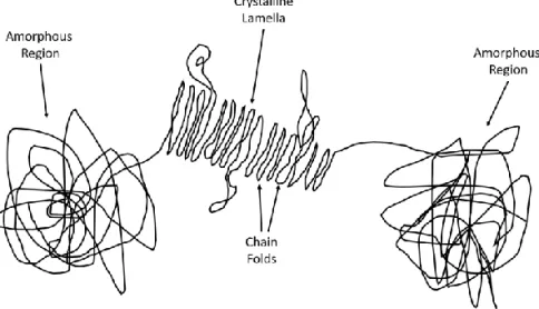
Processing and Thermal Transitions
Crystallization of polymers is a dynamic transition from an amor-phous state where the molecule chains are highly disordered, into a folded and ordered molecule state, called lamellae29. PEEK is a semi-crystalline polymer with relatively strong intermolecular forces that makes the material firm even at the glass transition temperature (143 °C). Producing a 100 % crystalline polymer is in practice impossible since the molecules have to fold into arrangement from the highly
Property Values Unit
Tensile Strength 90-100 MPa
Young's Modulus 3.6 GPa
Elongation @ break 50 %
Glass Temperatue (Tg) 143 °C
disordered state. The molecular chain is composed of crystalline la-mellas surrounded by amorphous regions and in straightened state the molecule can be several hundreds of meter long30 (Figure 4).
Figure 4. Schematic representation of polymer chains with repeating units of amorphous and crystalline regions.
PEEK has an advantage over other polymers due to its high degree of crystallinity which increases its mechanical strength and decreases the risk of stress cracking22. The crystalline structure of PEEK is built up by many spherulites. Dimensions and arrangement of these spherulites depends on the processing conditions and forgoing ther-mal history which affects the crystallinity of the polymer31-33. In gen-eral, the thickness of the lamellae is in the angstrom range (50-60 Å) and the spherulites are 25-40 µm in diameter30. PEEK as a semi-crystalline polymer can be processed with different level of crystal-linity depending on the processing conditions after the melt stage. If the melt is cooled rapidly, a glassy or amorphous state is achieved whereas a slow cooling develops a higher level of crystallinity30. When the melt is cooled the arrangement of the spherulites trans-forms from an amorphous state into a more ordered state. When reheated, the polymer will return to an amorphous state33. This oc-currences is illustrated in figure 5 where the rate of crystallization is dependent on the temperature.
Figure 5. Typical form of crystallization rate-temperature curve for a crystalline polymer.
Mechanical properties
The mechanical properties and behavior of a polymeric material are complex and can be divided into many subgroups. In this chapter the focus will be on the properties that are favorable or harmful for the use in medicine.
The following material properties are commonly evaluated in bio-materials science:
1. Stiffness – Resistance to deformation like elongation, twist-ing, bending etc.
2. Strength – The ability to resist force or pressure before rup-turing
3. Toughness – The absorbed strain energy under static, impact and fatigue loading until material fracture
4. Wear resistance – The ability to withstand loss of material upon contact friction
One of the major reasons for the medical field to gain interest into PEEK was its closely matched stiffness to bone. Orthopedic appli-ances as spinal cages, compression bone plates and hip prostheses were usually made from metallic alloys where the stiffness was
10-20 times that of cortical bone34. This mismatch in stiffness may cause
a stress shielding and may lead to bone resorption35-37. The bone
re-sorption around the inserted device may cause loosening of the im-plant or weakening of the bone and eventually fracture. The use of polymer devices with a similar stiffness as cortical bone has, in com-parison to metallic alloys, demonstrated a more favorable stress
dis-tribution to the surrounding bone38,39. According to Wolff’s law, the
bone will adapt to the exposed load. Therefore, the density of the
bone will be dependent of the stress distribution40. PEEK can be
tai-lored to the stiffness required for the application with addition of carbon reinforced fibers (CFR, Figure 6). CFR-PEEK can with con-tinuous fibers achieve excellent stiffness, strength and durability in
order to match the implant bed41,42. In addition, CFR-PEEK in a
to-tal hip model with saline, have demonstrated excellent compression strength durability43.
Figure 6. Different clinical applications of PEEK that are currently used in dental, orthopedic and reconstructive applications.
To understand the failure mechanisms of PEEK implants in spinal and dental applications, the strength of the material in static and
cyclic loading conditions has to be assessed10. The strain rate and
temperature in general decide the mechanical behavior of neat PEEK but it can also be changed by the molecule weight and size and
ori-entation of the crystallized lamellae44-48. In addition, an increase in
crystallinity has been found to decrease the toughness49. In room
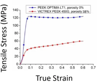
temperature, PEEK shows a linear elastic deformation at small strains and compressions (Figure 7). After the yield transition caused by strain or compression, the behavior varies depending on the tem-perature and rate of strain. Until material failure after large defor-mation, the behavior is difficult to characterize due to a change in molecular alignment. However, PEEK as a biomaterial is expected to operate in body temperatures (35-44°C) where the material is
pre-sent no structural deformation46,47.
Figure 7. Strain-stress plots for PEEK-OPTIMA and Victrex 450G with 0% and 38% porosity, respectively. Figure obtained from
Vaezi and YangII.
Neat PEEK has a high wear resistance and friction coefficient (µ ≥ 0.4 in dry sliding) in addition to a low thermal conductivity. These features has gain interest for PEEK arthroplasty bearing surfaces. In
total hip replacement a common reason to failure is a result of
asep-tic loosening50. Wear debris and stress shielding are believed to cause
the failure. PEEK has been evaluated in several studies with respect to contact pressure and cross-shear ratio in relation to the wear of
orthopedic materials51,52. Ultra-high-molecule-weight polyethylene
(UHMWPE) and cobalt-chromium-molybdenum (CoCrMo) pro-vided more wear particles in a wear-test model compared to
CFR-PEEK. A study by Howling et al.53 evaluated wear debris from
CFR-PEEK against alumina. The wear particles from CFR-PEEK in an in vitro culture model showed no cytotoxic effects on fibroblast cells (L-929) or a human hematopoietic cell line (U-937). Other authors suggested that PEEK wear particles have no adverse tissue reaction since it has
no cytotoxic effect on cells in culture10,53. An in vivo study of PEEK
applied in a total hip system (Stryker SA, Montreux, Switzerland) was evaluated in 30 patients after 3 years in function no implant was
replaced due to aseptic loosening54. However, this is not a strikingly
different outcome in comparison to the norm when placing ordinary types of hip arthroplasties.
Chemical and radiation stability
The chemical structure of PEEK is composed of aryl rings intercon-nected via ketone and ether groups opposite each other. This struc-ture creates an exceptional resistance and un-reactivity to chemicals but also against thermal and irradiation degradation. In addition, the PEEK structure can only be dissolved by sulfuric acid. Alto-gether, this may explain the excellent biocompatibility of PEEK. PEEK can endure extreme conditions with different liquids but as an implantable biomaterial the resistance in water is of particular inter-est. PEEK has a maximum solubility in water and brine of 0.5% w/w
at 75°C and is independent of the salinity of the exposed liquid45,55.
The results by Boinard and colleagues45 showed that the small water
uptake altered the degree of crystallinity at the surface which did not
re-form after the samples were dried. A study by Liebermann et al.56
evaluated the physicomechanical properties of PEEK among other polymers after aging in different liquid substances. The aim of the study was to elucidate the physicomechanical knowledge of PEEK
as a material to support fixed dental prostheses. The authors recom-mend the material for long-term oral restorations since it shows as low solubility and absorption rates as do other traditional PMMA-based materials.
The thermal stability of PEEK in a clinical application is of no
con-cerns since the temperature never exceeds the body temperature57,58.
However, when the material is sterilized and exposed to pressurized steam (autoclave) the temperatures exceeds the body temperature. However, no significant reduction in flexural strength was revealed
on CFR-PEEK after 100 autoclave cycles59. Minor changes in the
microstructure have been demonstrated but the bulk material
re-mains unaffected following steam sterilization60. If steam
steriliza-tion is to be avoided another sterilizasteriliza-tion process is the gamma ra-diation. Some polymers (UHMWPE) are sensitive to gamma radia-tion where breaking of chemical bonds may cause a formaradia-tion of
highly reactive free radicals61. Even after radiation, these free
radi-cals may react and influence the surrounding material and tissue. However, several aromatic polymers (such as PEEK) were exposed to gamma radiation under oxygen pressure to evaluate the changes
in tensile properties62. The effect of the radiation dose was estimated
by calculating the elongation at breakage which was found to be as low as 10-20% of the dose from electron-beam radiation. The au-thors concluded that the altered mechanical properties of the deteri-oration was caused by degradation of the polymer main chain (chain scission).
To summarize, PEEK has been found to perform extremely well in many environments thanks to its stable chemical structure. Radia-tion and aqueous exposure are of no concern and in applicaRadia-tions as a biomaterial where the temperature is around 37°C PEEK performs impeccably.
Clinical applications
The mechanical drawbacks of metal alloys inspired researchers in
the late 1980s to conduct studies on PEEK polymers27,28,34,63. PEEK
was by that time used in the automotive- and aerospace industry due to its chemical resistance, light weight and mechanical advantages
compared to metals. In 1998, Victrex® launched its
PEEK-OPTI-MATM for medical implant use and since then research and
develop-ments have been in progress in almost every area of application where metal alloys are presently being used. Interest was from the beginning specially gained in orthopedics, spine and trauma surgery but are currently under evaluation in new fields such as dentistry and cardiac surgery42,64-66.
Spinal implants
Patients diagnosed with a degenerative disc in the lumbar spine can be treated by a spinal fusion, a surgery designed to end the low back pain caused by motions of the vertebra. There are two main ap-proaches to spinal fusion; posterolateral interbody fusion (PLIF) and
anterior lumbar interbody fusion (ALIF)67-69. The main differences
between the two approaches are the access point and the site where the bone graft is inserted in the spine.
The PLIF technique used a pedicle screw fixation (Figure 8). Two screws are placed within the pedicle of each vertebral segment, one of each side of the spine. Each vertebra is connected with a metal rod. The bone graft is inserted between the transverse process in pos-terior side of the spine which allows bone fusion of the processes
Figure 8. Lateral radiograph at 12 months post-operative showing a PEEK cage implanted between the vertebras to enable a bone fusion.
Figure adapted from Lin et alIII.
The ALIF procedure is similar to the PLIF except that the spine is approached through the abdomen. This technique provides access to the vertebra body and the disc. After the disc is removed, the inter-vertebral space is filled with a spinal implant and bone graft (Figure 9). Compared to the PLIF, this technique provides an increased sur-face area with a more favorable bone fusion but the morbidity of the patient is higher due to the risk of injuries of the nearby large blood vessels (Vena Cava and Aorta).
Figure 9. Anterior PEEK cage in combination with posterior
Titanium and stainless steel have provided satisfying long-term re-sults due to their stiffness and rate of osseointegration. However, the stiffness also becomes a disadvantage because the mismatch to bone which may lead to adjacent segment degeneration (ASD). Post-oper-ative examination by conventional X-ray is difficult to interpret be-cause the metal streak artifact be-caused by the implants.
In 2001 the Food and Drug Administration (FDA) approved the CFR-PEEK for lumbar fusion. At this time, from surgical experience, the spinal fusion was performed with stand-alone threaded cages and the protocol of CFR-PEEK was described in a monograph by a
surgeon inventor, John Brantigan70. CFR-PEEK provided similar
elastic modulus as the human bone and the in the post-operative
evaluation the transparency to X-ray enhanced the interpretation
71-73. The Brantigan cage was intended to be inserted with an ALIF
approach together with posterior instrumentation in order to en-hance the stability, a so-called 360° fusion (Figure 10). Studies have confirmed the superiority of the Brantigan cage approach compared
to the posterolateral lumbar fusion74,75. According to several studies,
the radiolucency of PEEK has facilitated the radiographic assessment
of the fusion76-78. However, there are potential complications for
PEEK cages, as with any implant in load-bearing situations79,80.
Sub-sidence caused by wear debris and fracture of the implant cage has
been documented by some authors81,82.
Figure 10. The original design of an interbody spine implant made
Total joint arthroplasties
As mentioned before, PEEK is resistant to fatigue strain, retains me-chanical properties in the range of human bone while it is radiolog-ically transparent which makes it suitable for a range of orthopedic applications. In the 1990s, PEEK and CFR-PEEK were introduced
as load bearing implants for total joint arthroplasties (TJA)83. In a
TJA the load is applied to a femoral stem and the forces are spread to the surrounding bone and tissue (Figure 11). If the femoral stems are made of metal, the load to the surrounding tissue may be de-creased. This is called stress shielding and the biological response is a slow resorption and re-distribution of the adjacent bone. This may
potentially lead to implant loosening84. However, an implant failure
usually occurs by a combination of several mechanisms85. Wear
par-ticles from the implant surface may induce an inflammation which
may cause an aseptic loosening of the implant86,87. The biological
response to wear particles from PEEK in different joint replacements have been studied in several publications and it is generally accepted that phagocytozable particles (0.1-10 µm) are the ones with
poten-tial cytotoxic effect88,89. The particles can stimulate macrophages to
produce pro-inflammatory and osteolysis mediators that may cause aseptic implant loosening. Not until the characteristics of the PEEK particles are identified and repeatable analyses would it prove
Figure 11. CFR-PEEK has gained interest in the use for orthopedic implants stems due to its tailored mechanical properties, chemical stability and less tendency of wear debris. A: Cemented CFR-PEEK cup. B: Cementless CFR-PEEK cup. C: Cementless CFR-PEEK stem. D: Cemented CFR-PEEK stem. Image obtained from Nakahara et al.VI
Trauma implants
The clinical outcomes over the last decades of trauma implants and
fracture fixation has motivated a paradigm shift91-93. From a total
fixation with no mobilization to a more flexible fixation. A too rigid fixation may cause an incomplete bone healing with prolonged re-habilitation time. Consequently, a more flexible fixation has been introduced where the stress from the loading is distributed more evenly to the surrounding structures. PEEK holds all the qualifica-tion that makes it suitable for fracture fixaqualifica-tion. It is biocompatible, resistant to solvents and the stiffness is close to that of human bone. The stiffness can be further adjusted by adding carbon fibers to the compound (CFR-PEEK). Since PEEK is a thermoplastic polymer, the
shape can be designed and adjusted at the surgical procedure10.
PEEK also provides radiographic evaluation without artifacts which
these advantages, only a few clinical reports of the performance of PEEK have been published. A biomechanical study by Schliemann et
al.95 found that PEEK did not provide satisfactory fixation strength
when compared to titanium. The same authors published a 2-year prospective clinical study, comparing PEEK and conventional
lock-ing plates for humeral fractures94. The fixation with PEEK plates
was found comparable to the conventional locking plates.
Micro-motion is nowadays accepted, but only in the axial direction96. Due
to the flexibility of PEEK, the bending, torsional and shear forces are not sufficiently stabilized in fracture fixation. Furthermore, PEEK is under consideration in cranioplasty where CT-guidance and CAD/CAM (computer-aided design and computer-aided manufac-turing) techniques provide a precise and time-saving
reconstruc-tion97. However, metal locking plates are still dominant in the
trauma field, therefore PEEK needs further studies and clinical re-ports to increase its practice.
Dental applications
Titanium has since the 1960s been the most common choice of ma-terial in oral endosseous implants due to its superior mechanical properties and biocompatibility and titanium constructions have
re-sulted in increased quality of life for patients98-101. However,
tita-nium and titatita-nium alloys (such as Ti-6Al-7Nb and Ti-6Al-4V) have
over the years demonstrated several clinical complications. One problem is the potential hypersensitivity or allergy to titanium from
degradation products102-105. Another problem is the divergent elastic
modulus to the surrounding bone which may cause adverse stress at
the bone interface106. In esthetic anterior cases where the patient
have a high smile-line, the dark color of the titanium implant may
shine through the tissue107,108. All in all, there may be a need for an
introduction of an alternative to metal implants. Ceramic implants were introduced as an alternative to titanium with tooth-like color and good biocompatibility. However, zirconia suffers with similar
PEEK may be another alternative with superior elastic modulus for bone applications, excellent biocompatibility and tooth-like appear-ance27,110. Suggested dental applications for PEEK includes; dental
implants, implant abutments, fixed bridges and crowns and remov-able dentures and components (Figure 12).
Figure 12. Using subtractive process, full and partial dentures and overdentures, crowns and bridges can be manufactured. Image from Juvora Dental Innovations.
As for dental implants, studies have presented different conclusions
regarding stress shielding and biocompatibility. Lee et al.64
con-cluded that PEEK implants compared to titanium may reduce stress
shielding while Sarot et al.42 suggested that there is no difference
be-tween the stress distributions. Therefore, no general conclusions on stress shielding can be made on the present knowledge. Neat PEEK
is hydrophobic and bioinert111,112, even though some studies have
found no differences between the osseointegration between PEEK
and titanium or zirconia113,114. Numerous modifications have been
applied to improve the biological properties of PEEK. Further read-ings on this topic will be described later in this introduction. How-ever, as far as the author knows, there are no PEEK dental implant systems marketed.
Implant abutments made of PEEK have been evaluated in a con-trolled clinical trial (RCT) by Koutouzis et al.115 and revealed no
significant difference on inflammation and bone resorption com-pared to titanium abutments. In terms of bacterial adhesion, PEEK abutments has demonstrated comparable results as those made of
titanium and zirconia116. Santing and co-workers117 compared
tem-porary PEEK abutments with 2 commercial systems on fracture strength and failure mode. No differences between the systems were found except for anterior maxilla where PEEK showed significantly lower fracture strength. Similar results were discovered by Rubén et al.118 with less fracture resistance compared to titanium but the elas-tic deformation was significantly less than titanium.
The properties of PEEK has inspired dental innovators to improve the components in removable and fixed dental prostheses and crowns. Since PEEK can be manufactured by using CAD/CAM there are no design limitations. A study comparing retention clasps of PEEK and Cobalt Chromium (CoCr) demonstrated less retention for
PEEK cf. the metal119. However, the authors believes the retention
of PEEK clasps to be sufficient for clinical use. A clinical report on a maxillary obturator prostheses made of PEEK for a patient with maxillary defect provided the patient with a more functional and
lighter prostheses compared to the conventional alternatives120.
Using PEEK as cemented crowns requires a surface treatment. A study evaluating shear bond strength after sulfuric acid etching found more differences due to cement type than the etching time. However, the results seems to be promising but the authors
sug-gested further studies on other cement systems121. A fixed dental
bridge (FDP) comparison of CAD/CAM manufactured and conven-tional pressed from granulae was evaluated based on the
load-bear-ing capacity122. The study summarize that the CAD/CAM
manufac-tured FDPs showed advantages over the pressed granular PEEK FDPs.
In conclusion, PEEK is a newcomer in the dental field and the pub-lished data is limited. However, the results show promising abilities
and possible future applications. Therefore, more research and clin-ical trials are needed to discover indications for the material in future dental applications.
Definitions
Immediately when a biomaterial is inserted into a living host, inter-actions take place between the host and the biomaterial. The word “biocompatibility” refers to the ability of a material to perform with
an appropriate host response in a specific situation”123.
Biocompat-ibility is not a property of a material itself. It should be seen more like an interaction between the host and the biomaterial. Within the same host, a biomaterial could be biocompatible in one application but cytotoxic in another. Minimum requirements for implanted bi-omaterials is that they should be toxic, mutagenic and non-carcinogenic.
The biological performance of materials and living systems may
be viewed in two ways124:
1. The host response – the local or systemic response to the im-planted material such as macrophage proliferation or fibrous encapsulation
2. The material response – the material reaction to the living sys-tem such as corrosion or water absorption
Cytotoxicity and genotoxicity of bulk PEEK
There has been a considerable amount of research on bulk PEEK-OPTIMA and CFR-PEEK-PEEK-OPTIMA to meet the FDA requirements for biomaterials34,63,125-127.
Wenz et al.63 were the first to recommend PEEK for further in vivo
studies after showing the in vitro cellular response to PEEK to be insignificant in terms of cytotoxicity. Using a MMT assay and an ALP kit the metabolic activity of cells can be measured and related to the level of cytotoxicity of a given material. In a study by Macnair
and collegues128, PEEK showed insignificant toxicity on preserved
and all concluded PEEK to be a suitable material for bone
applica-tions126. The attachment and proliferation of osteoblasts and
fibro-blasts were assessed by Hunter et al.129. Present and prospective
or-thodontic biomaterials as PEEK, titanium, cobalt chrome molyb-denum alloy and UHMWPE were evaluated and compared in a se-ries of culture experiments. The results demonstrates that PEEK had no deleterious effect on the exposed cells and the attachment as well as proliferation was significantly enhanced compared to UHMWPE.
Katzer et al.127 evaluated the cytotoxicity and mutagenicity of PEEK
using Ames test on different bacterial Salmonella typhimurium strains. The bacteria produced no mutagenic or cytotoxic activity on PEEK. Therefore the authors suggested further experimental testing on animals.
Animal studies of bulk PEEK
Orthopedic implant system may generate wear debris after years in function which is believed to be one potential reason for aseptic
im-plant loosening130. To evaluate a new spinal system, made of metal
and PEEK, a worst case scenario was created where 50 million
par-ticles per site was injected into the spinal canal of a rabbit spine131.
This experimental model allows evaluation of the inflammatory re-sponse through histology of the neural tissues of the rabbit at 1, 4 and 12 weeks post-surgery. The results showed normal vasculariza-tion in the spine and the initial inflammavasculariza-tions was decreased over a 12-week period. At the late time-point an increase in connective thickness was discovered as a particle encapsulation. In conclusion by the authors, the PEEK particles was well tolerated by the neural tissues.
PEEK interaction with soft tissue was evaluated by Jockisch et
al.132 where PEEK and CFR-PEEK cylinders were inserted into the
vertebral muscles of a rabbit. Histopathological evaluation was car-ried out after 8 and 12 weeks of healing. In proximity of the cylin-ders no visible infection or adverse tissue responses were observed. The histological sections revealed a mild chronic inflammation and few macrophages and giant cells.
In orthopedic applications, implant debris is generally released af-ter years in function and the particles are believed to be one reason for aseptic loosening. A comparative study on PEEK-OPTIMA and
UHMWPE with respect to macrophage response to three different
particle sizes was performed by Hallab and co-workers133. No
sig-nificant difference was found on macrophage viability or prolifera-tion but a greater cytotoxicity responses to UHMWPE than PEEK-OPTIMA was observed. Conclusion was made that PEEK-PEEK-OPTIMA particles were more biocompatible than UHMWPE for this applica-tion.
The biocompatibility of a biomaterial in bone, osteocompatibility, is currently the most common situation for PEEK evaluations. In a sheep model, PEEK interbody fusion devices were inserted in the spine and subjected to radiographic and histologic evaluations at 5
months post-surgery134. There were no signs of degradation or wear
debris. The degree of bone mineralization inside the fusion chamber was found to be normal and comparable with the conventional metal cages. The authors suggested PEEK to be a useful material for interbody fusion cages due to the radiolucency, excellent
biocompat-ibility and decreased stiffness134.
Bioactive modifications of PEEK
PEEK is considered to be a relatively bioinert material but cannot achieve adequate osseointegration after implantation for load
bear-ing applications135. As mentioned earlier, PEEK holds excellent
me-chanical properties for orthopedic and dental applications but it lacks sufficient bioactive properties. Therefore, during in the last decades there have been large number of studies to evaluate diverse methods to improve PEEK with respect to its ability to osseointe-grate. This chapter is divided into the 2 methods of improving the bioactivity, surface modification and altering the composition of the bulk material.
Bioactive PEEK composites
A composite material is made from two or more constituent materi-als where the properties combined provide a new material charac-teristics, different from the original materials. In polymer science with the intention to improve the bioactivity of a material, calcium phosphates such as HA, β-tricalcium phosphate (β-TCP), calcium
Strontium-substituted HA has also been investigated139. These additives are
se-lected due to their ability to bond to living bone and are commonly
used in bone augmentation with successful clinical outcomes140-143.
Alone, these additives possess insufficient mechanical properties as an implant but incorporated with PEEK new favorable mechanical
properties can be created136,140. The past and current research on
PEEK composites have utilized beads and powder from the FDA ap-proved polymers by Victrex (150XF, 150PF and 450G) and
PEEK-OPTIMA® (LT1PF and LT3UF)138,139,144-158.
Bioactive PEEK composites with an impregnating particle size >100nm is not on the nano scale and, therefore classified as “con-ventionally sized particles”. Since PEEK possesses excellent mechan-ical properties, several studies have evaluated the mechanmechan-ical effects
of HA incorporated into PEEK composite10,146,159. The mechanical
properties were evaluated after varying the volume percentage of HA. By progressively adding more HA, the tensile modulus and mi-crohardness increased while strength and fracture resistance de-creased. Findings indicated that 30 vol. % is the optimal ratio where
the mechanical properties are similar to human bone159 (Figure 13).
Another study from the same research group found evidence of a microscopic interfacial failure between the HA filler and PEEK
ma-trix which may initiate and cause fractures146. Similar findings of
poor interfacial adhesion have been published by several au-thors138,145,155.
Figure 13. Typical strain-stress curve of PEEK reinforced with 20,
30, 40 and 50% vol. % HA whiskers. Figure from Converse et al.VII
An in vitro study, Zhang and co-workers160 evaluated the
biocom-patibility of HA reinforced polymers by selective laser sintering. The osteoblast cell attachment, morphology, proliferation and differen-tiation was monitored on primary human osteoblast cells. The amount of HA was found to positively affect the osteogenic response to the composite. The same technique evaluating different vol. % of
HA was applied by Tan et al.147,148 The microstructure and the
bio-activity of the formed composite was evaluated and the technique showed promising results. Several studies have confirmed cell at-tachment (fibroblasts, human osteoblasts and human fetal
osteo-blasts) on bioactive PEEK composite151-153. However, Wong et al.139
found no differences in the proliferation or differentiation of osteo-blasts for PEEK, HA-PEEK and Sr-PEEK. In contrast, another in vitro study demonstrated that an HA-coated surface compared with a plastic surface, increased osteoblast differentiation but suppressed their growth161.
In simulated body fluid (SBF), the ability to form apatite can be examined using ion concentrations close to human blood plasma. A
study by Kokubo et al.162 demonstrated a material that formed