EOR|volume 3|may 2018
DOI: 10.1302/2058-5241.3.170062 www.efortopenreviews.org
Post-traumatic and post-operative stiffness of the elbow joint is relatively common and may in pronounced cases markedly interfere with normal upper extremity function.
Soft-tissue contractures and heterotopic bone formation
are two major causes of limited movement.
Extensive recent research has elucidated many of the
pathways contributing to these conditions, but the exact mechanisms are still unknown.
In the early phase of soft-tissue contractures conservative treatment may be valuable, but in longstanding cases operative treatment is often necessary.
Several different options are available depending on the severity of the condition and the underlying offending structures. Surgical treatment may allow significant gains in movement but rarely complete restoration, and compli-cations are not uncommon.
The following presentation reviews the recent literature on pathomechanisms and treatment alternatives.
Keywords: stiff elbow; post-traumatic contracture; hetero-topic bone formation; treatment; contracture release Cite this article: EFORT Open Rev 2018;3
DOI: 10.1302/2058-5241.3.170062
Introduction
Stability, mobility and alignment are essential prerequi-sites for elbow function. The elbow is one of the most mobile joints of the body and unrestricted movement is necessary to allow free positioning of the hand in space. Average normal range of movement is approximately 0° to 145° of flexion and extension but individual variations may be quite considerable. The proximal forearm joint is also an integral part of the elbow and normal rotation of the radius is on average close to 160°. A minimal range of movement for unlimited use of the arm in everyday activi-ties has been described as 30° – 130° of flexion and exten-sion and a minimum of forearm rotation of 50° + 50° of pro- and supination. Certain activities may, however, call for larger ranges of movement and the limitations that a
patient will perceive as a functional deficit will vary depending on the level of activity.
Unfortunately, the elbow joint is particularly prone to post-traumatic and post-operative stiffness. To some extent this is probably due to the highly congruent con-struction that is necessary for stability and the ability to sustain loads via the long lever arm that is constituted by the forearm. The skeletal anatomy of the humero-ulnar joint in combination with the collateral ligaments allows very little laxity under normal circumstances and the prox-imal radio-ulnar joint is tightly stabilized by the lateral col-lateral ligament complex. The relatively confined joint space provided by the capsule and the close relationship of the muscles, working as secondary stabilizers, makes the elbow susceptible to contracture and stiffness follow-ing a trauma, be it accidental or surgical.
Loss of elbow movement may also ensue after, for example, neurological or congenital conditions, but this is not within the scope of this presentation.
Pathomechanisms of joint stiffness
Following an injury, bleeding and release of inflamma-tory agents involved in the repair process will induce activation of several pathways necessary for bone and soft-tissue healing. For unknown reasons the response to the trauma may, however, result in excessive scar forma-tion and contracture of the joint capsule or formaforma-tion of bone in the capsule or adjacent musculature. Hetero-topic bone formation (HO) is induced by activated stem cells producing osteoid which matures into lamellar bone without a periosteal envelope.1 This is believed to be caused by a combination of the inflammatory response to the trauma including an upregulated expres-sion of bone morphogenetic protein (BMP).2 Some evi-dence suggests a genetic predisposition.3 Recent studies have also indicated the role of peripheral nerve injury in inducing neuro-inflammatory mechanisms that seem to be involved both in fibrogenesis and heterotopic ossifica-tion.4,5 Increased risk of HO formation is reported in con-junction with burns, head trauma, delay of surgery and prolonged post-operative immobilization.6-8 The risk of developing HO which interferes with joint movement was approximately 20% according to a study by Foruria
Post-traumatic stiff elbow
Lars Adolfsson
POST-TRAUMATIC STIFF ELBOw
et al9 on fracture dislocations involving the proximal radius and ulna.
Capsular contractures have been shown to develop due to a markedly increased number of myofibroblasts, a cell type with contractile and synthetic properties. Based on the studies by Mattyasovszky et al,10 Hildebrand et al,11,12 and Kopka et al13 among others, it appears that an increased number of activated mast cells can be found in the devel-opment of a capsular contracture and that this in turn acti-vates the myofibroblasts and alters the balance of the matrix metalloproteinase system and collagen synthesis leading to collagen hyperplasia and fibrosis. Several growth factors and cytokines have been identified as involved in this pro-cess, transforming growth factor-beta 1 being the most investigated – this has been shown to be an important reg-ulator of connective tissue homeostasis.14-16
The exact mechanisms are, however, still to be eluci-dated.13 According to a recent report by Doornberg et al,17 excessive numbers of myofibroblasts are not present in long-standing contractures and were not seen later than five months after the trauma. This may prove to have implications for treatment since any procedure triggering an upregulation of myofibroblast activity in the early phase may aggravate fibrosis and contracture while simi-lar measures in the late stage may be well tolerated.
Classification of elbow stiffness
Morrey18 divided causes of elbow stiffness into intrinsic, extrinsic and mixed, based on the aetiology and location: intrinsic, implying an intra-articular origin such as loose bod-ies, osteophytes, arthritis, malunion and intra-articular adhe-sions; extrinsic contractures are typically extra-articular, for example capsular or muscular contracture, HO, extra- articular malunions and burn contractures; and mixed forms may encompass variations of both extrinsic and intrinsic forms.
Another classification proposed by Kay19 is based on the primarily causative structure: type 1, soft-tissue contrac-ture; type 2, soft-tissue contracture with ossification; type 3, undisplaced articular fracture with soft-tissue contrac-ture; type 4, displaced intra-articular fracture with con-comitant soft-tissue contracture; type 5, post- traumatic bony bars.
Clinical assessment
Restrictions of mobility of the elbow joint must be carefully analysed and related to the individual functional require-ments of the patient. Pain and instability are poorly toler-ated and if a stable joint cannot be ensured, stability usually has priority over mobility. In case a treatment carries a risk of leading to chronic instability some remaining stiffness is to be preferred. The condition of the surrounding soft
tis-the skin may itself sometimes cause or aggravate loss of movement. If surgical treatment is considered, meticulous planning of approach should be related to skin quality, placement of previous incisions and skin circulation. Severe scarring may call for plastic surgery with the use of local flaps or skin transplantation.
Neurological assessment is imperative and the ulnar nerve is particularly susceptible to injury associated with the initial trauma. In cases with severe scarring, the normal gliding and stretching of the nerve during elbow move-ments may be disturbed and any procedure increasing elbow mobility may create ulnar nerve symptoms if the nerve is embedded in scar formation. In many instances, an ulnar nerve release is therefore an integral part of the proce-dure and some even advocate a prophylactic routine ante-rior transposition to prevent post-operative symptoms.20 The radial and median nerves are less often disturbed by regaining elbow movement but may also be affected by surrounding scar formation following the initial trauma, and if symptomatic will also need surgical release.
The muscles around the elbow are frequently affected by an elbow injury, and total or partial tears, HO and scar formation with ensuing tightness and shortening are fre-quently seen. A procedure aimed at regaining movement may be limited by the incapacity of the muscles to stretch out to normal length and ruptures may need repair.
The collateral ligament complexes and the joint cap-sule should be assessed to identify the major components of restricted movement but also to ascertain competence of the ligaments to ensure stability.
Elbow function must also be related to the general medical condition of the patient and the entire function of the upper extremity since disorders of the shoulder girdle and the hand may add to the demands of the elbow and forearm.
Indications for treatment
As a general rule, the indications for treatment of a stiff elbow are relative and dependent on the patients’ appre-ciation of the functional deficit. Any given restriction measured in degrees may have different implications for different individuals depending on discomfort and the desired level of activity. whatever treatment is chosen, a severely injured elbow will never be completely normal and reasonable patient expectations are mandatory for a successful outcome. Marked limitation of mobility and conditions causing pain and instability are usually strong indicators for treatment.
Conservative treatment
standing cases the success of conservative treatment is, however, often disappointing and is not often recom-mended with conditions present for six to 12 months.21 within the early phase, splinting, either static or dynamic, has proven helpful.22-24 Schwartz,25 in a literature review, and Müller et al,26 in a recent meta-analysis, both found support for the use of static-progressive splinting when used in the early phase of post-traumatic and post- operative stiffness. Non-operative treatment also usually includes physiotherapy, guided range of movement exer-cises and passive mobilizations. Continuous passive move-ment has also been tried and advocated but is controversial, since no beneficial effects have been proven and some have even argued that the method might increase the risk of bleeding, swelling and ulnar nerve problems.27-30 There is little or no evidence to support the potential contribu-tions of the individual components of the commonly pre-scribed methods and the ideal conservative treatment for a post-traumatic elbow stiffness is yet to be proven.31
Operative treatment
The literature is replete with case series describing gener-ally satisfactory outcomes after surgical release of elbow contractures. There are different methods described to achieve an operative release, ranging from more or less extensive open release, arthroscopic release and open arthrolysis combined with external fixation, with or with-out concomitant distraction.32 The different methods have been combined with post-operative regimes of various kinds and the investigated series have included patients of different ages, varying severity of contracture and some-times with associated HO and other complicating factors. Furthermore, apart from reporting range of movement, various outcome measures have been used and there is no consensus on the definition of complications. All of this makes comparisons difficult and the literature cannot be trusted in finding the ideal treatment in each individual case. In general, the reported results almost unanimously describe gain in elbow movement ranging between approximately 40° and 80°, regardless of the method used. Slightly less total gain may seem to result following arthroscopic release but this is probably influenced by the selection of patients amenable for arthroscopic operation who usually have less pronounced contractures to begin with and rarely have pronounced HOs or skeletal deformi-ties. Reported complications, ranging from minor infec-tion and recurrence of stiffness to severe complicainfec-tions such as permanent nerve damage, fractures and deep infections, vary considerably. In their systematic review Kodde et al32 found that the rate of complications seemed to increase in relation to the extent of the surgical
Cai et al reported a complication rate of 24% and a re-operation rate of 34%. They also found that less pre- operative range of movement and female gender tended to be associated with an increased complication rate.
Open arthrolysis
Most publications on the surgical management of the stiff elbow have reported the outcome of an open release. Sev-eral different approaches have been described and the preferred method must be related to the type of contrac-ture, location of any heterotopic bone, need for skeletal corrections and nerve releases. Previous operations and scarring may limit the options for new incisions. The lat-eral column procedure as described by Mansat and Mor-rey,34 or variants thereof, seems to be the most frequently used.21,35-37 From a lateral approach, the lateral, anterior and posterior parts may be accessed but not the medial side around the collateral ligament complex and the ulnar nerve. This area frequently needs to be addressed for nerve release and incision of the posterior part of the medial collateral ligament complex, which requires a sep-arate incision or a longer posterior incision from which both the medial and lateral aspects may be approached. Other approaches are more rarely indicated.21
Arthroscopic arthrolysis
Patients with soft-tissue contractures, but without marked skeletal deformities needing to be addressed, may be can-didates for arthroscopic arthrolysis. Major HOs, malunions requiring correction or indications for other extra-articular procedures call for open surgery, in which case arthros-copy is seldom worthwhile. In such situations the neuro-vascular anatomy may also be altered, making arthroscopic procedures contraindicated for safety reasons. There may, however, be situations where a combination of an arthro-scopic intra-articular procedure can be used together with a limited open approach. Mostly an arthroscopic arthroly-sis includes partial synovectomy, debridement of intra-articular adhesions and capsular release or capsulotomy. Loose bodies may be removed and resection of intra- articular osteophytes can be simultaneously performed. The main benefits of the arthroscopic method are the obviously limited surgical trauma, which in turn usually allows for a more rapid course of rehabilitation, and the possibility of detailed examination of intra-articular pathol-ogy. Elbow arthroscopy in post-traumatic cases is consid-ered technically demanding and the close proximity of neurovascular structures makes it potentially hazard-ous.38,39 Severe complications seem, however, to be rare and, if the above-mentioned indications and contraindica-tions are respected, an arthroscopic elbow arthrolysis is regarded as a safe procedure.40
POST-TRAUMATIC STIFF ELBOw
External fixation
The use of external fixation in the treatment of elbow con-tractures has been advocated for different reasons. Zhou et al41 found that after an extensive open release the elbow was unstable, and they protected a ligament repair with a hinged external fixator. A good outcome and stable joints were reported. Pennig et al42 and wang et al43 used exter-nal fixation either as a stand-alone procedure or in conjunc-tion with an open arthrolysis, with the purpose of distracconjunc-tion of the soft tissues, and reported results quite similar to most other publications. In cases with post-traumatic arthritic changes the combination of open release, capsulectomy and interposition arthroplasty either with biological tissue or allografts, protected by a hinged external fixator, has reportedly produced reasonable results.44-46 This procedure has been recommended in younger, high-demand patients as an alternative to prosthetic replacement.
Manipulation under anaesthesia
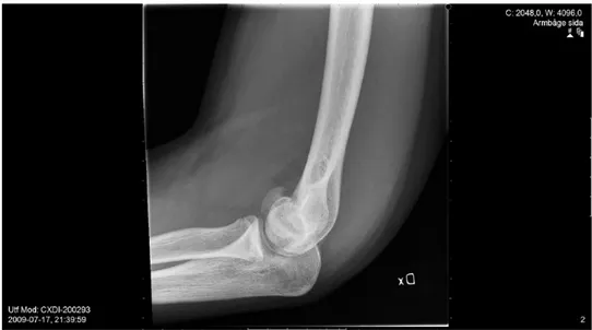
There are few reports describing results after this proce-dure and we have seen referred cases with iatrogenic frac-tures caused by aggressive manipulation under anaesthesia without previous surgical release (Fig. 1). If, however, the causes of a contracture are surgically removed, passive mobilization with gentle force is often beneficial at the end of the procedure. Araghi et al47 reported the outcome of series of 51 patients that had a mobilization under anaesthesia performed at a mean of 40 days after a previous open release and found the pro-cedure valuable. Ek et al48 reported the same experience having used the same procedure in a series of 12 paediat-ric patients.
Total or partial elbow arthroplasty
In elderly and low-demand patients with an elbow con-tracture associated with severe arthritic changes of the joint surfaces, a prosthetic replacement may be consid-ered.49-51 In such situations a complete soft-tissue release, ulnar nerve transposition and resection of joint surfaces as well as any impinging heterotopic bone is needed and a semi-constrained prosthesis is usually recommended. Anecdotal cases with hemiarthroplasty have been reported in younger patients with stable joints and relatively pre-served joint architecture but clinical results have to date been sparse.
Adjuvant therapies
Non-steroidal anti-inflammatory drugs such as celecoxib and indomethacin have been sparsely investigated in their role of preventing HO around the elbow. In a recent retro-spective study Sun et al52 found that administration of celecoxib for 28 days after open arthrolysis significantly reduced formation of HO. Costopoulos et al,53 also in a retrospective study on distal biceps repairs, found a sig-nificant reduction of HO in patients treated with indo-methacin post-operatively for ten to 42 days. Also, single-dose radiation therapy has been suggested and used both as prophylaxis following a trauma and after excision of manifest HO. The only randomized controlled study on acute injuries, by Hamid et al,54 was terminated before completion due to a high number of nonunions in patients receiving radiation therapy. The role of radiation as a post-operative adjunct following HO resection is con-troversial and although often recommended, there is a lack of evidence supporting its use.55
Fig 1. Iatrogenic coronal shear fracture caused by manipulation under anaesthesia without previous surgical release in a 16-year-old
Summary
Post-traumatic and post-operative stiffness of the elbow joint constitutes a significant problem since the elbow is prone to develop soft-tissue contractures and HO. Recent research has increased the knowledge of the biomechani-cal and biochemibiomechani-cal processes causing post-traumatic elbow stiffness but the exact mechanisms are still largely unknown. A large range of movement is essential for upper extremity function and restrictions may cause severe functional limitations. In patients with a pro-nounced limitation of movement, treatment may consist of physiotherapy and splinting in the early phase, while manifest contractures may require surgery. with soft- tissue contractures without extra-articular deformities or HO, arthroscopic release is often amenable but the tech-nique is demanding. Combinations of intrinsic and extrin-sic injuries such as bony abnormalities and associated injuries affecting nerves or skin are usually best addressed by an open approach, with or without adjunctive external fixation, and in general improvements of at least 50% of the movement loss can be expected (Table 1). The poten-tial effect of adjuvant therapies such as non-steroidal anti-inflammatory drugs and radiation are yet to be proven.
ICMJE ConflICt of IntErEst statEMEnt
L. Adolfsson declares payment for lectures for Acumed, DePuy/Johnson & Johnson, AO Foundation, Wright Medical and Swemac Education; royalties from Köigsee implantate GmBh, activities outside the submitted work.
fundIng statEMEnt
No benefits in any form have been received or will be received from a commercial party related directly or indirectly to the subject of this article.
lICEnCE
© 2018 The author(s)
This article is distributed under the terms of the Creative Commons Attribution-Non Commercial 4.0 International (CC BY-NC 4.0) licence (https://creativecommons.org/ licenses/by-nc/4.0/) which permits non-commercial use, reproduction and distribu-tion of the work without further permission provided the original work is attributed.
rEfErEnCEs
1. Jackson WM, aragon aB, Bulken-Hoover Jd, nesti lJ, tuan rs. Putative
heterotopic ossification progenitor cells derived from traumatized muscle. J Orthop Res 2009;27:1645-1651.
2. lounev VY, ramachandran r, Wosczyna Mn, et al. Identification of progenitor
cells that contribute to heterotopic skeletogenesis. J Bone Joint Surg [Am] 2009;91-A:652-663.
3. shore EM, Xu M, feldman gJ, et al. A recurrent mutation in the BMP type I
receptor ACVR1 causes inherited and sporadic fibrodysplasia ossificans progressiva. Nat
Genet 2006;38:525-527.
4. Monument MJ, Hart da, salo Pt, Befus ad, Hildebrand Ka. Posttraumatic
elbow contractures: targeting neuroinflammatory fibrogenic mechanisms. J Orthop Sci 2013;18:869-877.
5. davis El, davis ar, gugala Z, olmsted-davis Ea. Is heterotopic ossification
getting nervous?: the role of the peripheral nervous system in heterotopic ossification. Bone 2017;S8756-3282(17)30242-9.
6. abrams gd, Bellino MJ, Cheung EV. Risk factors for development of heterotopic
ossification of the elbow after fracture fixation. J Shoulder Elbow Surg 2012;21:1550-1554.
7. Bauer as, lawson BK, Bliss rl, dyer gsM. Risk factors for posttraumatic
heterotopic ossification of the elbow: case-control study. J Hand Surg Am 2012;37:1422-1429.
8. douglas K, Cannada lK, archer Kr, et al. Incidence and risk factors of heterotopic
ossification following major elbow trauma. Orthopedics 2012;35:e815-e822.
Procedure Preferred indication (as reported) Reported gain Comment
Splinting Soft-tissue contractures in early stage (6 mths) up to 40° Arthroscopic release Intrinsic contractures; capsular contracture,
arthrofibrosis, osteophytes and loose bodies 30° to 60° Not for extra-articular bony procedures but may be combined. Not in case of altered neurovascular anatomy
Open release Extrinsic and mixed contractures, heterotopic
bone formation excision 35° to 85° In late stages and when osteotomies or extra-articular procedures needed. Increased safety of neuro-vascular structures
Open release and external fixator when release of collateral ligaments has been
performed. Complete ankylosis 30° to 85° (116°) To ensure reduction and to protect ligament healing Distraction arthroplasty In combination with open release or isolated
for arthrofibrosis 30° to 90° Continuous passive movement Post-operative management after surgical
release Efficacy controversial
Manipulation under anaesthesia Peri-operative following surgical release Not recommended as a stand-alone procedure. Iatrogenous injuries reported
Interposition arthroplasty Pain relief in younger patients with secondary
osteoarthritis Up to 55° Primarily for pain reduction Total elbow arthroplasty For severe post-traumatic osteoarthritis in
low-demand patients Up to 90° Pain reduction and increased range of movement
autHor InforMatIon
Department of Orthopaedics, University Hospital of Linköping, Sweden. Correspondence should be sent to: L. Adolfsson, Department of Orthopaedics, University Hospital of Linköping, S-581 85 Linköping, Sweden.
POST-TRAUMATIC STIFF ELBOw
9. foruria aM, augustin s, Morrey Bf, sánchez-sotelo J. Heterotopic
ossification after surgery for fractures and fracture-dislocations involving the proximal aspect of the radius or ulna. J Bone Joint Surg [Am] 2013;95:e66, 1-7.
10. Mattyasovszky sg, Hofmann a, Brochhausen C, et al. The effect of the
pro-inflammatory cytokine tumor necrosis factor-alpha on human joint capsule myofibroblasts.
Arthritis Res Ther 2010;12:R4.
11. Hildebrand Ka. Posttraumatic elbow joint contractures: defining pathologic
capsular mechanisms and potential future treatment paradigms. J Hand Surg Am 2013;38:2227-2233.
12. Hildebrand Ka, Zhang M, Befus ad, salo Pt, Hart da. A myofibroblast-mast
cell-neuropeptide axis of fibrosis in post-traumatic joint contractures: an in vitro analysis of mechanistic components. J Orthop Res 2014;32:1290-1296.
13. Kopka M, Monument MJ, Befus ad, et al. Serum mast cell tryptase as a marker
of post-traumatic joint contracture in a rabbit model. J Orthop Trauma 2017;31:e86-e89.
14. Mu X, Bellayr I, Walters t, li Y. Mediators leading to fibrosis - how to measure
and control them in tissue engineering. Oper Tech Orthop 2010;20:110-118.
15. Vaughan MB, Howard EW, tomasek JJ. Transforming growth factor-beta1
promotes the morphological and functional differentiation of the myofibroblast. Exp Cell Res 2000;257:180-189.
16. Hildebrand Ka, Zhang M, Hart da. Myofibroblast upregulators are elevated in
joint capsules in posttraumatic contractures. Clin Orthop Relat Res 2007;456:85-91.
17. doornberg Jn, Bosse t, Cohen Ms, et al. Temporary presence of myofibroblasts
in human elbow capsule after trauma. J Bone Joint Surg [Am] 2014;96:e36, 1-8.
18. Morrey Bf. The posttraumatic stiff elbow. Clin Orthop Relat Res 2005;431:26-35. 19. Kay nr. Arthrolysis of the post-traumatic stiff elbow. In: Stanley D, ed. Surgery of the
elbow, K.N. London: Arnold, 1998:228-234.
20. shuai C, Hede Y, shen l, et al. Is routine ulnar nerve transposition necessary in
open release of stiff elbows? Our experience and a literature review. Int Orthop 2014;38:2289-2294.
21. Mellema JJ, lindenhovius al, Jupiter JB. The posttraumatic stiff elbow: an
update. Curr Rev Musculoskelet Med 2016;9:190-198.
22. doornberg Jn, ring d, Jupiter JB. Static progressive splinting for posttraumatic
elbow stiffness. J Orthop Trauma 2006;20:400-404.
23. lindenhovius al, doornberg Jn, Brouwer KM, et al. A prospective
randomized controlled trial of dynamic versus static progressive elbow splinting for posttraumatic elbow stiffness. J Bone Joint Surg [Am] 2012;94-A:694-700.
24. Veltman Es, doornberg Jn, Eygendaal d, van den Bekerom MP. Static
progressive versus dynamic splinting for posttraumatic elbow stiffness: a systematic review of 232 patients. Arch Orthop Trauma Surg 2015;135:613-617.
25. schwartz da. Static progressive orthoses for the upper extremity: a comprehensive
literature review. Hand (NY) 2012;7:10-17.
26. Müller aM, sadoghi P, lucas r, et al. Effectiveness of bracing in the treatment
of nonosseous restriction of elbow mobility: a systematic review and meta-analysis of 13 studies. J Shoulder Elbow Surg 2013;22:1146-1152.
27. o’driscoll sW, giori nJ. Continuous passive motion (CPM): theory and principles of
clinical application. J Rehabil Res Dev 2000;37:179-188.
28. lindenhovius al, van de luijtgaarden K, ring d, Jupiter J. Open elbow
contracture release: postoperative management with and without continuous passive
29. Higgs ZC, danks Ba, sibinski M, rymaszewski la. Outcomes of open
arthrolysis of the elbow without post-operative passive stretching. J Bone Joint Surg [Br] 2012;94-B:348-352.
30. Carpenter CV, amirfeyz r. Continuous passive motion following elbow arthrolysis.
J Hand Surg Am 2014;39:350-352.
31. Jones V. Conservative management of the post-traumatic stiff elbow: a
physiotherapist’s perspective. Shoulder Elbow 2016;8:134-141.
32. Kodde If, van rijn J, van den Bekerom MP, Eygendaal d. Surgical treatment
of post-traumatic elbow stiffness: a systematic review. J Shoulder Elbow Surg 2013;22:574-580.
33. Cai J, Wang W, Yan H, et al. Complications of open elbow arthrolysis in
post-traumatic elbow stiffness: a systematic review. PLoS One 2015;10:e0138547.
34. Mansat P, Morrey Bf. The column procedure: a limited lateral approach for
extrinsic contracture of the elbow. J Bone Joint Surg [Am] 1998;80-A:1603-1615.
35. gundlach u, Eygendaal d. Surgical treatment of posttraumatic stiffness of the
elbow: 2-year outcome in 21 patients after a column procedure. Acta Orthop 2008;79:74-77.
36. Kruse KK, Papatheodorou lK, Weiser rW, sotereanos dg. Release of the
stiff elbow with mini-open technique. J Shoulder Elbow Surg 2016;25:355-361.
37. Pettersen PM, Eriksson J, Bratberg H, et al. Increased ROM and high patient
satisfaction after open arthrolysis: a follow-up-study of 43 patients with posttraumatic stiff elbows. BMC Musculoskelet Disord 2016;17:74.
38. tucker sa, savoie fH III, o’Brien MJ. Arthroscopic management of the
post-traumatic stiff elbow. J Shoulder Elbow Surg 2011;20(suppl):S83-S89.
39. adams JE, King gJ, steinmann sP, Cohen Ms. Elbow arthroscopy: indications,
techniques, outcomes, and complications. J Am Acad Orthop Surg 2014;22:810-818.
40. Cefo I, Eygendaal d. Arthroscopic arthrolysis for posttraumatic elbow stiffness.
J Shoulder Elbow Surg 2011;20:434-439.
41. Zhou Y, Cai JY, Chen s, et al. Application of distal radius-positioned hinged
external fixator in complete open release for severe elbow stiffness. J Shoulder Elbow Surg 2017;26:e44-e51.
42. Pennig d, Heck s, Möhring r. External fixation with motion capacity and radius
fractures. Methods and results. Unfallchirurg 2011;114:105-113.
43. Wang J, li H, Zheng Q, et al. Distraction arthrolysis of posttraumatic elbow
stiffness with a hinged external fixator. Orthopedics 2012;35:e1625-e1630.
44. larson an, Morrey Bf. Interposition arthroplasty with an Achilles tendon allograft
as a salvage procedure for the elbow. J Bone Joint Surg [Am] 2008;90-A:2714-2723.
45. nandi s, Maschke s, Evans PJ, lawton Jn. The stiff elbow. Hand (NY)
2009;4:368-379.
46. Erşen a, demirhan M, atalar aC, salduz a, tunalı o. Stiff elbow: distraction
interposition arthroplasty with an Achilles tendon allograft: long-term radiological and functional results. Acta Orthop Traumatol Turc 2014;48:558-562.
47. araghi a, Celli a, adams r, Morrey B. The outcome of examination
(manipulation) under anesthesia on the stiff elbow after surgical contracture release. J
Shoulder Elbow Surg 2010;19:202-208.
48. Ek Et, Paul sK, Hotchkiss rn. Outcomes after operative treatment of elbow contractures
in the pediatric and adolescent population. J Shoulder Elbow Surg 2016;25:2066-2070.
49. Mansat P, Morrey Bf. Semiconstrained total elbow arthroplasty for ankylosed and
stiff elbows. J Bone Joint Surg [Am] 2000;82-A:1260-1268.
ankylosed or fused elbow. J Bone Joint Surg [Br] 2008;90-B:1198-1204.
52. sun Y, Cai J, li f, et al. The efficacy of celecoxib in preventing heterotopic
ossification recurrence after open arthrolysis for post-traumatic elbow stiffness in adults. J
Shoulder Elbow Surg 2015;24:1735-1740.
53. Costopoulos Cl, abboud Ja, ramsey Ml, et al. The use of indomethacin in the
prevention of postoperative radioulnar synostosis after distal biceps repair. J Shoulder Elbow
Surg 2017;26:295-298.
ossification prophylaxis acutely after elbow trauma: a prospective randomized study. J Bone
Joint Surg [Am] 2010;92-A:2032-2038.
55. Ploumis a, Belbasis l, ntzani E, tsekeris P, Xenakis t. Radiotherapy for
prevention of heterotopic ossification of the elbow: a systematic review of the literature. J
Shoulder Elbow Surg 2013;22:1580-1588.