*Corresponding author: Mikael Sonesson, Department of Orthodontics, Malmö University, Malmö, Sweden, Tel: +46 406658447; E-mail:
Mikael.Sonesson@mau.se
Citation: Sonesson M, Karlander EL, Astemo A, Lindh C (2018) Reliability of Assessments of Apical Root Resorption during Treatment with Removable Or-thodontic Appliance in Children. J Dent Oral Health Cosmesis 3: 010. Received: November 14, 2018; Accepted: December 07, 2018; Published: December 20, 2018
Copyright: © 2018 Sonesson M, et al. This is an open-access article distributed under the terms of the Creative Commons Attribution License, which permits un-restricted use, distribution, and reproduction in any medium, provided the original author and source are credited.
Introduction
External root resorption seems to be an inevitable adverse effect of the mechanical forces produced by orthodontic appliances [1,2]. The Swedish Council on Health Technology Assessment (SBU) in health care concluded in a previous systematic review on risks and compli-cations of orthodontic treatment that Apical Root Resorption (ARR) occurs in 97-100% of the patients and resorption up to one-third of the root length occurs in 11-28% of the patients [3]. The scientific evidence of the conclusions was however stated low due to study de-sign. Apical root resorption in patients with fixed appliances seems to aggravate during treatment in those patients who presented resorption early in treatment [4,5]. Thus, to identify patients at risk of more se-vere root resorption during treatment, an additional radiographic ex-amination after six months of treatment has been recommended [4]. There is limited numbers of investigations on children treated with removable orthodontic appliance and development of ARR compared to studies on adolescents treated with fixed appliance and ARR. In a retrospective study on children with removable functional appli-ance, approximately 9% of the patients had minor resorption of the maxillary incisors after treatment [6]. In another retrospective inves-tigation, apical root resorption was studied on patients treated with removable or a combination of fixed and removable appliance. Even though, treatment with fixed appliance showed significantly more apical root resorption than removable appliance, the latter caused mi-nor root resorption [7]. Janson et al. [8], investigated the amount of apical root resorption that occur after treatment with two removable
Dentistry: Oral Health & Cosmesis
Research Article
Mikael Sonesson1*, Eva Lilja Karlander1, Angelica Astemo2
and Christina Lindh3
1Department of Orthodontics, Malmö University, Malmö, Sweden 2National Health Service, County of Skåne, Sweden
3Department of Oral and Maxillofacial Radiology, Malmö University, Sweden
Reliability of Assessments of
Apical Root Resorption during
Treatment with Removable
Orthodontic Appliance in
Children
Abstract Aims
Apical Root Resorption (ARR) of the maxillary incisors is an ad-verse effect in patients during treatment with orthodontic appliance. Limited number of prospective investigations on ARR in patients with Removable Orthodontic Appliance (ROA) is available and the reliability of assessments of ARR in periapical radiographs has not been evaluated. The aims were to investigate 1) Observer agree-ment regarding assessagree-ment of ARR in radiographs obtained during treatment with ROA in children and 2) The extent and severity of ARR in children during treatment with ROA.
Material and Methods
One hundred ten children, with Angle class II malocclusion or an-terior cross bite suitable for treatment with ROA were consecutively recruited. Periapical radiographs of maxillary incisors were obtained before (T0) and after six month of treatment (T1). The contour of the roots was scored as 0, 1, 2, 3 or 4 according to a modified Levander and Malmgren index, by five observers. Score 0 represent normal root contour, score 4 severe root resorption. Inter- and intra-observer agreement was calculated as overall agreement and kappa values.
Results
In the 110 patients, 49 females, 61 males, the mean age was 10.6 years. Totally 439 roots were available for assessment. Kappa val-ues for pair wise inter-observer agreement during treatment ranged between -0.01 and 0.27. Kappa values for intra-observer agreement ranged between -0.09 and 0.45 before treatment and -0.22 and 0.48 after treatment.
Conclusion
Reliability in assessing root morphology/resorption in periapical radiographs obtained in children during treatment with removable appliance is low. ARR seems to be of minor extent, however, the low reliability indicates difficulties in evaluating the severity and thereby the clinical importance in this young patient group.
Keywords: Apical root resorption; Assessment reliability; Children;
• Page 2 of 7 •
J Dent Oral Health Cosmesis ISSN: 2473-6783, Open Access Journal DOI: 10.24966/DOHC-6783/100010
Volume 3 • Issue 1 • 100010 appliances and compared the amount of resorption with an untreated
control group. They found that apical root resorption was significant-ly more common in children treated with one of the removable appli-ances (Fränkel function regulator) compared to the controls. Never-theless, Apajalathi and Peltola [9], didn’t find any correlation between root resorption and treatment with removable appliances. Thus, avail-able results regarding treatment with removavail-able appliance and apical root resorption in children seems to be contradictory.
In most clinical studies, root resorption is assessed by means of periapical radiography of the maxillary incisors. This imaging meth-od has certain shortcomings regarding image distortion and tissue overlapping, even when efforts are made to ensure accurate technique concerning receptor positioning and tube angulations. Furthermore, irrespective if root resorption has been assessed during treatment with fixed or removable appliances, in many studies not more than one or two observers performed the assessments [6-10]. However, the validi-ty of an imaging method is dependent on both accuracy and reliabilivalidi-ty [11]. Investigating the agreement between and within observers pro-vides information about the amount of error inherent in a diagnosis or score and the observer agreement may represent an “upper boundary” for diagnostic accuracy efficacy [12].
The aim of this study was therefore to investigate observer agree-ment in the assessagree-ment of root resorption in periapical radiographs of upper maxillary incisors during treatment with removable orthodon-tic appliance in children. An additional aim was to investigate if any apical root resorption of maxillary incisors occurs and if it does the frequency and severity during six months of treatment.
Material and Methods
One hundred ten healthy children, (49 females, 61 males) with a high objective treatment need, referred to the department of Ortho-dontics at Malmö University, Sweden, were consecutively recruited during a two year period. The patients were suitable for treatment with; a) Removable appliance to correct an Angle class I malocclu-sion with large over jet (>6 mm with insufficient lip closure) or b) Anterior cross bite. The sample size was based on the occurrence of apical root resorption of clinical interest (>2 mm <one-third of the root length) reported in previous studies on patients with removable appliances [6,8]. Individuals with a history of known trauma to the anterior maxilla, nails biting and patients not cooperating from start were excluded. All guardians of the patients signed an informed con-sent form before the study and in addition, the patients had to give their oral consent to take part in the study. The Ethical Review Board in Lund, Sweden, approved the study (Dnr.2008/245).
The patients with the Angle class I malocclusion were treated with a van Beek activator with a buccal shield of acrylic covering the en-tire clinical crowns of the maxillary incisors. The children and their custodians were informed to use the activator for 12 to 14 hours a day [13]. The patients with anterior cross-bite were treated with an expan-sion plate equipped with buccal spring and protruexpan-sion elements on the lingual surfaces of the maxillary incisors. The participants were informed to use the plates for 22 to 24 hours a day. Treatment progres-sion and adherence to given instructions were checked every 4 to 6 weeks.
The Guidelines for Reporting Reliability and Agreement Studies (GRRAS) was used [12].
Radiographic Examination
Two or three periapical radiographs of the maxillary incisors were obtained at two occasions; before the start of treatment (T0) and fol-lowing treatment of six months (T1). The X-ray unit used was a Pro-style Intra Planmeca (Planmeca Oy, Helsinki, Finland) with exposure settings 60 kV and 16 mAs. A digital sensor (Planmeca ProSensor, Planmeca Oy, Helsinki, Finland) was used for image capture. Care was taken to place the sensor as parallel as possible to the incisor roots with the X-ray beam perpendicular to the long axis of the teeth as well as to the sensor (right-angle technique). The X-ray unit was equipped with a long spacer cone. Standard quality criteria for peri-apical radiography were used and, wherever possible, inadequate radiographs were repeated. All radiographic examinations were per-formed at the Department of Oral Radiology, Faculty of Odontology, Malmö University, Malmö, Sweden.
Assessment of Root Resorption
The two series (T0, T1) of radiographs from each patient were projected on a monitor (Hewlett Packard ProBook 6650b, Palo Alto CA, USA) in a dark room and interpreted under standardized con-ditions. Five observers independently assessed and scored the root contour and, when present, the degree of apical root resorption of the maxillary lateral and central incisors according to a modified index by Levander and Malmgren [14], score; 0 = intact root contour; 1 = irregular root contour; 2 = slight loss of dental tissues (<2 mm); 3 = root resorption (>2 mm <one-third of the root length); 4 = root resorp-tion (>one-third of the root length). When in doubt, the lower score was chosen. Two of the observers were specialists in orthodontics with professional experiences of six and 23 years respectively, two observers were specialists in oral and maxillofacial radiology with professional experiences of three and 26 years, respectively. Finally, one observer was a resident in oral and maxillofacial radiology. None of the observers was involved in the treatment of the patients. Prior to the assessment, the observers had a joint discussion and calibration on how to assess the radiographs. The assessment criteria were discussed and written down in a protocol that the observers had at hand during their assessments. To simulate the everyday clinical situation, serie 0 (T0) was primarily assessed followed by serie 1 (T1). The observers were blinded regarding type of the two appliances used in the treat-ments during the assesstreat-ments of the root resorption.
In order to calculate intra-observer agreement all observers as-sessed a random sample of 20 series of radiographs about four weeks after the first observation in order to minimize observer recall bias. Observer agreement was calculated in two ways; 1) for the exact scoring of 0 to 4 of all roots and 2) grouped scoring when the five scores (0 to 4) were cut-off and divided into two groups. One group comprised assessments of score 0, 1 and 2 representing regular/ir-regular contour or minor resorption. The other group, scores 3 and 4, representing severe and extreme resorption of the roots.
In addition, the individual observer assessment of progression of root resorption exceeding more than one score during treatment (T0 to T1), was calculated.
Statistical Evaluation
Inter and intra-observer agreement were calculated as overall agreement and Kappa value and interpreted according to Landis and
Volume 3 • Issue 1 • 100010 Koch [15,16]. The assessment of progression of root resorption
ex-ceeding more than one score during treatment, was calculated and presented as frequency of root resorption in percent and tooth catego-ry (tooth 12, 11, 21, 22).
Results
In the 110 children, recruited to the study, the mean age was 10.6 years at start of treatment (T0). Additional demographic characteris-tics and treatment information are presented in Table 1. All patients ad-hered to the given instructions how to use the appliances, and checked clinically to evaluate treatment progression. In total, 439 teeth in 110 patients were available for assessment (one patient had one missing lateral incisor) resulting in 2195 assessments for all observers togeth-er, including before start of treatment (T0) as well as during treatment (T1). For all observers together and all available sites (roots) for as-sessments there were 56 roots in 24 patients that were not measurable before start of treatment (T0) and the corresponding figures during treatment (T1) was 27 roots in 13 patients. These figures represent 2.5% and 1.2%, respectively, of the total number of available assess-ments (Table 2).
Before start of treatment (T0) score 0, 1, and 2 were observed in 96.2% of the assessments, when taking all observers assessments into account. One point four per cent was assessed as score 3 and no root was assessed as score 4. During treatment (T1), 93.4% of the roots were assessed as score 0, 1, and 2 and 5.5% as score 3 and 4 (Table 2).
Similar numbers of root resorptions were observed in patients during treatment with van Beek activator as well as in patients with expansion plate. Eight roots in seven patients were assessed as having score 3 by three of the five observers. Four of these patients were treated with van Beek activator and three with expansion plate. In one patient treated with van Beek activator one tooth were assessed as score 4 by two observers.
Observer Agreement Exact Scoring
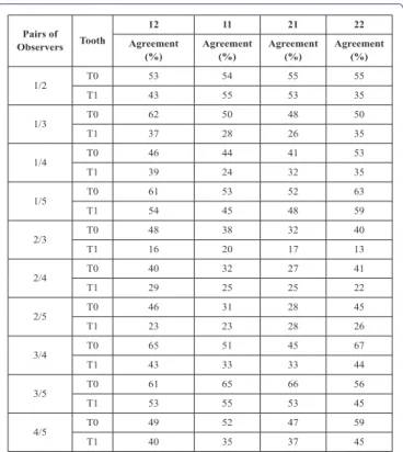
Pairwise inter-observer agreement for scoring 0, 1, 2, 3, and 4 varied between 27% and 67% before start of treatment (T0) and be-tween 13% and 59% during treatment (T1) (Table 3). Agreement was generally lower in the assessments during treatment (T1). Due to too few assessments of scores 3 and 4 it was not possible to calculate kappa values for pair wise inter-observer agreement before start of treatment (T0). Kappa values for pair wise inter-observer agreement during treatment (T1) varied between -0.01 and 0.27 interpreted as no to fair agreement [15].
Intra-observer agreement for scoring 0 to 4 varied between 30% and 80% before start of treatment (T0). The percentages during treat-ment (T1) varied between 37% and 75%. Kappa values ranged be-tween -0.09 and 0.45 (T0) and -0.22 and 0.48 (T1), interpreted as no to moderate agreement [15].
Observer Agreement Grouped Scoring
Pair wise observer agreement expressed as overall agreement in percentage was noticeably higher when the cut-off was made, varying
Mean Age (Range)
(T0) Mean Age (Range) (T0) Type of Appliance (n)
Female 10.7 (7.4 - 13.8) 11.4 (8.1 - 14.2) Van Beek Activator (31),Expansion plate (18) Male 10.4 (8.3 - 13.8) 11.1 (8.9 - 15.4) Van Beek Activator (38),Expansion plate (23) All 10.6 (7.4 - 13.8) 11.2 (8.1 - 15.4) Van Beek Activator (69),Expansion plate (41) Table 1: Gender and mean age before start of treatment (T0), during treatment (T1)
and type of removable appliance.
Tooth Assess-ment
Assessments According to Score 0 to 4 N= Number of Assessments (%)
Score 0
N (%) Score 1 N (%) Score 2 N (%) Score 3 N (%) Score 4 N (%) N.P.A.*
12 T0 292 (53.1) 190 (34.5) 53 (9.6) 2 (0.4) 0 (0.0) 13 (2.4) T1 153 (27.8) 237 (43.1) 131 (23.8) 19 (3.5) 1 (0.2) 9 (1.6) 11 T0 206 (37.5) 231 (42.0) 94 (17.1) 12 (2.2) 0 (0.0) 7 (1.3) T1 80 (14.5) 229 (41.6) 193 (35.1) 45 (8.2) 3 (0.5) 0 (0) 21 T0 202 (36.7) 238 (43.3) 84 (15.3) 16 (2.9) 0 (0) 10 (1.8) T1 76 (13.8) 225 (40.9) 206 (37.4) 32 (5.8) 3 (0.5) 8 (1.4) 22 T0 289 (52.5) 174 (31.6) 60 (10.9) 1 (0.2) 0 (0.0) 26 (4.2) T1 137 (24.9) 244 (44.3) 141 (25.6) 16 (2.9) 2 (0.4) 10 (1.8) Total T0 989 (45.0) 833 (37.9) 291 (13.3) 31 (1.4) 0 (0.0) 56 (2.5) T1 446 (20.3) 935 (42.5) 671 (30.6) 112 (5.1) 9 (0.4) 27 (1.2) Table 2: Inter observer agreement, all observers, presented as percentage of
agree-ment (%) for the assessagree-ment of apical root morphology/resorption in periapical ra-diography of maxillary incisors before start of treatment (T0) and during treatment (T1).
Note: *N.P.A. Not possible to assess
Pairs of Observers Tooth
12 11 21 22 Agreement
(%) Agreement (%) Agreement (%) Agreement (%)
1/2 T0 53 54 55 55 T1 43 55 53 35 1/3 T0 62 50 48 50 T1 37 28 26 35 1/4 T0 46 44 41 53 T1 39 24 32 35 1/5 T0 61 53 52 63 T1 54 45 48 59 2/3 T0 48 38 32 40 T1 16 20 17 13 2/4 T0 40 32 27 41 T1 29 25 25 22 2/5 T0 46 31 28 45 T1 23 23 28 26 3/4 T0 65 51 45 67 T1 43 33 33 44 3/5 T0 61 65 66 56 T1 53 55 53 45 4/5 T0 49 52 47 59 T1 40 35 37 45
Table 3: Pairwise inter observer agreement presented as percentage of agreement
(%) for the assessment of root morphology/resorption in periapical radiographs of the maxillary incisors before start of treatment (T0) and during treatment (T1). The scoring represents an exact score, 0 to 4.
• Page 4 of 7 •
J Dent Oral Health Cosmesis ISSN: 2473-6783, Open Access Journal DOI: 10.24966/DOHC-6783/100010
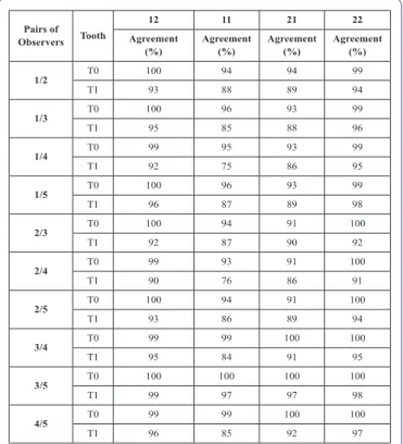
Volume 3 • Issue 1 • 100010 between 91% and 100% before start of treatment (T0) and between
75% and 98% during treatment (T1) (Table 4). Kappa values during treatment (T1) varied between -0.01 and 0.66, interpreted as no to substantial agreement [15].
During treatment (T1) the agreement values varied between 85% and 100% and the Kappa values between -0.18 and 1.0, interpreted as no to almost perfect [15].
It was not possible to calculate kappa values for all observer pairs prior to treatment or intra-observer agreement in percentage and kap-pa values before start of treatment (T0), due to too few assessments in the group at scores 3 and 4.
The individual observers’ assessments of root resorption exceed-ing more than one score before start of treatment (T0) and durexceed-ing treatment (T1) varied between 2% and 23% for tooth 12, between 5% and 25% for tooth 11, between 4% and 24% for tooth 21 and finally between 3% and 29% for tooth 22. One observer assessed the change in root morphology to be noticeably more than the other observers. Another observer assessed this change to be noticeably less than the other observers did.
Discussion
The overall finding of the present multi-observer study on api-cal root resorption assessed in periapiapi-cal radiographs performed before and during treatment with removable orthodontic appliance, demonstrated that inter- and intra-observer agreement varied truly.
The rational for the five-observer performance approach was to create conditions similar to an everyday clinic situation, thus the observ-ers represented different professional experiences as well as differ-ent time lengths of expertise. Inter-observer overall agreemdiffer-ent varied substantially, even though the observers jointly discussed and wrote down assessment criteria to serve as a calibration (Table 2). Special-ly, pair wise inter-observer agreement for exact scoring was low (no agreement to fair agreement). The highest disagreement was found between one senior oral-and maxillofacial radiologist and one senior orthodontist (observer 2/3) (Table 3). This might be explained by the different professional experience and different expectations to detect apical root resorptions. The largest difference could also be seen be-tween the same two observers in assessment of progression of root resorption.
The intra-observer agreement was low when using the five exact scores (0, 1, 2, 3, and 4) to assess apical root resorptions, score 1 and 2 were especially difficult to assess. It is important to remember that radiography is an imprecise tool for detection of small differences in tissue loss, which might be one explanation for the low agreement. However, the higher intra-observer agreement when dividing the five scores into the two groups (scores 0 to 2 and scores 3, 4), suggests that the index used in this study might be even further modified when root resorptions are less pronounced in the population. The higher intra-observer agreement when dividing the five scores into the two groups indicate however that no apical root resorption of major clin-ical importance of the maxillary incisors occurred, as no single tooth was assessed by all five observers in agreement as having severe api-cal root resorption (scores 3 or 4) during treatment.
When assessing root morphology/resorption the diagnostic effica-cy of periapical radiographs is dependent both on accuraeffica-cy and ob-server performance [11]. In this study, using periapical radiographs to assess root resorption during treatment, it was not possible to obtain any reference standard. However, the reliability estimations are use-ful in determining the extent to which the inaccuracy of a diagnostic method is due to decision-making errors. Unfortunately, reliability studies are generally neglected and do not appear in the different stag-es of evaluating studistag-es of diagnostic methods [16] or in studistag-es where diagnostic methods are used to evaluate treatment outcomes [17]. Other radiological methods like Cone Beam Computed Tomography (CBCT) may result in more reliable assessments, however, this meth-od cannot in the current situation be justified due to higher radiation dosage.
The clinical consequences of root resorption can be discussed [18]. It has been stated that the degree of root loss in most situations is clinically insignificant while others believed that roots with severe resorption could be at risk for tooth mobility or never resist the stress of function as well as un-resorbed roots [19,20]. Mohandesan et al. [10], used external apical root resorption of 1 mm at 12 months of active treatment period as the cut-off to determine the clinical impor-tance of root resorption. In present study we have assumed that loss of root tissue exceeding 2 mm (scores 3 and 4) is of clinical importance in the way that it might change how the treatment will continue. Several studies on root resorption during treatment with remov-able orthodontic appliance are performed retrospectively by limited numbers of observers [6-9]. The patient history might then be un-known as well as that the image quality might not have been carefully taken into account when the radiographs were obtained. The strength
Pairs of Observers Tooth
12 11 21 22 Agreement
(%) Agreement (%) Agreement (%) Agreement (%) 1/2 T0 100 94 94 99 T1 93 88 89 94 1/3 T0 100 96 93 99 T1 95 85 88 96 1/4 T0 99 95 93 99 T1 92 75 86 95 1/5 T0 100 96 93 99 T1 96 87 89 98 2/3 T0 100 94 91 100 T1 92 87 90 92 2/4 T0 99 93 91 100 T1 90 76 86 91 2/5 T0 100 94 91 100 T1 93 86 89 94 3/4 T0 99 99 100 100 T1 95 84 91 95 3/5 T0 100 100 100 100 T1 99 97 97 98 4/5 T0 99 99 100 100 T1 96 85 92 97
Table 4: Pairwise inter observer agreement presented as percentage of agreement
(%) for the assessment of root morphology/resorption in periapical radiographs of the maxillary incisors before start of treatment (T0) and during treatment (T1). The scoring 1 to 5 were grouped into two groups of scores, 1 to 3 and scores 4 and 5. 1: Observer 1; 2: Observer 2; 3: Observer 3; 4: Observer 4; 5: Observer 5.
Volume 3 • Issue 1 • 100010 with a prospective study is that the diagnostic quality of the
radio-graphs will be under control with optimal vertical angulation to depict the apex clearly, a prerequisite for assessment of root contours and resorption. Despite the ambition to obtain images of optimal quality in this study, for some roots the apical part was not possible to assess (2.5% at T0 and 1.2% at T1). A possible explanation is that in some patients the apical part of the root of a maxillary incisor is curved in the bucko-palatinal direction and consequently blurred in the im-age. Treatment with removable appliance results in more intermittent loading on the maxillary incisors compared to treatment with fixed appliance [6]. In present study two different types of removable ap-pliances was used, the expansion plate and the van Beek activator. The plate was provided with active protrusive elements tipping the anterior incisors in a labial direction.
The activator, equipped with headgear, force together with the muscle tension transferred to maxillary teeth the incisors in palatal direction. In both types of appliances, the size of the forces on the maxillary incisors was unknown. The force of the springs of the pansion plate will certainly decrease after a couple of weeks, thus ex-posing less compression of the periodontal membrane. The intermit-tent loading created by the appliances might cause less damage to the most compressed areas of the periodontal membrane. Applications with less force create smaller hyaline zones and a more favorable en-vironment for bone remodeling without involving root damages [21]. These intermissions in loading might explain why apical root resorp-tion was less evident in the present study compared with studies on fixed appliances. An interesting finding was also that similar numbers of apical root resorptions were assessed, by three of the five observ-ers, in patients treated with the van Beek activator and the expansion plate. Thus, the direction of the forces, palatal or labial, might be of minor importance. An additional explanation for the low occurrence of apical root resorption in the present study might be that the roots of children have wider apical foramina of the maxillary incisors with ar-eas of high vascularity, which prevent damages of the roots compared to adolescents, having a more developed and narrow apical foramina [22,23]. Factors as gender, age, habits, trauma and certain ethnicities as predispose factors for root resorption could be interesting to inves-tigate, but was out of topic of the present study. These factors have been highlighted in some previous studies on fixed appliance [23-27].
Conclusion
Reliability in assessing root morphology/resorption in periapical radiographs obtained in children during treatment with removable ap-pliance is low. ARR seems to be of minor extent; however, the low reliability indicates difficulties in evaluating the severity and there by the clinical importance in this young patient group.
Funding
This survey was funded by the authors’ academic institution.
Acknowledgement
The authors are grateful to Drs. Kristina Hellén-Halme and Fariba Boustanchi at the Faculty of Odontology, Malmö University, Malmö,
Sweden for acting as observers. The authors declare no competing interestst.
References
1. Reitan K (1974) Initial tissue behavior during apical root resorption. Angle Orthod 44: 68-82.
2. Artun J, Van´t Hullenaar R, Doppel D, Kuijpers-Jagtman AM (2009) Iden-tification of orthodontic patients at risk of severe apical root resorption. Am J Orthod Dentofacial Orthop 135: 448-455.
3. Swedish Council on Health Technology Assessment (2005) Malocclusions and orthodontic treatment in a health perspective: A systematic review. SBU Systematic Review Summaries SBU Yellow Report No. 176. 4. Levander E, Bajka R, Malmgren O (1998) Early radiographic diagnosis of
apical root resorption during orthodontic treatment: A study of maxillary incisors. Eur J Orthod 20: 57-63.
5. Artun J, Smale I, Behbehani F, Doppel D, Van’t Hof M, et al. (2005) Api-cal root resorption six and 12 months after initiation of fixed orthodontic appliance therapy. Angle Orthod 75: 919-926.
6. Högberg M, Rosenqvist L (1974) Root resorption following activator treatment. Odontol Foren Tidskr 38: 185-192.
7. Linge BO, Linge L (1983) Apical root resorption in upper anterior teeth. Eur J Orthod 5: 173-183.
8. Janson G, Nakamura A, de Freitas MR, Henriques JF, Pinzan A (2007) Apical root resorption comparison between fränkel and eruption guidance appliances. Am J Orthod Dentofacial Orthop 131: 729-735.
9. Apajalahti S, Peltola JS (2007) Apical root resorption after orthodontic treatment -- a retrospective study. Eur J Orthod 29: 408-412.
10. Mohandesan H, Ravanmehr H, Valaei N (2007) A radiographic analysis of external apical root resorption of maxillary incisors during active ortho-dontic treatment. Eur J Orthod 29: 134-139.
11. Fryback DG, Thornbury JR (1991) The efficacy of diagnostic imaging. Med Decis Making 11: 88-94.
12. Kottner J, Audige L, Brorson S, Donner A, Gajewski BJ, et al. (2011) Guidelines for Reporting Reliability and Agreement Studies (GRRAS) were proposed. Int J Nurs Stud 48: 661-671.
13. van Beek H (1984) Combination headgear-activator. J Clin Orthod 18: 185-189.
14. Levander E, Malmgren O (1988) Evaluation of the risk of root resorption during orthodontic treatment: A Study of upper incisors. Eur J Orthod 10: 30-38.
15. Landis JR, Koch CG (1977) The measurement of observer agreement for categorical data. Biometrics 33: 159-174.
16. Cohen J (1960) A coefficient of agreement for nominal scales. Educ Psy-chol Measurment 20: 37-46.
17. Swets JA, Pickett RM (1982) Evaluation of Diagnostic Systems: Methods from Signal Detection Theory. Academic Press, New York, USA. 18. Copeland S, Green LJ (1986) Root resorption in maxillary central incisors
following active orthodontic treatment. Am J Orthod 89: 51-55.
19. Phillips JR (1955) Apical root resorption under orthodontic therapy. Angle Orthod 25: 1-22.
20. Levander E, Malmgren O (2000) Long-term follow-up of maxillary inci-sors with severe apical root resorption. Eur J Orthod 22: 85-92.
• Page 6 of 7 •
J Dent Oral Health Cosmesis ISSN: 2473-6783, Open Access Journal DOI: 10.24966/DOHC-6783/100010
Volume 3 • Issue 1 • 100010
21. Roscoe MG, Meira JB, Cattaneo PM (2015) Association of orthodontic force system and root resorption: A systematic review. Am J Orthod Den-tofacial Orthop 147: 610-626.
22. Lilja E (1983) Tissue reactions following different orthodontic forces in rat and man. Department of Oral Pathology, School of Dentistry, Karolinska Institutet, Huddinge, Sweden.
23. Mavragani M, Vergari A, Selliseth NJ, Bøe OE , Wisth PJ (2000) A radio-graphic comparison of apical root resorption after orthodontic treatment with a standard edgewise and a straight-wire edgewise technique. Eur J Orthod 22: 665-674.
24. Odenrick L, Brattström V (1983) The effect of nailbiting on root resorption during orthodontic treatment. Eur J Orthod 5: 185-188.
25. Malmgren O, Goldson L, Hill C, Orwin A, Petrini L, et al. (1982) Root resorption after orthodontic treatment of traumatized teeth. Am J Orthod 82: 487-491.
26. Linge L, Linge BO (1991) Patient characteristics and treatment variables associated with apical root resorption during orthodontic treatment. Am J Orthod Dentofacial Orthop 99: 35-43.
27. Sameshima GT, Sinclair PM (2001) Predicting and preventing root re-sorption: Part I. Diagnostic factors. Am J Orthod Dentofacial Orthop 119: 505-510.
Herald Scholarly Open Access, 2561 Cornelia Rd, #205, Herndon, VA 20171, USA.
Submit Your Manuscript: http://www.heraldopenaccess.us/Online-Submission.php
Journal of Agronomy & Agricultural ScienceJournal of AIDS Clinical Research & STDs
Journal of Alcoholism, Drug Abuse & Substance Dependence Journal of Allergy Disorders & Therapy
Journal of Alternative, Complementary & Integrative Medicine Journal of Alzheimer’s & Neurodegenerative Diseases Journal of Angiology & Vascular Surgery
Journal of Animal Research & Veterinary Science Archives of Zoological Studies
Archives of Urology
Journal of Atmospheric & Earth-Sciences Journal of Aquaculture & Fisheries
Journal of Biotech Research & Biochemistry Journal of Brain & Neuroscience Research Journal of Cancer Biology & Treatment Journal of Cardiology: Study & Research Journal of Cell Biology & Cell Metabolism Journal of Clinical Dermatology & Therapy Journal of Clinical Immunology & Immunotherapy Journal of Clinical Studies & Medical Case Reports Journal of Community Medicine & Public Health Care Current Trends: Medical & Biological Engineering Journal of Cytology & Tissue Biology
Journal of Dentistry: Oral Health & Cosmesis Journal of Diabetes & Metabolic Disorders Journal of Dairy Research & Technology
Journal of Emergency Medicine Trauma & Surgical Care Journal of Environmental Science: Current Research Journal of Food Science & Nutrition
Journal of Forensic, Legal & Investigative Sciences Journal of Gastroenterology & Hepatology Research Journal of Gerontology & Geriatric Medicine
Journal of Internal Medicine & Primary Healthcare Journal of Infectious & Non Infectious Diseases Journal of Light & Laser: Current Trends Journal of Modern Chemical Sciences Journal of Medicine: Study & Research
Journal of Nanotechnology: Nanomedicine & Nanobiotechnology Journal of Neonatology & Clinical Pediatrics
Journal of Nephrology & Renal Therapy Journal of Non Invasive Vascular Investigation
Journal of Nuclear Medicine, Radiology & Radiation Therapy Journal of Obesity & Weight Loss
Journal of Orthopedic Research & Physiotherapy Journal of Otolaryngology, Head & Neck Surgery Journal of Protein Research & Bioinformatics Journal of Pathology Clinical & Medical Research
Journal of Pharmacology, Pharmaceutics & Pharmacovigilance Journal of Physical Medicine, Rehabilitation & Disabilities Journal of Plant Science: Current Research
Journal of Psychiatry, Depression & Anxiety
Journal of Pulmonary Medicine & Respiratory Research Journal of Practical & Professional Nursing
Journal of Reproductive Medicine, Gynaecology & Obstetrics Journal of Stem Cells Research, Development & Therapy Journal of Surgery: Current Trends & Innovations Journal of Toxicology: Current Research Journal of Translational Science and Research Trends in Anatomy & Physiology
Journal of Vaccines Research & Vaccination Journal of Virology & Antivirals
Archives of Surgery and Surgical Education Sports Medicine and Injury Care Journal