Synthesis and investigation of
bacterial effector molecules
Michael Franz Albers
Doctoral Thesis, Department of Chemistry Umeå University, 2016
Responsible publisher under swedish law: the Dean of the Faculty of Science and Technology
This work is protected by the Swedish Copyright Legislation (Act 1960:729) ISBN: 978-91-7601-411-0
Electronic version available at http://umu.diva-portal.org/ Tryck/Printed by: VMC-KBC Umeå
Table of Contents
Table of Contents i
Abstract iii
List of Abbreviations iv
List of Publications vii
Author contributions viii
Papers by the author, but not included in this thesis viii
Enkel sammanfattning på svenska ix
Introduction 1
Post-translational modifications 1
Nucleotidylylation and phosphocholination 2
Small GTPases 7
Pathogens modify host cells at a molecular level 9
Quorum sensing in Legionella pneumophila 12
Proteomics towards PTMs 13
Chapter 1: Towards the identification of adenylylated proteins and
adenylylation-modifying enzymes (Paper I – III) 19
Previous work 19
Outline: From building blocks to antibodies 22
Synthesis of a tyrosine-AMP building block 23
Synthesis of a threonine- and serine-AMP building block 26
Synthesis of adenylylated Peptides 28
Generation of AMP specific antibodies 30
Mass fragmentation patterns of adenylylated peptides 37 Immunoprecipitation of adenylylated proteins 42 Non-hydrolysable mimics for the study of deadenylylating enzymes 47
Future work 52
Ongoing Work – Covalent trapping of substrates of adenylyl transferases 53
Conclusions 60
Chapter 2: Tools for the investigation of phosphocholination
(Paper IV – V) 61
Outline 61
Synthesis of PC building blocks 61
Synthesis of phosphocholinated peptides 63
Site-directed chemoenzymatic labelling 64
Future work 65
Conclusions 66
Chapter 3: Small molecule signalling of Legionella pneumophila
(Paper VI – VII) 67
Introduction 67
LAI-1 regulates Lqs-dependent signalling 69 Inter-kingdom signalling 72 Future work 74 Conclusions 76 Acknowledgements 77 References 79 Appendix 93
Chapter 1: General procedure for immunoprecipitation experiments 93 Chapter 1: Synthesis of non-hydrolysable mimics of adenylylated motifs 97 Chapter 1: Organic synthesis of ATP and NAD+ derivatives 101
Abstract
During infections, bacterial microorganisms initiate profound interactions with mammalian host cells. Usually defense mechanisms of the host destroy intruding bacteria in rapid manner. However, many bacterial pathogens have evolved in a way to avoid these mechanisms. By use of effector molecules, which can be small organic molecules or proteins with enzymatic activity, the host is manipulated on a molecular level. Effectors mediating post-translational modifications (PTMs) are employed by many pathogens to influence the biological activity of host proteins. In the presented thesis, two related PTMs are investigated in detail: Adenylylation, the covalent transfer of an adenosine monophosphate group from adenosine triphosphate onto proteins, and phosphocholination, the covalent transfer of a phosphocholine moiety onto proteins. Over the past years, enzymes mediating these modifications have been discovered in several pathogens, especially as a mechanism to influence the signaling of eukaryotic cells by adenylylating or phosphocholinating small GTPases. However, the development of reliable methods for the isolation and identification of adenylylated and phosphocholinated proteins remains a vehement challenge in this field of research.
This thesis presents general procedures for the synthesis of peptides carrying adenylylated or phosphocholinated tyrosine, threonine and serine residues. From the resulting peptides, mono-selective polyclonal antibodies against adenylylated tyrosine and threonine have been raised. The antibodies were used as tools for proteomic research to isolate unknown substrates of adenylyl transferases from eukaryotic cells. Mass spectrometric fragmentation techniques have been investigated to ease the identification of adenylylated proteins. Furthermore, this work presents a new strategy to identify adenylylated proteins. ATP-derivatives containing an electrophilic trap were used in conjunction with modified adenylyl transferases, to isolate and analyse the covalent complex between enzyme and protein substrate.
Furthermore, small effector molecules are involved in the regulation of infection mechanisms. In this work, the small molecule LAI-1 (Legionella autoinducer 1) from the pathogen Legionella pneumophila, the causative agent of the Legionnaire’s disease, was synthesised together with its amino-derivatives. LAI-1 showed are a clear pharmacological effect on the regulation of the life cycle of L. pneumophila, initiating transmissive traits like motility and virulence. Furthermore, LAI-1 was shown to have an effect on eukaryotic cells as well. Directed motility of the eukaryotic cells was significantly reduced and the cytoskeletal architecture was reorganised, probably by interfering with the small GTPase Cdc42.
List of Abbreviations
A adenine
aa amino acid
ab antibody
ABC ammonium bicarbonate ADP adenosine diphosphate AMP adenosine monophosphate
ARTD diphtheria toxin-like ADP-ribosyl transferase Atase adenylyl transferase
ATP adenosine triphosphate BCA bicinchoninic acid BSA bovine serum albumin BTT 5-benzylthio-1H-tetrazole
Bz benzoyl
CAI-1 Cholerae autoinducer-1
cAMP cyclic adenosine monophosphate CDP cytidine diphosphate
CID collision induced dissociation CMP cytidine monophosphate
CoA Coenzyme A
DAPI 4’,6-diamidino-2-phenylindole
DCM dichloromethane
DEAE diethylethanolamine
DIAD diisopropyl azodicarboxylate DMAD dimethyl azodicarboxylate DMF dimethyl formamide DNA deoxyribonucleic acid DIPEA diisopropyl ethyl amine DTT dithiothreitol
EDTA ethylene diamine tetraacetate
eq equivalent
ER endoplasmic reticulum ETD electron transfer dissociation
FACE filter aided antibody capturing and elution FASP filter assisted sample preparation
Fic filamentation induced by cAMP GAP GTPase activating protein GDI GTP dissociation inhibitor GDP guanosine diphosphate
GEF guanosine nucleotide exchange factor GFP green fluorescent protein
GlcNAc N-acetylglucosamine GPI glycosylphosphatidylinositol GS glutamine synthetase GTP guanosine triphosphate
HATU 1-[bis(dimethylamino)methylene]-1H-1,2,3-triazolo[4,5-b]pyridinium 3-oxid hexafluorophosphate
HBTU 2-(1H-benzotriazol-1-yl)-1,1,3,3-tetramethyluronium hexafluorophosphate
HCD high-energy collisional dissociation HOAt 1-hydroxy-7-azabenzotriazole HOBt hydroxybenzotriazole
HPLC high-performance liquid chromatography HRMS high-resolution mass spectrometry HRP horseradish peroxidase
HYPE Huntingtin interacting protein E IEF iso-electronic focussing
IMAC immobilised metal-ion affinity chromatography IMPRS International Max Planck Research School
IP immunoprecipitation
KLH keyhole limpet hemocyanin
KNT kanamycin nucleotidylyl transferase LAI-1 Legionella autoinducer 1
LAMP lysosome-associated membrane glycoprotein LCMS liquid chromatography-mass spectrometry LCV Legionella-containing vacuole
Lqs Legionella quorum sensing
MBS 3-maleimidobenzoyl-N-hydroxysuccinimide ester MICAL molecules interacting with CasL
MS mass spectrometry
MWCO molecular weight cut-off
NAD+ nicotinamide adenine dinucleotide
NADH nicotinamide adenine dinucleotide, reduced nhAd non-hydrolysable adenosyl sulfonamide NHS N-hydroxysuccinimide
NMN nicotinamide mononucleotide NMR nuclear magnetic resonance P4M phosphatidylinositol-4-phosphate PAC polyaminocarboxylate
PAGE polyacrylamide gel electrophoresis PBS phosphate buffered saline
Pi inorganic phosphate POI protein of interest
PPM metal-dependent protein phosphatase PTM post-translational modification PVDF polyvinylidene fluoride
PyBOP benzotriazol-1-yl-oxytripyrrolidinophosphonium hexafluorophosphate
Q-TOF quadrupole-time-of-flight RIPA radioimmunoprecipitation assay Rf retention factor
RNA ribonucleic acid
rt room temperature
SAB (S)-2-amino-butyrate SDS sodium dodecyl sulfate
SEC size-exclusion chromatography SG side chain group
SPPS solid-phase peptide synthesis STAGE stop-and-go extraction
TBAF tetra-n-butylammonium fluoride TBDPS tert-butyldiphenylsilyl
TBHP tert-butylhydroperoxide TBME tert-butylmethyl ether TBS tris buffered saline TCA trichloroacetic acid
TCEP tris(2-carboxyethyl)phosphine TFA trifluoroacetic acid
THF tetrahydrofuran TIPS triisopropylsilane
TLC thin-layer chromatography TMP trimethyl phosphate tRNA transfer RNA UTP uridine triphosphate
List of Publications
The following publications form the basis of this dissertation. In text, the publications are referred to by use of the corresponding roman numbers.
I. Smit, C., Blümer J., Eerland, M. F., Albers, M. F., Müller, M. P., Goody, R. S., Itzen, A., & Hedberg, C. Efficient synthesis and applications of peptides containing adenylylated tyrosine residues. Angew. Chem. Int. Ed. 2011, 50, 9200–9204.
II. Albers, M. F., van Vliet, B. & Hedberg, C. Amino acid building
blocks for efficient Fmoc solid-phase synthesis of peptides adenylylated at serine or threonine. Org. Lett. 2011, 13, 6014–7. III. Hansen, T., Albers, M., Hedberg, C. & Sickmann, A. Adenylylation,
MS, and proteomics - Introducing a ‘new’ modification to bottom-up proteomics. Proteomics 2013, 13, 955–963.
IV. Albers, M. F. & Hedberg, C. Amino acid building blocks for Fmoc
solid-phase synthesis of peptides phosphocholinated at serine, threonine, and tyrosine. J. Org. Chem. 2013, 78, 2715–2719.
V. Heller, K., Ochtrop, P., Albers, M. F., Zauner, F. B., Itzen, A. & Hedberg, C. et al. Covalent protein labeling by enzymatic phosphocholination. Angew. Chem. Int. Ed. 2015, 54, 10327–10330. VI. Schell, U., Simon, S., Sahr, T., Hager, D., Albers, M. F., Kessler, A.,
Fahrnbauer, F., Trauner, D., Hedberg, C., Buchrieser, C. & Hilbi, H. The α-hydroxyketone LAI-1 regulates motility, Lqs-dependent phosphorylation signalling and gene expression of Legionella pneumophila. Mol. Microbiol. 2015, doi:10.1111/mmi.13265.
VII. Simon, S., Schell, U., Heuer, N., Hager, D., Albers, M. F., Matthias, J., Fahrnbauer, F., Trauner, D., Eichinger, L., Hedberg, C. & Hilbi, H. Inter-kingdom signaling by the Legionella quorum sensing molecule LAI-1 modulates cell migration through an IQGAP1-Cdc42-ARHGEF9-dependent pathway. PLOS Pathog. 2015, 11(12), e1005307 (2015).
All papers have been reprinted with the permission from the corresponding publisher.
Author contributions
Paper I: Contributed to organic synthesis of the tyrosine-AMP building block, synthesis of the adenylylated and non-adenylylated peptides, synthesis and analysis of the BSA-peptide conjugates and immunosorbent assay, organic synthesis of biotin-PEG-NHS and biotin-PEG-antibody conjugate, minor writing and translation of the paper for Angewandte Chemie, German edition.
Paper II: All of the organic synthesis of the threonine- and serine-AMP building blocks, synthesis of adenylylated and non-adenylylated peptides, writing.
Paper III: Synthesis of adenylylated and non-adenylylated peptides as reference material for the MS experiments.
Paper IV: All of the organic synthesis of the tyrosine-, threonine- and serine-PC building block, synthesis of phosphocholinated and non-phosphocholinated peptides, writing.
Paper V: Synthesis of the phosphocholinated peptides, minor writing, contribution to translation of the paper for Angewandte Chemie, German edition.
Paper VI: Organic synthesis of LAI-1 and Am-LAI-1.
Paper VII: Organic synthesis of LAI-1 and Am-LAI-1.
Papers by the author, but not included in this thesis
(*) Müller, M. P., Albers, M. F., Itzen, A. & Hedberg, C. Exploring adenylylation and phosphocholination as post-translational
Enkel sammanfattning på svenska
Vid infektion av humana celler initierar bakteriella mikroorganismer en mängd interaktioner och modifikationer på molekylär nivå. Vanligen förstör värdcellen invaderande bakterier kvickt och effektivt, men vissa bakterier har vänt cellens försvarssystem till sin egen fördel. Bakterierna utnyttjar effektormolekyler, vilka kan vara små sekundära metaboliter, eller proteiner med enzymatisk aktivitet, som pumpas in i värdcellen och påverkar direkt dess kemi och signalvägar. Effektor-proteiner som medierar posttranslationella modifieringar på värdcellens proteiner utnyttjas av många patogener. I den här avhandlingen undersöks den detaljerade kemin runt adenylylering och fosfokolinering, vilka är två av dessa posttranslationella modifieringar. Adenylylering består i kovalent fastsättande av adenosin monofosfat (AMP) på värdcellens proteiner, medan fosfokolinering innebär kovalent transfer av fosfokolin (PC) till proteiner på serin, threonin och tyrosin. Under de senaste åren har enzymer som katalyserar ovanstående reaktioner identifierats från ett antal obligata intracellulära bakterier. Deras huvudsyfte är att under infektionscykeln påverka värdcellens signalvägar genom att adenylylera och fosfokolinera små GTPaser. Att effektivt kunna anrika och isolera dessa modifierade proteiner är en stor utmaning och en nödvändighet för att tränga djupare in i mekanismerna bakom intracellulär infektion, vilket har varit huvudsyftet med avhandlingsarbetet.
Här presenteras generella metoder för syntes av adenylylerade och fosfokolinerade peptider modifierade på serin, threonin och tyrosin. Från dessa peptider har mono-selektiva polyklonala antikroppar tagits fram genom immunisering i kanin, följt av affinitetsupprening. Dessa antikroppar har sedan använts för att detektera och isolera adenylylerade proteiner. Vidare har peptiderna använts som referensmaterial för att utveckla selektiva masspektrometrimetoder för adenylylerade proteiner. Detta arbete innehåller också en ny metod för att detektera substrat för adenylyltransferaser som bygger på kovalent fastsättande av en ATP-analog i transferaset via en elektrofil sidokedja. Detta är första exemplet där ett transferas konverteras till ett kemiskt trappingreagens.
Även små molekyler från bakterien påverkar värdcellen. Ett exempel är LAI-1 från Legionella pneumophila, vilken har syntetiserats som (R) och (S) enantiomerer inom ramen för detta arbete, så även dess biosyntetiska prekursor amino-LAI-1. Dessa molekyler visar effekt på Legionellabakteriens livscykel och påverkar dess mobilitet och virulens. I tillägg har effekten av LAI-1 med enantiomerer och prekursormolekyler undersökts i avseende på värdcellen. Här finns det en klart påvisbar effekt på cytoskelettet via Ccd42. Den exakta mekanismen är fortsatt under utredande.
Introduction
Post-translational modifications
The central dogma of molecular biology, as stated by Francis Crick, is that for the biosynthesis of proteins, DNA is transcribed into RNA, which is subsequently translated into the amino acid sequence composing a given protein (scheme 1).[1,2] While this admittedly over-simplified statement terminates the protein biosynthesis at this point, we know nowadays that most proteins undergo a maturing process after (or in some cases during) the translation of RNA into the protein. A major role in the maturing process play post-translational modifications (PTMs) that are often essential for the protein to fulfil its physiological function in the cell. These modifications are usually introduced by catalytic reactions of other enzymes, but can also originate from other environmental influences.[3]
Scheme 1. Updated central dogma of molecular biology. The solid lines indicate information transfer between biopolymers that occurs in all living cells.[2] Dashed lines show specialised
transfer found in some organisms. For the biosynthesis of proteins, DNA is transcribed into RNA, which is translated into the corresponding proteins. Once a protein is synthesised, it cannot be reversed into the RNA or DNA sequence anymore. While the original concept ends after the translation, it is now known that most proteins undergo maturing processes by PTMs.
Although PTMs can be very diverse, often nucleophilic amino acid side chain functionalities or the N- or C-termini of a protein undergo addition of chemical functionalities, as in the case of phosphorylation,[4] glycosylation,[5] acetylation[6] or prenylation.[7] In this way, the set of the 20 natural amino acids can be extended, increasing the limited repertoire of chemical functionalities. Tight spatial and temporal control of PTM-mediating enzymes makes these modifications ideal regulatory components in cellular signalling pathways. For example, phosphorylation,[4] mediated by kinases,
plays a key role in many signalling pathways, activating or deactivating proteins dependent on their phosphorylation state. In other examples, phosphorylation induces degradation of proteins.[8] Next to the addition of chemical functionalities, the addition of whole peptides or even proteins is also included under the term PTM, e.g. ubiquitination[9] or SUMOylation.[10] Other PTMs result from non-enzymatic origins and can be rather unspecific, like in the case of aggregation-inducing carbonylation as a reaction to oxidative stress.[11] Furthermore, proteolysis (the cleavage of peptide bonds) can be considered a PTM. Prominent examples include the removal of the initiating methionine after translation or the generation of the active peptide hormone insulin from the propeptide proinsulin.[12]
Nucleotidylylation and phosphocholination
Many PTMs are mediated by transferases, which are enzymes that catalyse the transfer of a chemical group from a precursor onto the target protein, creating a new covalent bond. To make these reactions feasible, the precursors need to have a high transfer potential. This requires high-energy bonds to make the reactions thermodynamically favourable upon breakage. In this process, the transferase facilitates the reaction by bringing the nucleophile of the target protein in close proximity to the precursor. Cleaving the energy-rich bond upon nucleophilic attack creates the necessary driving force for the transferase reaction. Ribonucleotides represent such precursors with high transfer potential, as they feature several suitable electrophilic positions. These positions include up to two phosphoanhydrides, one phosphoester and one glycosidic bond, which are all used by different transferases (or hydrolases) to catalyse PTMs, for example in phosphorylation, nucleotidylylation, phosphocholination and ribosylation.
The work presented in this thesis mainly focusses on nucleotidylylation and more specifically adenylylyation * (also termed AMPylation) and
phosphocholination. Protein adenylylation consists of the transfer of an adenosine monophosphate moiety onto a nucleophilic amino acid of a protein substrate, using adenosine triphosphate (ATP) as a precursor (scheme 2).[13] Enzymes mediating this reaction are referred to as adenylyl transferases and they catalyse the nucleophilic attack of the hydroxyl functionality of a tyrosine or threonine at the α-phosphate of ATP, releasing pyrophosphate as a leaving group. Additionally, enzymatic hydrolysis of the pyrophosphate under physiological conditions accelerates the reaction. The resulting phosphodiester is stable under physiological conditions but, like in
the case of many other PTMs, the reaction can be reversed enzymatically. Adenylyl hydrolases generate the catalytic environment enabling hydrolysis of the phosphodiester bond, releasing adenine monophosphate and the unmodified protein substrate (scheme 2).
Scheme 2. Representation of reversible adenylylation. ATP is used as a precursor to transfer an AMP moiety onto protein substrates. In addition to tyrosine, adenylylation has been observed on threonine residues. The AMP modification can be removed again by adenylyl hydrolases.
Adenylylation was first described in 1967 by Stadtman et al.,[14–16] as a process for the metabolic regulation of the glutamine synthetase (GS) in Escherichia coli. If high levels of glutamine are present in the cell, the glutamine synthetase adenylyl transferase (GSAtase) adenylylates GS at a specific tyrosine residue, thereby inhibiting the enzyme. The corresponding adenylyl hydrolase was discovered shortly after,[17] adding to the understanding of the environment-dependent regulation mechanism of the glutamine synthesis and the corresponding nitrogen metabolism. In another example related to this PTM, bacterial resistance in Staphylococcus aureus to the antibiotic kanamycin can originate from kanamycin nucleotidylyl transferase (KNT). Here, this nucleotidylylation of the aminoglycoside kanamycin with ATP, GTP or UTP eliminates the bacteriocidal effect of the antibiotic.[18,19] Interestingly, GSAtase and KNT share a common DNA-polymerase β-like fold.[20,21]
Decades later, the interest in adenylylation as a PTM was revived when several groups reported the involvement of this PTM in the regulation of host cell proteins by pathogenic intracellular bacteria.[22,23] The adenylyl transferases VopS from Vibrio parahaemolyticus and IbpA from Histophilus somni adenylylate specific tyrosine residues of small Rho GTPases. Intriguingly, this modification blocks certain protein-protein-interactions of
the small GTPases, leading to major interference with the cytoskeletal reorganisation of the host cell (for more details on the importance of small GTPases, see section “small GTPases”). The newly discovered adenylyl transferase activities are part of another structural protein motif, namely the Fic (filamentation induced by cAMP) family. The Fic family consists of more than 3000 members and is present in all three kingdoms of life.[24] However, their catalytic purpose consists of not only adenylylation, but also includes other PTMs, like phosphorylation.[25,26] Furthermore, adenylylation mediated by pathogens is not limited to Fic domains. For example DrrA, a protein from Legionella pneumophila, contains an adenylyl transferase domain with a DNA-polymerase β-like fold that adenylylates Rab GTPases and manipulates the vesicular transport (for an overview of adenylyl transferases and related proteins, see table 1).[27,28] For adenylyl hydrolases, only one additional protein has been identified so far, namely Legionella pneumophila’s SidD, which features a metal-dependent protein phosphatase-like (PPM) fold.[29–31]
DNA-polymerase β-like adenylyl transferases share the conserved motif Gx11[D/E]x[D/E]. Mechanistically, the second aspartate residue in the motif of DrrA deprotonates the tyrosine hydroxyl functionality of the protein substrate, enabling nucleophilic attack at the α-phosphate of ATP (scheme
3, A).[32] Fic domains on the other hand consist of a characteristic helical
bundle of six α-helices and the conserved motif HxFx[D/E]GN[G/K]R.[33,34] While the basic histidine residue deprotonates the nucleophilic hydroxyl function of the protein substrate, the remaining residues of the catalytic motif facilitate positioning and activation of the phosphates for the nucleophilic attack (scheme 3, B).[35,36] Fic domains and DNA-polymerase β-like adenylyl transferases employ magnesium cations as a co-factor for coordination of the phosphates of the nucleotides. It should also be noted that not only ATP is accepted as a substrate for VopS, IbpA or DrrA.[37,38] However, catalytic efficiency for GTP, UTP or CTP is generally lower and, additionally, ATP concentrations are much higher under physiological conditions compared to the other nucleoside triphosphates.[39]
R ef [14] [18] [27] [22] [23] [36 ] [4 0 ] [4 1] [2 5] [4 2] [2 9 ,3 0 ] [4 2] Ta b le 1 . Ove rv ie w of a d en ylyl a n d p hosp hoc hol in e t ra n sf er ase s an d r elate d e n zyme s. F u n ct io n me tab ol ic r egu lati on K an am yc in r esi sta n ce ve si cle r ed ir ec ti on d isr u p ti on o f c ytoske le to n d isr u p ti on o f c ytoske le to n u n kn ow n u n kn ow n ba cte ri osta si s ( ?) In hi bi ti on o f t ra n slati on ve si cle r ed ir ec ti on re gu lati on o f D rr A re gu lati on o f A n kX T ar g e t m o le cu le (s ) GS Kan am yc in Rab G T Pa se s Rho G T P ase s Rho G T P ase s u n kn ow n in v ivo u n kn ow n in v ivo DN A g yr ase , to p oi some ra se I V EF -Tu Rab G T Pa se s ad en ylyl ate d Ra b G T Pa se s p hosp hoc hol in ate d Rab G T Pa se s O rg anis m va ryi n g St ap hy loc oc cu s a u re us L eg ion ella p n eu mo p hila V ibrio p ara ha emol yt ic u s H is to p hil u s s om n i H omo sa p ie n s Ba rton ella he n se la e Ba rton ella sc hoe n bu ch en sis E n te roba ct eri a p ha ge P1 L eg ion ella p n eu mo p hila L eg ion ella p n eu mo p hila L eg ion ella p n eu mo p hila R e ac ti o n ad en ylyl at ion n u cle ot id ylyl ati on ad en ylyl at ion ad en ylyl at ion ad en ylyl at ion ad en ylyl at ion ad en ylyl at ion ad en ylyl at ion p hosp hor ylati on p hosp hoc hol in ati on d ea d en ylyl ati on d ep hosp hoc hol in at io n E n zyme G SAtas e K N T Dr rA V op S Ib p A H Y PE Bep A V bhT (F ic T ) Doc An kX Si d D L em3 F ami ly DN A -pol y-me ra se β-li ke Fi c PP M li ke
Scheme 3. A. Catalytic mechanism of DNA-polymerase β-like adenylyl transferase domain of DrrA.[32] Three acidic aspartate residues hold two magnesium ions in place, which coordinate to
the triphosphate group of ATP. A fourth aspartate residue acts as base to deprotonate the hydroxyl functionality of the tyrosine from the protein substrate Rab1. B. Catalytic mechanism of the Fic domain containing protein VopS with the Fic motif HGFTDGNGR.[35] One magnesium
ion and the asparagine and arginine residue coordinate triphosphate of ATP. The histidine residue activates the hydroxyl functionality of the protein substrate, which subsequently attacks the α-phosphate.
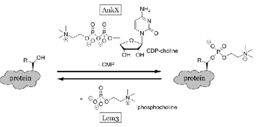
Recently, a new Fic domain-mediated PTM, termed phosphocholination, was discovered. This modification by the Legionella protein AnkX consists of the transfer of an phosphocholine moiety to either threonine or serine residues (scheme 4).[42,43] In this case, CDP-choline is used as a precursor, which is positioned in an inverted orientation in the catalytic pocket when compared to the adenylylation.[44] This way, the CMP moiety functions as a leaving group and the β-phosphate with the attached choline is transferred to the protein substrate. Interestingly, AnkX targets the small GTPases Rab1 and Rab35 in a similar fashion to the Legionella protein DrrA. Another analogy to adenylylation of DrrA was revealed by the description of the dephosphocholinase Lem3 from Legionella.[42]
While the abundance of Fic domain containing enzymes in pathogens is intriguing, adenylylation and related PTMs emerge as a general principle for the regulation of protein activities. During infections, these mechanisms exploit host cell signalling pathways to the benefit of the intruder. However, even the human Fic domain containing protein HYPE (Huntingtin interacting protein E) has been shown to have adenylylating activity in vitro,[45,46] indicating that these PTMs are most likely not limited to
Scheme 4. Schematic presentation of phosphocholination. Phosphocholinase AnkX uses CDP-choline to transfer phosphoCDP-choline onto threonine or serine residues of protein substrates. Dephosphocholinase Lem3 hydrolytically removes the modification.
Small GTPases
Small GTPases (more accurately termed “small G proteins”, because they have both GDP/GTP-binding and GTPase activities) are a protein super-family that consists of more than 100 members.[47] The term “small” refers to the relatively small protein size of 20-40 kDa. According to their function, the members can be divided into at least five families: Ras, Cdc42/Rho/Rac, Rab, Sar/Arf and the Ran. The important biological functions controlled by these families include cell mobility, organisation of the cytoskeleton, vesicular transport, signal transduction and nuclear import and export (table 2).
Table 2. Summary of the small GTPase families and their function.
Family Function Reference
Ras Signal transduction/gene expression [48,49]
Cdc42/Rho/Rac Cytoskeletal reorganisation/gene expression [50,51]
Rab Vesicle trafficking [52–54]
Sar/Arf Vesicle budding, celia formation [55,56]
Ran Nuclear import/export, microtubule organisation [57,58]
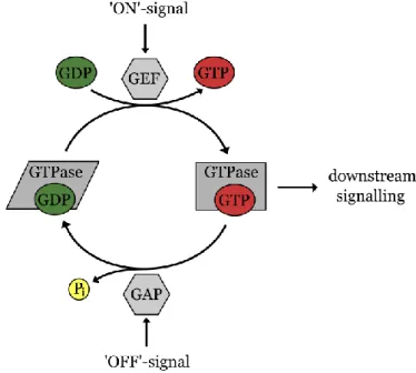
All small GTPases feature a binding site for guanosine nucleotides. A defining characteristic of small GTPases is the significant change in conformation, dependent on which nucleotide is bound to the protein.[59] If guanosine diphosphate (GDP) is bound, the small GTPase is in its inactive state, in which it is not interacting with downstream signalling effectors. If
the GDP is exchanged with guanosine triphosphate (GTP), the active conformation is attained and downstream signalling with effector proteins is possible (figure 1).[60] Two flexible regions of the small GTPases, referred to as switch I and switch II region, undergo large conformational changes upon nucleotide exchange.[61] Guanosine nucleotide exchange factors (GEFs) catalyse the reversible exchange of GDP to GTP, which is driven by the higher excess of GTP compared to GDP in the cell.[39,59,62,63] On the other hand, GTPase-activating proteins (GAPs) catalyse the inactivation of the small GTPase. The slow intrinsic GTPase activity of the small GTPase is thereby accelerated by several orders of magnitude upon binding of the
GAP.[59,62,63]
Because of this distinct, conformation-induced separation of active and inactive state, small GTPases are often referred to as molecular switches.[64] Many effector and regulatory proteins are involved in the interplay of small GTPases. However, the central role of small GTPases in many signalling pathways and regulatory mechanisms of a cell make them a popular target of many pathogens[65,66] and genetic mutations are often of oncogenic character.[67]
Figure 1. Small GTPases act as molecular switches in mammalian cells. An inactive GDP-bound state can be converted to the active state by nucleotide exchange induced by guanosine nucleotide exchange factors (GEFs). In the active state, the GTPases interacts with downstream
Pathogens modify host cells at a molecular level
When a mammalian host encounters a microorganism, processes like phagocytosis[68] and lysosomal disruption[69] are usually capable of destroying the intruding organisms. Intracellular pathogenic microorganisms[70] on the other hand have evolved in a way that allows them to survive these defence mechanisms and to establish themselves inside of the affected host cells.[71] The location inside of mammalian cells protects the microbe from the detection by the immune system and the effect of many antibiotics. Normally, intruding pathogens are engulfed by macrophages, the frontline defence of the innate immune system. After ingestion, the pathogen-containing phagosome matures sequentially to an endosome-like entity and fuses with the lysosomal network, creating the phagolysosome where harsh digestive conditions destroy the pathogen (figure 2, A).[68,72] For the survival of the intracellular pathogen, it is important to prevent the phagosome-lysosome interaction. Therefore, many intracellular pathogens employ infection mechanisms to enter the cell, creating specialised vacuoles that do not interact with the lysosomal system. Here, a large number of proteins are injected into the host cell, which allow the manipulation of the host at a molecular level.[73,74] After establishment in the cell, the pathogen is largely dependent on nutrients from the host cell and its parasitic lifestyle becomes apparent. After a phase of extensive proliferation of the pathogen, lysis of the host cell is induced and the pathogens are released into the surrounding environment.
The infection mechanism of Legionella pneumophila, the causative agent of the legionnaires’ disease in humans,[75] is a prime example. Legionella bacteria are not exclusively intracellular pathogens and can be found for example in cooling towers[76], swimming pools or air-conditioning units[77], where they usually infest protozoa.[78] Upon uptake in the human body via infected aerosols, a Legionella bacterium enters an alveolar macrophage and injects approximately 300 effector proteins into the host cell by the Dot/Icm type IV secretion system (figure 2, B).[79] Some of these effector proteins are essential for the creation of the Legionella-containing vacuole (LCV), a membrane-surrounded niche for the pathogen that resists fusion with the lysosomes.[80,81] In the usual endocytic pathway, shortly after phagocytosis, maturation to the phagosome is induced by recruitment of the small GTPases Rab5, Rab7 and, at a later stage, lysosome-associated membrane glycoproteins (LAMPs) which ultimately lead to phagolysosome formation. This development is impaired significantly by Legionella.[82] Instead, vesicles from the endoplasmic reticulum (ER) are recruited to the LCV shortly after phagocytosis and the LCV membrane is enriched with ribosomes.[81,82] High-jacking of the vesicle transport from the ER to the Golgi apparatus is partially achieved by interference with the small GTPases Arf1 and Rab1.[83]
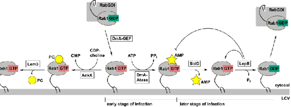
On a molecular level, modification of the latter by activation and adenylylation by the Legionella protein DrrA, leads to constantly active Rab1 on the membrane of the LCV (figure 3).
Figure 2. A. Phagocytosis of a non-pathogenic bacterium by a macrophage. After uptake of the bacterium, the phagosome is sequentially matured to an endosome-like entity and transferred to the lysosome. The harsh conditions of the endosomes and phagolysosomes digest the intruding bacterium. B. Infection of a macrophage with Legionella pneumophila and modulation of vesicle trafficking. Upon contact and uptake of the Legionella bacterium, the Dot/Icm type IV secretion system injects effector proteins into the host cell, leading to an inhibition of the transfer of the LCV to the lysosome network. Instead, vesicle trafficking from the ER to the Golgi complex is redirected to the LCV, forming a stable ribosome studded intracellular vacuole. After replication of the pathogen to high numbers, the host cell is lysed and the bacteria released (not shown). Figure modified from reference 81.
DrrA consists of three domains: one adenylyl transferase domain, one GEF domain and one phosphatidylinositol-4-phosphate binding domain (P4M) domain. The DrrA-P4M domain is responsible for the localisation of DrrA on the outer side of the LCV membrane.[84] Here, the DrrA-GEF domain activates Rab1:GDP from the cytosol by exchange of the nucleotide GDP to GTP (figure 3).[85] Cytosolic inactive Rab1:GDP is bound by Rab GDP dissociation inhibitor (RabGDI), which complexes to the C-terminal geranyl-geranyl lipid anchor.[86] However, upon activation, the small GTPase is released from the complex and the lipid anchor of Rab1 consequently attaches to the nearby membrane of the LCV.[87] In this way, the DrrA-GEF domain effectively recruits Rab1 to the LCV. The active Rab1:GTP is then adenylylated at a specific tyrosine residue in the switch II region.[27] The switch II region is an essential interaction site for many host cell effector proteins, like MICAL (molecules interacting with CasL) proteins,[88] and adenylylation effectively blocks these interactions. Furthermore, the protein
Interestingly, Legionella also contains its own GAP for Rab1 (LepB) that can be found on the LCV at later stages of infection.[90] The existence of SidD and LepB indicates that the pathogen allows inactivation and removal of Rab1 from the LCV at a certain stage of infection. After hydrolysis of GTP to GDP, RabGDI extracts Rab1 from the LCV membrane (figure 3).
Figure 3. Modification of Rab1 by Legionella enzymes. The GEF domain of DrrA activates Rab1:GDP by GDP to GTP exchange and thereby recruits the enzyme to the membrane of the LCV. Here, Rab1:GTP undergoes adenylylation by DrrA, blocking the binding site for host cell effector proteins and GAPs. At a later stage of infection, SidD removes the adenylylation and the
Legionella enzyme LepB (and potentially other GAPs) deactivates Rab1:GTP by hydrolysis to
Rab1:GDP. Rab1:GDP can be extracted from the membrane by Rab1GDI. Alternatively, Rab1:GTP can undergo phosphocholination by AnkX at the LCV. Lem3 acts as a dephosphocholinase and removes the modification. Figure modified from reference 91.
Surprisingly, the switch II region of Rab1 is not only undergoing adenylylation, but also phosphocholination during the infection process. The Legionella enzyme AnkX, which is secreted into the host cell, phosphocholinates a specific serine residue by using CDP-choline as a co-substrate (figure 3).[42–44] As in the case of adenylylation, some protein-protein interactions are affected by phosphocholination, e.g. the interaction with Rab1 GDI and with the GEF Connecdenn.[43,92] AnkX alone is sufficient for inducing a specific phenotype in transfected cells, showing Golgi disruption and inhibited alkaline phosphatase secretion.[93] However, how the induction of this phenotype works in detail has not yet been described. Furthermore, Legionella features the corresponding dephosphocholinase, making the reaction reversible.[92]
How the adenylylation and phosphocholination are regulated in detail, and how exactly these modifications have an impact on the whole eukaryotic system during infection, is still a matter of ongoing scientific investigation. However, many pathogens apply similar mechanisms to manipulate the host cell behaviour to their benefit. Understanding these mechanisms on a
molecular level is essential for the advancement of infection biology. As a consequence, new targets for the development of infection-inhibiting drugs might arise. Identifying the substrate scope of the related PTM-mediating enzymes is of major importance because these interactions are the key to understanding the effect and relevance of pathogenic proteins.
Quorum sensing in Legionella pneumophila
While the interactions with the host cell are of major importance for the survival of the pathogen, it also has to switch between a intracellular growth phase, characterised by excessive replication, and a virulent phase for the extracellular transmission to a new host. The virulent phase consists of opposing traits compared to the stationary growth phase: while the replication is repressed, motility, competence, extracellular filaments and expression of genetic fitness islands are induced. Different signals can induce switching, for example starvation and increasing intracellular alarmone concentrations.[94]
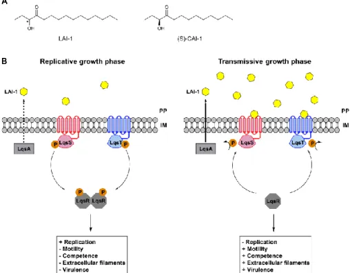
The pivotal biphasic life style[95] is regulated by quorum sensing, a population-density based bacterial cell-cell communication.[96] Small molecules, termed autoinducers, are produced by the bacteria and the increase in concentration based on the density of bacteria is detected by corresponding sensor systems.[97] In Legionella, quorum sensing is regulated by the Lqs (Legionella quorum sensing) system (figure 4, B). The Lqs system consists of the two sensor histidine kinases LqsS and LqsT,[98] located in the inner membrane of the cell, one response regulator LqsR[99] and the autoinducer synthase LqsA,[100] which synthesises the autoinducer 3-hydroxypentandecane-4-one (LAI-1, figure 4, A).[101] LAI-1 is produced by the biosynthesis gene-cluster LqsA and is enriched in the cell environment, depending on the population density. At low LAI-1 concentrations, LqsS and LqsT are autophosphorylated at conserved histidine residues.[98] The phosphorylation converges with the binding partner LqsR, which dimerises upon phosphorylation. The Lqs-dependent signalling is therefore switched off and the transmissive growth state is repressed. At high LAI-1 concentrations (meaning high cell density), LAI-1 inhibits the autophosphorylation of LqsS and LqsT, leading to unphosphorylated and therefore monomeric LqsR. Consequently, LqsR induces the transition from the stationary, replicative to the virulent, transmissive growth phase.
Many intracellular pathogens employ similar mechanisms for the regulation of their growth. A closely related example is found in Vibrio cholerae, where the autoinducer synthase CqsA produces
(S)-3-Figure 4. Legionella quorum sensing (Lqs) system controls the switch between exponential and stationary growth phase. The phosphorylated membrane-bound sensor kinases LqsS and LqsT phosphorylate the regulator LqsR. LqsR dimerises upon phosphorylation and induces replication, while traits like motility, virulence and fitness are repressed. When concentrations of the autoinducer LAI-1, which is synthesised by LqsA, increase at the periplasm, phosphorylation of LqsS and LqsT is inhibited. Consequently, the bacteria shifts from the replicative growth phase to the transmissive growth phase, which is characterised by repression of replication and promotion of pathogenic competence and fitness. PP = periplasm, IM = inner membrane. Figure modified from reference 104.
Proteomics towards PTMs
In analogy to genomics, proteomics has risen as central scientific field around the large-scale study of proteins and their functionalities.[105] The proteome consists of the expressed proteins of a genome, thus proteomics can be considered as functional genomics on a protein level.[106] In contrast to the genome, which might undergo epigenetic changes but is usually not altered in its sequential order of nucleobases, the proteome is highly dynamic. Environmental factors like stress or differentiation of cell types can induces significant changes to the composition and amount of the proteins content in a cell. As a reporter for the situation of a cell, proteomics is
therefore a more suitable approach than the study of the genome or the transcriptome, due to its consideration of the regulation of gene expression and translation.[107]
Another level of complexity to the proteomic analysis of cells is added by the presence of PTMs. Proteins can undergo a wide variety of PTMs, which often are crucial for the understanding of the physiological function of a protein. Conventionally, three different strategies have been applied for the identification of post-translationally modified proteins: radioisotope labelling (e.g. 32P for phosphorylation, 3H and 14C for acetylation and methylation), western blotting (e.g. phospho-tyrosine and lysine-acetylation and –methylation) and peptide/protein arrays (e.g. phosphorylation and methylation).[108–110] While isotopic labelling and western blot analysis are excellent tools for the detection and the validation of PTMs, they lack the feasibility for high-throughput identifications. Peptide and protein arrays on the other hand allow for a larger amount of samples analysed in parallel but sensitivity and specificity of employed PTM-mediating proteins under the experimental conditions are often a drawback.
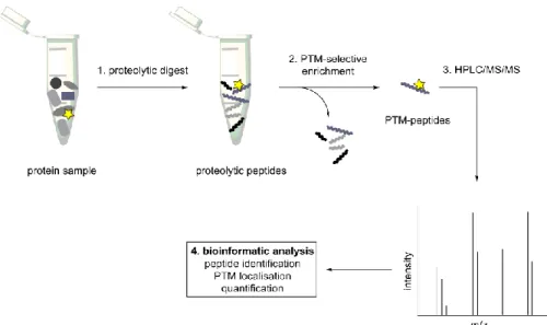
With the rise of MS-based proteomics, a new powerful bioanalytical tool has become available for the study of PTMs. The high sensitivity and specificity allows the global, proteome-wide identification of PTMs. However, because proteomic samples from crude cell lysates are often too complex for direct MS analysis, strategies for the selective enrichment of the post-translationally modified proteins are a necessity.[111] A general workflow for the modification-specific MS analysis of a cell lysate is illustrated in figure 5. In the first step, a protein sample, usually a cell lysate, is submitted to proteolytic digestions. For this step, trypsin is the most commonly used protease, because it specifically cleaves after positively charged amino acids, generating a more homogenous charge-distribution among the peptides. Secondly, an enrichment technique is employed to separate the post-translationally modified peptides from the rest of the peptides. In the next step, the resulting peptide samples are submitted to nano-HPLC/MS/MS. The chromatographic separation by HPLC splits the peptides based on their hydrophobicity and the MS/MS technique allows the identification of the peptide and the localisation of the PTM, based on the fragmentation pattern. Finally, the generated data is bioinformatically processed, to verify the results statistically and to identify the associated proteins, by comparison with an in silico proteolytically digested sample.[112] In an alternate approach, the samples can be digested after the enrichment step, creating proteomic samples on a protein rather than a peptide level. Furthermore, sample complexity might be reduced prior to the lysis or
Figure 5. MS-proteomic workflow for the identification of PTM-sites. A complex protein sample, like a cell lysate, is proteolytically digested. The resulting peptides undergo a PTM-selective enrichment step, to separate modified from unmodified peptides. The resulting samples with PTM-carrying peptides are analysed by HPLC/MS/MS and the identified masses are processed bioinformatically. As a result, peptides are assigned to the corresponding proteins, the PTM sites are identified precisely and eventually a quantification of the PTM is possible.
The low abundance of PTM-bearing peptides in the huge amount of unmodified peptides in a lysate makes an enrichment step crucial for a successful identification. It has become apparent that this enrichment is a major bottleneck for the high-throughput identification of proteome-wide PTMs and, consequently, several techniques have been developed to achieve a selective sample separation. These techniques include enrichment based on antibody-affinity, chemical derivatisation, ionic interactions and PTM-specific enzymatic reactions.
Antibodies pose excellent tools for the detection, as well as the enrichment of PTMs (scheme 5, A). The generation of a general, high-quality pan-PTM antibody, ideally completely independent from the surrounding peptide backbone, is a requirement for this approach. This enrichment technique has been successfully employed for the identification of sites for lysine acetylation,[113,114] serine, threonine, tyrosine and histidine phosphorylation,[115–117] arginine and lysine methylation[118] and tyrosine nitration.[119]
Chemical derivatisation includes strategies like metabolic labelling, where small chemical groups are introduced into the co-substrates of the PTM reaction.[120] Most often, azides and alkynes are employed, due to their small
size and addressability by bioorthogonal Huisgen cycloaddition (click-reaction, scheme 5, C).[121] Almost all PTMs have been explored based on this approach, e.g. farnesylation,[122] palmitoylation,[123] myristoylation [124] and glycosylation.[125] Similarly, some PTMs can be converted in vitro to reactive groups, which can be chemically captured. For example, phospho-serine or -threonine can undergo β-elimination under basic conditions.[126] In a subsequent Michael-like addition, the site can be linked to an affinity tag, like biotin or a perfluoroalkyl residue.[127] The advantage of the chemical derivatisation approach is the highly stable covalent linkage between tag and modification, which allows for easy isolation of the tagged proteins. However, the underlying chemical reaction needs to be of high efficiency and the additional reaction step always introduces the possibility of undesired side reactions.
Especially for phosphopeptides, ionic interaction-based enrichment using immobilised metal-ion affinity chromatography (IMAC) has been highly successful. Originally, Fe3+ immobilised on beads was used to coordinate the phosphate group of phosphorylated peptides (scheme 5, B).[128,129] Other metal ions have been investigated as well for this purpose, illustrating Ga3+ as the best candidate.[130] In addition to IMAC, TiO2-based surfaces have demonstrated vast potential and surpass IMAC in efficiency and robustness for the separation of phosphopeptides.[131,132] While these methods are highly interesting for PTMs with ionic character, uncharged PTMs elude enrichment based on these techniques.
The application of PTM-specific enzymes for the purpose of identifying PTM-bearing moieties is another highly selective approach. For example, glycosylphosphatidylinositol(GPI)-anchored proteins can be specifically released from cell surfaces by treatment with phosphatidylinositol-specific phospholipases. The released proteins could be identified by MS analysis.[133,134] In another example, O-β-N-acetylglucosamine specific galactosyl transferase introduced a ketone functionality to GlcNAc-modifed proteins, making it addressable by a reaction with O-functionalised hydroxylamines (scheme 5, D).[135]
While vast advances over the past years have been realised in the proteomic field, the selective enrichment of post-translationally modified peptides and proteins remains one of the major bottlenecks for a proteome-wide mapping of PTMs. New techniques and improved sensitivities are needed for the further development of the field and a necessity for a further elucidation of the intriguing PTM-based regulatory network in eukaryotic cells.
Scheme 5. Examples of the PTM-oriented isolation of peptides and proteins. A. Enrichment of lysine-acetylated peptides based on affinity to an antibody immobilised on solid support. B. IMAC of phosphorylated peptides. Fe3+ is immobilised on solid support and forms a complex
with the phosphate group of the modified peptides. C. Chemical derivatisation of myristoylated proteins. Alkynyl-myristic acid is added to the samples and replaces myristic acid in the PTM transfer reaction. The alkynyl functionality can be addressed by Cu(I) catalysed click chemistry to introduce a tag, e.g. an affinity tag like biotin. Subsequently, the modified proteins can be enriched, digested and analysed by MS. D. PTM-specific enzymatic modification of O-β-N-acetylglucosamine modified proteins. Engineered β-1,4-galactosyl transferase catalyses the addition of a galactose derivative, carrying a ketone functionality. The corresponding UDP-galactose derivative is used as a precursor for the reaction. Subsequently, the ketone can be reacted with hydroxylamine carrying an affinity tag, allowing isolation and identification of the modified proteins.
Chapter 1: Towards the identification of
adenylylated proteins and
adenylylation-modifying enzymes (Paper I – III)
To date, several adenylylating enzymes have been revealed. The striking abundance of Fic-domain-containing proteins, especially in intracellular pathogens, indicate that nucleotidylylation, phosphocholination and other modifications play a major role during infections. In the case of adenylylation, one key necessity for a deeper, molecular understanding of these modifications and their consequences is the detection of the substrates of adenylyl transferases. The knowledge of which protein is adenylylated inside a cell, allows conclusions to be drawn regarding the functions and mechanisms of adenylyl transferases.
Previous work
The identification of modified proteins from a highly complex sample (such as a cell lysate) has proven to be a major challenge for the PTM-devoted research. For adenylylation, two approaches have been investigated so far: on the one hand, metabolic labelling, which consists of the introduction of radioactive isotopes or “chemical handles” to the co-substrate of the PTM reaction, and, on the other hand, the use of antibodies targeting the modification selectively.
Metabolic labelling has been employed for the investigation of almost all known PTMs.[120] Consequently, the strategy has been applied to the studies of adenylylation. In its simplest form, the ATP co-substrate is replaced by its radioactive analogue, 32P-α-labelled ATP. If an adenylylation reaction occurs, the 32P-α-AMP modified protein substrates can be visualised by autoradiography.[22] However, this method is not suited for the enrichment of adenylylated proteins, but was used in combination with other stable isotopes of ATP for a proteomics based approach to identify the substrates of the adenylyl transferase BepA.[136]
Grammel et al.[137] developed a chemical reporter, which allows covalent functionalisation of adenylylated substrates with a propargyl group on the N6 position of the AMP moiety. This propargyl group can consequently undergo bioorthogonal click chemistry,[121] to introduce fluorescent dyes or affinity tags by triazole formation. The use of affinity tags, like biotin, allows the isolation of the adenylylated proteins out of lysates and subsequent identification by MS (scheme 6). This method was applied to identify targets of VopS[137], HYPE[46] and FicT toxins,[41] although in the latter case severe difficulties with the enrichment are reported. Furthermore,
N6-fluorescein-labeled ATP was used to generate both fluorescent and enrichable adenylylated proteins, following an immunoprecipitation protocol with the use of fluorescein antibodies.[138]
Recently, the chemical handle approach has been employed in combination with high density protein arrays (nucleic acid programmable protein arrays, NAPPA) for the high-throughput identification of a range of mammalian substrates of adenylyl transferases like VopS, IbpA,[139]
HYPE[140] or DrrA.[141]
Scheme 6. Approaches for the identification of unknown substrates of adenylyl transferases using chemical handles or antibodies. In the chemical handle approach, a propargyl group is introduced at the N6 position of adenine and transferred to the substrates by the adenylyl transferase. Selective click reactions with an affinity tag (e.g. biotin) carrying an azide allows the isolation of modified proteins. In the antibody-based approach, adenylylated proteins are isolated by affinity purification over immobilised AMP-antibodies.
The high selectivity of the biorthogonal click reaction between alkynes and azides and the stability of the generated covalent bond between the tag and the PTM are the main advantages of this method. However, in case of adenylylation, several drawbacks have to be considered: The introduced chemical handle at the N6 position of adenine, although sterically small (in case of a propargyl group), creates an additional spatial requirement in the nucleotide binding site of the adenylyl transferase. While some adenylyl transferases, like VopS, seem to accept this substrate without complications, steric clashes are to be expected for other adenylyl transferases.[142] For example, kinetic data for DrrA shows the importance of the free and
this problem, a large excess of enzyme is needed,[137] which is most likely accompanied by less specific reactions. One approach to solve this problem would be to vary the position of the chemical handle on the adenosine, like it is reported for position C2.[143] Furthermore, the ATP derivative has to compete with endogenous ATP, which is present in cells at milli molar concentrations.[144] As the native substrate (ATP) is likely a better substrate for adenylyl transferases than the corresponding N6-modified ATP, large excesses of the probe are required for successful transfer of the chemical handle. Given the various roles of ATP in biological processes, the high concentrations of modified ATP can lead to unwanted side reactions.
Antibodies on the other hand represent a different approach for the detection of adenylylated proteins. For many PTMs, antibodies have been a powerful tool for the enrichment of modified proteins and their subsequent identification by MS techniques (see section “proteomics towards PTMs”). Furthermore, antibodies allow for the easy detection of modified proteins by western blot procedures. One major advantage of the enrichment method is that the structure of the PTM itself is not altered, like in the case of chemical derivatisation. This allows a direct method to detect the modification site by MS/MS, giving a first validation of the identified protein as a substrate. Another advantage is that antibodies can be very selective and usable in low concentration, making unspecific reactions less likely.
In general, adenylylation (and other nucleotidylylations) seem to be well suited for antibody binding due to the polar and hydrophobic characteristics and the size of the modification. While adenylylated proteins are unlikely to raise antibodies specific for the modification, but rather for the protein itself, adenylylated peptides present a more suitable antigen. In parallel to our work, Hao et al.[145] used the adenylylated peptide of the switch I region of the small GTPase Rac1 (EYIPT*(amp)VF, synthesised by Fmoc-SPPS according to Al-Eryani et al.[146]), to raise threonine-AMP specific polyclonal antibodies, which were used for the detection and immunoprecipitation of in vitro adenylylated VopS substrates. However, how these antibodies perform against adenylylated proteins, which are not substrates of VopS, and their performance in immunoprecipitations of in vivo adenylylated proteins has not been demonstrated.
Outline: From building blocks to antibodies
The primary goal of the work presented in this thesis was the generation and use of a generic antibody against adenylylated proteins to facilitate the identification of substrates of bacterial adenylyl transferases. The first step was to develop a suitable strategy for the synthesis of adenylylated peptides, fully compatible with the Fmoc-SPPS protocol. The adenylylated peptides would function as reference material for MS experiments, to facilitate the identification of adenylylated proteins by proteomics. Furthermore, the adenylylated peptides would be used as antigens for the generation of generic antibodies against adenylylated motifs. In the next step, AMP-antibodies could be used for the immunoprecipitation of adenylylated proteins. With such an enrichment method at hand, proteomic investigations on adenylylated proteins could be greatly promoted (scheme 7).
Synthesis of a tyrosine-AMP building block
Previously reported strategies for the synthesis of adenylylated peptides include an assembly and a building block approach. In the inter-assembly approach (scheme 8, A),[145,146] the peptide is first synthesised on solid support by standard Fmoc synthesis. The hydroxyl functionality of the amino acid which is going to be modified (threonine or serine) is left unprotected and is phosphonylated after completion of the peptide synthesis. The H-phosphonate-adenosine is generated with 2’,3’-isopropylidene protected adenosine and PyBOP, followed by oxidation with iodine and acidic cleavage to yield the final product. While this is a quick and easy approach, adenylylation on tyrosine remains inaccessible and the method has strict requirements on the peptide sequence, e.g. excluding oxidation sensitive amino acids like tryptophan, methionine and cysteine.
A building block approach, where the adenylylated motif is introduced into the peptide chain via a preformed adenylylated amino acid, would allow a more flexible composition of the peptide. Filippov et al.[147] have shown that tyrosine-nucleotidylylated peptides could be synthesised from nucleotidylylated building blocks by use of an acyl-based protective group strategy (scheme 8, B). However, the use of 2’, 3’-diester protection on the adenosine promotes depurination under the acidic conditions during SPPS, consequently reducing the yields dramatically in the case of tyrosine-AMP. Mechanistically, it is hypothesised that depurination is promoted by anchimeric assistance from carbonyl functionality of the 2’-ester group.
Scheme 8. Previously described methods for the synthesis of adenylylated peptides. A. On-resin phosphonylation of the completed peptide sequence, followed by coupling with adenosine, oxidation and deprotection. B. Building block approach for Fmoc-SPPS with ester protection on the sugar. The yields are dramatically decreased by depurination under acidic conditions.
We envisioned a building block approach for the synthesis of tyrosine-adenylylated peptides that is not hampered by the drawbacks described. Building on the work of Filippov et al., we intended to avoid depurination by use of cyclic isopropylidene protection at position 2’ and 3’ of the adenosine. The choice of bis-Boc-protection at the N6 position of the adenosine would further deactivate the adenine ring system and decrease the rate of depurination. These considerations result in building block 1, which can be retrosynthetically disconnected to two alternate phosphoramidate precursors and the corresponding coupling partners (route A and route B, scheme 9). Both routes have been investigated.
Scheme 9. Ester protection on the 2’ position gives rise to depurination under acidic conditions. Protection with isopropylidene should prevent this side reaction. A change from benzoyl to bis-Boc protection at the N6 position should further deactivate the ring system. Envisioned building block 1 can be disconnected either according to route A or B.
The straightforward synthesis of Fmoc-tyrosine allyl ester (4)[148] and N6 -bis-Boc-2’,3’-isopropylidene adenosine[149] (3) provides the necessary precursors for the formulation of the phosphoramidate.
Diisopropylamino-Fmoc-tyrosine allyl ester 4 was reacted with 6 and, after quick purification under anhydrous and oxygen-free conditions, was directly reacted with adenosine 3 via a tetrazole promoted coupling. Subsequent oxidation with TBHP gave phosphotriester 1 as 1:1 diastereomeric mixture in 45% isolated yield. Reversing the reaction sequence according to route B (scheme 10) increased the yield to 56% under otherwise identical reaction conditions. Further optimisation of the reaction conditions showed that using a 2:1 mixture of tetrazole/diisopropylammonium tetrazolide (7) as a coupling reagent increased the yield to 76% isolated yield. However, the potentially explosive character of the diisopropylammonium tetrazolide salt has to be considered. The building block synthesis was completed by Pd-mediated deallylation of the amino acid carboxyl terminus, employing phenylsilane as a nucleophile. The product 8 was obtained in good yields after reversed phase (C18 Sep Pak) purification. Subsequently, the building block 8 could be used directly for Fmoc-SPPS (scheme 13).
Scheme 10. Synthesis route A and B to adenylylated tyrosine-AMP building block 8 for SPPS. Route B gives higher yields after the tetrazole mediated coupling. Optimisation revealed that a 2:1 mixture of diisopropyl ammonium tetrazolide (7) with 1H-tetrazole gave higher yields than 1H-tetrazole alone. TBHP: tert-butyl hydroperoxide, DIPEA: diisopropylethyl amine.
Synthesis of a threonine- and serine-AMP building block
Initially, we intended to extrapolate the tyrosine-AMP building block approach to the generation of serine- and threonine-adenylylated peptides. However, it is known from the SPPS of phosphopeptides[150] that preformed phosphotriester building blocks of serine and threonine undergo β-elimination under basic conditions.[151,152] Applied to adenylylated serine and threonine, the use of a phosphotriester building block leads to competitive β-elimination as the adenosine monophosphate diester acts as a good leaving group upon abstraction of the proton at the α-position of the amino acid (scheme 11). To avoid this side reaction, we investigated the unprotected, mono-anionic phosphodiester as a building block for the peptide synthesis. Upon β-elimination, the di-anionic phosphomonoester would be generated, representing a much poorer leaving group and thereby inhibiting the elimination process. Synthesis of the mono-anionic phosphodiester can be easily achieved by replacing the 2-cyanoethyl protecting group at the phosphorus from the previous described synthesis with an allyl protecting group. In the final deallylation step, the mono-anionic diester is generated.Scheme 11. β-Elimination (E2 mechanism) on the threonine or serine side chain of the AMP building block during treatment with bases in SPPS (or during amino acid activation). A: With 2-CNE protection at the phosphorus. B: Without additional protection of the phosphorus. The generation of a di-anionic leaving group in B is less favourable, inhibiting the reaction. R = Me, H; B = Base; Pep = peptide chain during SPPS.