ACTA UNIVERSITATIS
UPSALIENSIS
Digital Comprehensive Summaries of Uppsala Dissertations
from the Faculty of Medicine
1171
Fetal Movements in late Pregnancy
Categorization, Self-assessment, and Prenatal
Attachment in relation to women’s experiences
MARI-CRISTIN MALM
ISSN 1651-6206 ISBN 978-91-554-9446-9
Dissertation presented at Uppsala University to be publicly examined in Föreläsningssal 6, Högskolegatan 2, Falun, Thursday, 25 February 2016 at 13:00 for the degree of Doctor of Philosophy (Faculty of Medicine). The examination will be conducted in Swedish. Faculty examiner: Docent Siw Alehagen (Institutionen för medicin och hälsa. Linköpings universitet).
Abstract
Malm, M.-C. 2016. Fetal Movements in late Pregnancy. Categorization, Self-assessment, and Prenatal Attachment in relation to women’s experiences. Digital Comprehensive
Summaries of Uppsala Dissertations from the Faculty of Medicine 1171. 73 pp. Uppsala: Acta Universitatis Upsaliensis. ISBN 978-91-554-9446-9.
Aim: To explore how pregnant women experience fetal movements in late pregnancy. Specific aims were: to study women’s experiences during the time prior to receiving news that their unborn baby had died in utero (I), to investigate women’s descriptions of fetal movements (II), investigate the association between the magnitude of fetal movements and level of prenatal attachment (III), and to study women’s experiences using two different self-assessment methods (IV).
Methods: Interviews, questionnaires, and observations were used.
Results: Premonition that something had happened to their unborn baby, based on a lack of fetal movements, was experienced by the participants. The overall theme “something is wrong” describes the women’s insight that the baby’s life was threatened (I). Fetal movements that were sorted into the domain “powerful movements” were perceived in late pregnancy by 96 % of the participants (II). Perceiving frequent fetal movements on at least three occasions per 24 hours was associated with higher scores of prenatal attachment in all the three subscales on PAI-R. The majority (55%) of the 456 participants reported average occasions of frequent fetal movements, 26% several occasions and 18% reported few occasions of frequent fetal movements, during the current gestational week. (III). Only one of the 40 participants did not find at least one method for monitoring fetal movements suitable. Fifteen of the 39 participants reported a preference for the mindfetalness method and five for the count-to-ten method. The women described the observation of the movements as a safe and reassuring moment for communication with their unborn baby (IV).
Conclusion: In full-term and uncomplicated pregnancies, women usually perceive fetal movements as powerful. Furthermore, women in late pregnancy who reported frequent fetal movements on several occasions during a 24-hour period seem to have a high level of prenatal attachment. Women who used self-assessment methods for monitoring fetal movements felt calm and relaxed when observing the movements of their babies. They had a high compliance for both self-assessment methods. Women that had experienced a stillbirth in late pregnancy described that they had a premonition before they were told that their baby had died in utero. Keywords: Fetal movements, pregnancy, prenatal attachment, self-assessment, stillbirth Mari-Cristin Malm, Department of Women's and Children's Health, Akademiska sjukhuset, Uppsala University, SE-75185 Uppsala, Sweden.
© Mari-Cristin Malm 2016 ISSN 1651-6206
ISBN 978-91-554-9446-9
List of Papers
This thesis is based on the following papers, which are referred to in the text by their Roman numerals.
I Malm M-C., Lindgren H., Rådestad I. (2010-2011). Losing contact with one’s unborn baby. Mother’s experiences prior to receiving news that their baby has died in utero. OMEGA, 62(4):353-367.
II Malm M-C., Hildingsson I., Rubertsson C., Rådestad I., Lindgren H. (2014). Development of a tool to evaluate fetal movements in full-term pregnancy. Sexual & Reproductive
Healthcare, 5(1):31-35.
III Malm M-C., Rubertsson C., Hildingsson I., Rådestad I., Lindgren H. (2015). Prenatal attachment is associated with fetal movements during pregnancy– a population based survey.
(Submitted)
IV Malm M-C., Rådestad I., Rubertsson C., Hildingsson I., Lindgren H. (2014). Women’s experiences of two different self-assessment methods for monitoring fetal movements in full-term pregnancy. A Crossover trial. BMC Pregnancy and
Childbirth, 14:349.
To all the women who participated in this research
Contents
INTRODUCTION ... 11
BACKGROUND ... 12
The fetal period ... 12
Placenta, amniotic fluid and the umbilical cord ... 12
Fetal movements ... 13
The fetus’s cycles of rest, sleep and activity ... 14
The fetal physical environment and setting ... 15
Technical procedures for recording fetal movement ... 15
Women’s perceptions of fetal movement ... 16
Fetal movements as an indicator of fetal wellbeing ... 17
Decreased fetal movements ... 17
Morbidity and mortality ... 19
Attachment ... 19
Maternal fetal attachment ... 19
Prenatal attachment ... 20
Self-assessment methods ... 20
Counting fetal movements ... 21
Mindfetalness ... 21
Antenatal care in a Swedish context ... 21
The antenatal standard visiting schedule ... 22
Rationale ... 23
AIMS ... 24
MATERIALS AND METHODS ... 25
Study design ... 25 Study I ... 27 Study design ... 27 Sample ... 27 Data collection ... 27 Data analysis ... 29 Study II ... 30 Study design ... 30 Sample ... 30 Data collection ... 30 Data analysis ... 32
Study III ... 33 Study design ... 33 Sample ... 33 Data collection ... 34 Data analysis ... 35 Study IV ... 35 Study design ... 35 Sample ... 35 Data collection ... 35 Data analysis ... 38 Ethical considerations ... 38 RESULTS ... 40 Study I ... 40
Not feeling in touch with their baby ... 41
Worry ... 41
Not understanding the unbelievable ... 42
Wanting information ... 42
Being certain that their baby had died ... 42
Feeling something is wrong ... 42
Study II ... 43
Powerful movements ... 43
Non-powerful movements ... 44
Study III ... 45
Study IV ... 45
The women’s emotions during self-assessment ... 46
Women’s preference of method ... 46
Researchers observations ... 47
DISCUSSION ... 48
Categorization of fetal movements ... 48
Fetal movements and prenatal attachment ... 49
Self-assessment methods ... 52 Methodological considerations... 53 Interviews ... 53 Questionnaires ... 54 Observations ... 56 CLINICAL IMPLICATIONS ... 57 FUTURE RESEARCH ... 58 CONCLUSIONS ... 59
TILLKÄNNAGIVANDEN ... 63 REFERENCES ... 66
ABBREVIATIONS
ANOVA Analysis of variance CTG Cardiotocography
IBM International Business Machines PAI The Prenatal Attachment Inventory
PAI-R The revised Prenatal Attachment Inventory
INTRODUCTION
Fetal movements are unique for each individual fetus as are women’s experi-ences of movements. Fetal movements have long been of interest as a possible measure of fetal wellbeing. The most common reasons for unscheduled visits to antenatal care centres are concerns regarding decreased fetal movements and an uncertainty of what is considered as normal fetal movements. For this reason, more knowledge of fetal movements in late pregnancy is necessary. Three different methods of data collection were used in this thesis: interviews, questionnaires, and observations, with the overall aim being to explore how pregnant women experience fetal movements in late pregnancy.
BACKGROUND
The fetal period
According to international convention the length of a pregnancy is counted in terms of completed weeks and days and is further divided into three periods known as the first, second, and third trimester. In this thesis, the third trimester will be named as late pregnancy. The first trimester lasts up to 13 weeks and 6 days (13+6), the second trimester lasts between 14+0 until 27+6 and the third trimester lasts from 28+0 until birth. Before 37+0 weeks of gestation the pregnancy is considered to be preterm and after 42+0 weeks of the gestation the pregnancy is referred to as a prolonged or post-term pregnancy. From the eight week of gestation,the developing baby is called a fetus. It is during this stage that the facial features become distinguishable, the genital organs start to develop, and the digits on the limbs start appearing. At 12 weeks of gesta-tion, the fingers and toes are distinct and the organ systems are completed. This is when the bones start to develop, as well as the circulatory system, and the sex of the fetus can be determined by ultrasonography. At this stage, the development of all body systems is completed. At full-term pregnancy, i.e. 37+0 weeks of gestation, the fetus has increased its weight to approximately 3500 grams and birth generally occurs after approximately 38-40 weeks of gestation (1-3).
Placenta, amniotic fluid and the umbilical cord
The second trophoblastic invasion, during which the muscular coats of the spiral arteries are stripped, ensures that the placental circulation causes the lowest level of impendence possible in the fetoplacental circulation. Growth restriction, attributed to uteroplacental insufficiency, occurs where this pro-cess is presumed to have failed and is usually detected during late pregnancy. Normal placental vascular maturation is usually completed by 20 weeks of gestation. The placenta is an incomplete endocrine organ as well as the means through which the fetus obtains its needs and the efficiency of many functions depends on adequate uterine blood flow. The placenta selects and transports the nutrients necessary for life and growth from the mother’s blood and changes the properties of the mother’s blood so that the fetus receives the nu-trients it requires. In a healthy placenta, the copious villous bed allows the
exchange of oxygen and metabolic products. The fetal sac consists of a double membrane; the outer, chorion, and the inner, amnion, where the fetus floats in the amniotic sac that is filled with a clear pale amniotic fluid that increases through a dynamic process. This process essentially involves diffusion of the fetal urine and lung fluid along with maternal fluid through the membranes into the amniotic cavity. By the time 38 to 39 weeks of gestation have been completed, the amount of fluid is 900-1000 ml, however, that amount will subsequently decrease. The umbilical cord extends from the fetal umbilicus to the fetal surface of the placenta. At birth, the mature cord is about 50–60 cm in length and 12 mm in diameter (4). It is composed of an embryonic form of connective tissue, intermingled with a gelatinous substance, and is covered with amnion. It carries two arteries containing impure blood to the placenta and one umbilical vein containing pure blood returning to the fetus after hav-ing been oxygenated and replenished in the placenta (1-3).
Fetal movements
The development of fetal movement patterns can by followed through preg-nancy with the use of obstetric ultrasound examinations of the fetus. The ear-liest spontaneous fetal movements can be observed at seven weeks of gesta-tion. Early fetal movements are sparse, simple, uncoordinated, or extremely quick twists. The movements gradually show a progressive development in their complexity, and become more sophisticated, stronger, regular, coordi-nated, complex, and sustained and they increase with gestational age (5). Re-searchers have described and classified the onset and development of fetal movements. The categories of movements show the development and matu-ration of the fetal central nervous system. The strength with which they are performed as well as the duration of the movements increases with the in-crease in muscle mass (6).
Eye movements are the physical expression of upper fetal brainstem function and at 28 weeks of gestation the pupillary membrane disappears and the eyes can open and close. Four fetal eye movement patterns were initially charac-terized based on early ultrasound observations (7) and have been further mod-ified (8). Fetal breathing is thought to originate from the medullary respiratory centers and is a part of the process of respiratory development. In early fetal life, breathing movements tend to be erratic but they develop a more regular pattern with advancing gestational maturity. At times these movements are perceptible to the mother (9). At 32 weeks of gestation the phenomenon known as fetal behavioral states represented by combinations of quiet or active sleep or quiet or active periods of being awake are possible to detect. In late pregnancy, fetal movement maturation is continuous until about 36 weeks of
gestation when behavioral states, according toNijhuis et al. (1982), are estab-lished in 80 percent of normal fetuses. The original article by Nijhuis et al. stated explicitly that gross movements consist of various patterns that are clas-sified into four behavioral states. This was introduced as a concept based on fetal heart rate and movement classification and information was discovered about the development of the autonomic nervous system during pregnancy. This is the coordination of state parameters as an expression of neurological maturation (10). Later studies indicate that this is a valid approach for fetal state detection. The definition is based on observations made with ultrasound regarding the occurrence and timing of the relationships of general movements of the body, eyes, as well as respiratory movements, with simultaneous regis-tration of variations in the fetal heartbeat as measured by CTG or biomagne-tometer (11).
As pregnancy advances, the type of fetal movement might change, weak movements decrease and are superseded by more vigorous movements, which increase over a period of several weeks. Reduced amniotic fluid volume and increased fetal growth appears to be one of the reasons for the change in fetal movements, but it is primarily caused by improved fetal coordination due to fetal brain maturation (12). The intensity and frequency of the movements in-crease from gestational week 22 until gestational week 32 after which a plat-eau phase is described (13, 14). In contrast, after the peak of increasing fre-quency in the second trimester, the type of movements changes as the preg-nancy processes (15-17).
The fetus’s cycles of rest, sleep and activity
Between 20 and 30 weeks of gestation, general body movements become rou-tinized and the fetus demonstrates rest and activity cycles that become pro-longed with gestation age (18). The normal range of movement incidence is unclear (19) since individual differences exist (20) but periods of alertness last about 20-40 minutes andthe average number of body movements observed at term has been reported to be in the range of 16-45 movements per hour (13). Periods of inactivity have been described to vary from 20-75 minutes with a mean length of 23 minutes. Sometimes, inactive periods last for over an hour, but they rarely exceeded 90 minutes for a healthy fetus (21-24). Fetal move-ments exhibit significant circadian patterns and diurnal rhythms have been found to develop in late pregnancy, with peak values occurring late in the evening and during the night (13, 25, 26). There is no evidence that fetal move-ments decline during the active periods in late uncomplicated pregnancy, alt-hough they may be experienced differently by different women (24).
The fetal physical environment and setting
Fetal movements are affected by aspects of the physical environment and they are essential for development (27). It is, however, unclear if gender influences fetal movements and there have been conflicting reports with regard to gender differences (28). The inherent neuromuscular functions of the fetus, the amount of free intrauterine space, the location of the amniotic fluid, and fetal positioning are the major physical influences that have been shown to affect fetal movement (29). The quality and quantity of general fetal movements re-flect the integrity of the nervous system and require that neuromuscular func-tions are intact. To provide this, adequate provisions of oxygen and nutrients to the central nervous system are necessary (30). Fetal movements change as the pregnancy advances due to improved fetal coordination and reduced am-niotic fluid volume, coupled with the increased size of the fetus (14, 31, 32). Amniotic fluid volume can be an important determinate of fetal movements as restricted intrauterine space due to diminished amniotic volumes might physically limit fetal movements (33). Furthermore, the fetus’s ability to move may be influenced by maternal and fetal diseases as well as factors related to maternal lifestyle (29, 34-37). The pressure on maternal blood vessels in-creases, accompanied by increased supply of blood to the fetus with subse-quent maternal cardiac output and fetal oxygen saturation, causing pressure on blood vessels if the woman lies on her back or on her right side during late pregnancy (38, 39). Fetal movements may be stimulated by acoustic stimula-tion, vibration (40-42), and light (43), as well as maternal ingestion of glucose (31). Ingestion of any kind of food or juice is the most frequently given prac-tice advice to stimulate fetal movements in an outpatient setting although it does not seem to have any effect (44).
Technical procedures for recording fetal movement
Many variables may influence fetal movements with gestational age being the most significant factor. However, one single test of the fetus’s wellbeing can-not predict with certainty if the fetus’s heath is compromised (45). Early re-search of fetal movements has been conducted by monitoring pressure changes transmitted through the maternal abdominal wall (46, 47). These complex registration procedures were replaced by the ultrasound evolution in the late 1960s. Initially, fetal movements were measured with a Doppler fetal activity monitor with a transducer designed to detect and register fetal move-ments as spikes on standard monitor paper. Measuremove-ments of fetal activity were conducted by passing the unprocessed Doppler signal through a band-pass filter that characterized different fetal activities. The development of ul-trasound techniques may detect fetal movements before the woman herself
perceives them and may provide an opportunity for the investigators to com-pare observed fetal moments by ultrasound with those that are perceivable by the women (48). After gestational week 20, it is no longer possible to visualize the entire fetus in its completeness with ultrasound and small movements might go unnoticed, however, advanced imaging via a fetal magnetic reso-nance imaging (MRI) offers a tool for visualizing the entire fetus and thus obtaining extended information (49).
Women’s perceptions of fetal movement
Maternal perception of fetal movements arises as a result of pressure against the body-wall and signs of fetal life that are perceivable by the woman. Ini-tially, movements are short and weak and can be difficult to distinguish from intestinal activity. In late pregnancy, the perceptions of fetal movements are related to the power of the movements (50, 51). Women expecting their first baby (primiparas) usually feel the first fetal movements in gestational week 18 to 20 (50). Women with prior pregnancies (multiparas) generally feel movements earlier, from about 16 weeks of gestation (52), but parity has not been found to affect maternal perception of fetal movements in late pregnancy (53). For women with an anterior placenta seen in early second trimester ul-trasoundscreenings, there are conflicting findings regarding if this causes any association with a perceived decrease in fetal movements (53-55). The major-ity of fetal movements are perceived when the woman lies down. Fewer move-ments are perceived while sitting, and the least number of movemove-ments are per-ceived when the woman is in a standing position (32, 56). The ability of women to perceive fetal movements is influenced by several physical, psy-chological, and social factors (19). The agreement between examinations con-ducted using ultrasound simultaneously with a woman’s recorded perception of movement has been examined in several studies, and ranges from 4 to 94 percent of movements that are detectable using current sonography scanning technology (57). When body parts were specified, women felt 82 percent of the movements. Trunk and limb movements lasting 1-3 seconds were felt 84 percent of the time (58) both when the movements encompassed the entire fetal body or only the extremities (50). Based on these criteria Rayburn et al. divided fetal movements into four categories; "general body movements", "single trunk and limb movements", "movements of the lower extremities", and "hiccups" (50). Maternal perception of fetal hiccups is of interest although hiccups are not considered to be a fetal movement. However, there seems to be a strong association (OR 3.25) between maternal perceptions of fetal hic-cups and a reduced risk of stillbirth in late pregnancy (39). Classification of fetal movements has partly been based on women’s perceptions and described by the researchers in terms of magnitude and speed of the movement, that is, if the movements are weak or strong, short-term or persistent. De Vries et al.
(1982) were the first research team to classify fetal movements in the first and second trimester using ultrasound observations (59). Rådestad and Lindgren (2012) suggest a classification according to the power level of the movements. Furthermore, they suggest a division of the data into seven different types of fetal movements for analysis according to the women's own real-time descrip-tions in late pregnancy (60).
Fetal movements as an indicator of fetal wellbeing
There is a previously known association of decreased fetal movements with a range of adverse pregnancy outcomes including the associated conditions of fetal growth restriction, and fetal death. These conditions are more probable if the fetus’s movements are reduced or weak (39, 61, 62). In contrast, a woman’s perception of the frequency and strength of fetal movements during the second and third trimester of pregnancy is a sign of the fetus’s wellbeing. The most important factor for neurological assessment is the quality of fetal movements, in particular of general movements (63). The presence of a vig-orous fetus is reassuring and an overall increased and sustained frequency and strength of movements in addition to the amount of movements during late pregnancy is associated with a healthy pregnancy outcome (39, 50). No re-markable decrease has been recorded in fetal movements, unless subsequent perinatal distress occurs. Each fetus has its own pattern and frequency which do not change significantly as gestation progress (64). Hence, there is no con-sensus with regard to what is considered as positive fetal movement rhythm and normal fetal movement patterns, nor is there is consensus regarding what can be considered as normal fetal movements (51, 65). Among caregivers, normal frequencies and types of movement include everything that falls be-tween 25 kicks per hour and three kicks per 24 hours and there is no clinical instrument for evaluation (35). There is limited information in relation to fetal tactile manipulation but the procedure is not recommended since its benefits have not been demonstrated (66). Still, a gentle variant through poking and nudging the belly is used in a clinical context by women to get response from and contact with their unborn babies (67).
Decreased fetal movements
A perception of decreased fetal movements is known to be significantly asso-ciated with adverse pregnancy outcomes such as restricted fetal growth, pre-term birth, and stillbirth (68). Decreased fetal movements, which are also known as reduced fetal movements, can be a sign of dysfunction as well as placental pathologies. The lack of fetal movements may be an important symptom that reflects changes in the central nervous system and fetal health,
and be a sign of fetal compromise (52, 69-71). The rationale is that a fetus will respond to reduced uteroplacental blood flow and hypoxia by decreasing the amount of movements. Fetal movement observations have been suggested as a screening tool to identify impaired placental function. The reactive fetus re-sponds to severe uteroplacental insufficiency by reducing flow to non-essen-tial areas such as the lower limbs and abdominal organs and increasing flow to the cerebral circulation, heart, and the adrenal gland at the expense of the lower body (72, 73).
There is no consensus on the definition of decreased fetal movements (51). Midwives and obstetricians have different ideas, and practices vary for women who express concern regarding their baby’s movements and decide to contact health care providers (65). It is difficult to specify a quantitative alarm limit for the minimum number of fetal movements (61). Signs of reduced fetal movements tend to be normalized by the pregnant women (67). Heazell and Frøen (2008) suggest that there is no evidence of a better definition for de-creased fetal movements than the mother’s own perception to identify a baby at risk (57). The fetus may be affected by a lack of oxygen, which may result in reduced fetal movements. Hence, women should receive information re-garding the importance of fetal movements in late pregnancy as well as paying daily attention to them. The most common procedure is to inform pregnant women that they need to be alert to changes in the nature and frequency of fetal kicks as the absence or reduction of fetal movements can indicate a risk of fetal hypoxia. However, this occurs late in the chronic oxygen starvation process and requires judgment along with other monitoring methods because the woman's subjective sense of decreased fetal movements often raises a false alarm. Reduced fetal movements can be an indicator of impending fetal death (47) and women should be instructed to contact an obstetric clinic if a reduc-tion in the number of movements is perceived or if they cease completely. Other studies have subsequently demonstrated similar results (51). Nijhuis (45) and Vindla and James (5), suggest that the perception of reduced fetal movements may be the first signals of ill health in the fetus and propose that the development of appropriate methods for the analysis of fetal behavior should be a high priority in clinical research. Overall, several studies show that the frequency and intensity of fetal movements can predict the outcome of a pregnancy. There is limited knowledge regarding the perception of fetal movements by women in late pregnancy prior to receiving news that their baby has died before birth. Additionally, there is a lack of knowledge in how women interpret any changes in fetal movements which may indicate that their expected baby is unwell (67, 74-76). There is a lack of research into pregnant women’s own descriptions of their experiences with regard to different types of fetal movements in late uncomplicated pregnancy (17, 77). Knowledge of pregnant women’s experiences of fetal movements might result in improved
opportunities to identify fetuses that are at risk and thus, in the long term, reduce the proportion of stillborn babies.
Morbidity and mortality
A reduction in fetal movements is associated with a wide variety of pregnancy pathologies as well as an increased risk of adverse pregnancy outcomes and incidences of fetal distress, pre-term labor and stillbirth (57, 68, 78).
While perinatal mortality rates have declined in high-income countries where the reduction is mostly attributed to advances in neonatal care, the rate of still-births worldwide has remained constant in recent decades (79). Approxi-mately 2.6 million stillbirths occur worldwide every year. Ninety-eight per-cent of these deaths take place in low-income countries (80). According to Statistics Sweden (SCB), 456 babies were stillborn in Sweden in 2014 (81). In high-income countries, maternal overweight and obesity is the highest-ranking modifiable risk factor for stillbirth. Primiparity and advanced mater-nal age contribute as pregnancy disorders, and smoking, pre-existing diabetes, and hypertension remain contributors to stillbirth (79). Fetal growth restriction is the most common known cause of stillbirth (80), however, a number of deaths have been reported as unexplained. In 55 percent of cases of stillbirth, women perceived a reduction in the number of fetal movements or an absence of fetal movements prior to stillbirth (74, 75).
Attachment
Attachment theory is based on the work of Bowlby and Ainsworth in the 1980s and is widely used across several disciplines (82). In this thesis, the term “prenatal attachment” is used to refer to the emotional tie between a preg-nant women and her unborn baby with particular reference to the increasing attachment following women’s experience of fetal movements during late pregnancy (83). Maternal awareness of fetal movements is desirable and may positively influence maternal-fetal interaction. Thus, when a positive influ-ence on maternal-fetal interaction occurs, the pregnant woman’s awareness of fetal movements might influence the outcome of the pregnancy (84, 85). In contrast, a low level of maternal awareness with regard to fetal movements is associated with an increased risk of a poor perinatal outcome (84).
Maternal fetal attachment
Maternal fetal attachment (MFA) has been defined by Cranley (1981) as “the extent to which women engage in behaviors that represent an affiliation and
interaction with their unborn child”. MFA is manifested in behaviors that demonstrate care and commitment to the unborn baby and include nurturance, comforting, and physical preparation. These maternal behaviors have been identified to indicate maternal fetal interaction. The woman’s ability to com-municate with her baby e.g. familiarity with and awareness of the characteris-tics of the unborn baby’s movements as well as sleep and wake cycles, is of importance for the maternal-fetal attachment. Cranley created the theoretical construct of MFA based on the first antenatal attachment scale, the Maternal-Fetal Attachment Scale (MFAS) (86).
Prenatal attachment
A pregnant woman’s relationship with her baby and the status of prenatal at-tachment can be influenced by a variety of factors (87, 88). Since its origin, prenatal attachment has been described as sensitivity in how a woman learns to recognize and develop love for her unborn baby and her relationship with the baby (82). Women can differ in the degree of attachment to their babies and most women have a complex mix of feelings towards the baby. This may vary at different stages of pregnancy and fluctuate at times in accordance with bodily experiences and external conditions. A woman with positive prenatal attachment invests herself in the baby. Some women have passive feelings or are preoccupied with negative thoughts and fantasies, a sense of internal emp-tiness and sensations that can take the form of worries about the normality or viability of the baby (89). Muller (1990) defined prenatal attachment as “the unique, affectionate relationship that develops between a woman and her fe-tus” and developed the Prenatal Attachment Inventory (PAI). The PAI is an instrument that measures prenatal attachment and the instrument was designed to measure affectionate attachment. Originally the instrument consisted of 21 items (90), but was later further developed and revised (the PAI-R) with an three-factor structure in 2014 by Pallant et al. The terms “Anticipation” (fan-tasy and imagination regarding the baby), “Differentiation” (a mother’s sense of differentiation from the unborn baby), and “Interaction” (a mother’s sense of interaction with the unborn baby) refer to the three subscales. Six of the items in the 18-item scale are clearly connected to thoughts, feelings, and sit-uations women may experience which are connected to fetal movements dur-ing pregnancy (91).
Self-assessment methods
Before the mid-1970s, maternal perception of fetal movements was the most common antepartum surveillance tool used for the assessment of fetal wellbe-ing durwellbe-ing a wakeful period of the fetus.The fact that qualitative monitoring
of fetal movements is considered as feasible is illustrated by the fact that al-most all women can, and without difficulty, feel fetal movements (92). How-ever, since there is uncertainty regarding the definition and management of self-reported assessments, this leads to a variation in clinical practices (93). Additional national guidelines and recommendations also vary (94, 95). After approximately 32 weeks of gestation, different types of fetal movements can be distinguished (12, 58). As with the quantification of fetal movement, the qualitative assessment is also subjective and difficult to evaluate due to the variety of different individual fetus movement patterns which are character-ized by a wide variability in speed, amplitude, force, and intensity (14).
Counting fetal movements
Two methods for the monitoring of fetal movements in order to measure fetal wellbeing were introduced in the 1970s. The first study was presented by Sa-dovsky and Yaffe in 1973. It found that recording and assessing fetal move-ment by counting fetal movemove-ments daily helped prevent adverse health events for the fetus (96). Subsequently, Pearson & Weaver’s (1976) method
Count-to-ten was regarded as more user-friendly, since it takes less time within
nor-mal pregnancies (34). Both methods were considered to be economical, easy, self-assessment methods. However, a weakness of these techniques was that the ability of women to accurately perceive fetal movements varies widely (97). A variety of subsequent studies were modified from the initial methods with different “alarm signals” that were designed to signify insufficient fetal movements. Heazell and Frøen (2008) considered these modified methods to be of questionable value that could even be potentially harmful, as they were never developed to be used as screening tools (57).
Mindfetalness
Rådestad (2012) suggested a concept for monitoring fetal movementsin a structured manner, and named the self-assessment method mindfetalness. The method is built on women developing an awareness of the fitness of their baby and taking notes of the quality of the movements perceived. Women in late pregnancy may devote 15 minutes daily, when the baby is awake, to focus on the characteristics, intensity, duration, and frequency of fetal movements without counting them (98)
Antenatal care in a Swedish context
The objective of antenatal care is to deliver effective appropriate screening as well as preventive or treatment interventions (99). A document to support the daily work of professionals in maternity care, regardless of the organization
of care, has been produced by experts from the Swedish association of mid-wives and the Swedish society of obstetrics and gynecology (100). Swedish registered midwives work in the area of sexual and reproductive health and perinatal care and the midwives’ medical responsibility is regulated under sep-arate agreements. According to such an agreement, physicians and midwives have a mutual mission to promote health care for women and their children (100). Each health care center that offers an antenatal clinic has a general prac-titioner whose responsibility includes the general medical health of pregnant women. A senior physician who is a specialist in obstetrics and gynecology is responsible for the overall antenatal care within a local county or district. The duties of the specialist include overseeing policies pertaining to maternity health, in collaboration with a coordinating midwife and together with the midwives working at the antenatal clinics. Midwives have an exclusive re-sponsibility for the health care of pregnant women as well as the on-going possibility to consult a physician. There is an obligation to give pregnant women the opportunity to contact the midwife at the antenatal clinic on short notice in the event of any worrying symptoms or other problems. The midwife makes the first assessment and, if it is deemed necessary, refers the pregnant woman to other health care professionals (100).
The antenatal standard visiting schedule
Every year, approximately 110 000 babies are born in Sweden (101) and all pregnant women are offered antenatal care free of charge – all the financial expenses are paid through general taxes. Almost all pregnant women attend. The purpose of the national proposal for the antenatal standard visiting sched-ule is to prevent serious consequences for mother and baby through the iden-tification of risk factors that may lead to complications. Visit frequency and a woman’s actual schedule are based on medical opinion and designed to mini-mize risk to the mother and baby. The recommended number of visits is six to eight during an uncomplicated pregnancy (100). Factors that could influence the number and frequency of visits include the woman’s medical and psycho-social risk factors, lifestyle issues, as well as any other needs specific to an individual.
On the basis of a lack of evidence referring to the NICE guidance from the United Kingdom (102), screening of fetal movements does not occur as part of a routine checkup within antenatal care. Still, due to regional guidelines it is common for midwives to write notes that fetal movements have been no-ticed by the women. However, this is not possible to explore without an in-depth review of the midwives’ written health records due to the fact that there is no specific area in which to record fetal movements on the standard form
that is used by the Swedish National Pregnancy Register. Women who expe-rience a decrease in perceived fetal movements are urged to contact a special-ized maternity care unit (100).
A decrease in perceived fetal movements is a common reason for unplanned obstetric service consultations. A bothersome situation occurs when women in some way perceive a reduction in fetal movements in late pregnancy since there is neither a widely accepted definition of normal fetal movements nor of “alarm limits” and pregnant women are given a wide range of non-evidence-based advice (93). A maternally sensed reduction in the number of fetal move-ments falls within a wide range from four to sixteen percent reported as being the reason for unplanned consultations with obstetric services in late preg-nancy (22, 62, 103). Still, Frøen (2004) reported that 50 percent of all women affected by stillbirth waited more than 24 hours before contacting healthcare providers and a third waited more than 48 hours without feeling any fetal movements (56). Similar results were reported by Neldam in 1978. The women reported that they had felt fewer fetal movements, and some no fetal movements, for two to five days before they contacted the unit. They ex-plained that they did not contact healthcare providers immediately because they wanted to “wait and see”, or because they were too busy (104). Normal-izing appears to be a containing mindset, many women do not want to be thought of as being unnecessarily worried despite having had a premonition that something was wrong with their baby (67, 74, 105).
Rationale
There is a link between a woman’s connection to her unborn baby, experience of decreased fetal movements, and negative pregnancy outcome. There is no consensus regarding what should be considered as normal fetal movements, how fetal movements should be measured both quantitatively and qualita-tively, and at which stage women should contact an obstetric service due to experiencing abnormal patterns of fetal movement. Different forms of self-assessment need to be developed. Self-self-assessment methods should be based on an increase in knowledge regarding both the intensity and frequency of fetal movements in late pregnancy. Such a development could provide support to women and health care professionals in the event that the fetal movements begin to differ from the usual pattern. Women’s perceptions of fetal move-ments in late pregnancy, fetal movement categorization, self-assessment, and prenatal attachment is still an unexplored arena, both from an epidemiological and medical point of view as well as the women’s own experience. Normal fetal movements, their clinical value, and possible practical applications are not yet established.
AIMS
The overall aim of this thesis was to explore how pregnant women experience fetal movements in late pregnancy. The specific aims were:
I. To study women’s experiences during the time prior to receiving news that their unborn baby had died in utero.
II. To investigate if women’s descriptions of fetal movements could be sorted with regard to intensity and type of movements, using a matrix under development to be a tool for evaluating fetal move-ments in clinical praxis.
III. To investigate the association between the magnitude of fetal movements and level of prenatal attachment within a 24-hour pe-riod in the third trimester.
IV. To study women’s experiences using two different self-assess-ment methods for monitoring fetal moveself-assess-ments and to determine if the women had a preference for one or the other method.
MATERIALS AND METHODS
Study design
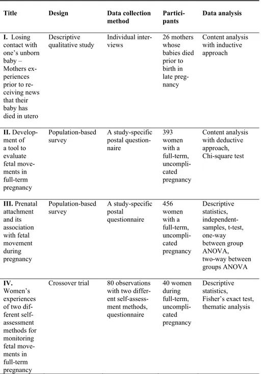
Three methods for collecting data were used in this thesis. Data from inter-views were used in study I, data from questionnaires were used in studies II and III, and data from observations were used in study IV. An overview of the studies is presented in Table 1.
Table 1. An overview of the studies within the thesis
Title Design Data collection method Partici-pants Data analysis I. Losing contact with one’s unborn baby – Mothers ex-periences prior to re-ceiving news that their baby has died in utero Descriptive qualitative study Individual inter-views 26 mothers whose babies died prior to birth in late preg-nancy Content analysis with inductive approach II. Develop-ment of a tool to evaluate fetal move-ments in full-term pregnancy Population-based
survey A study-specific postal question-naire 393 women with a full-term, uncompli-cated pregnancy Content analysis with deductive approach, Chi-square test III. Prenatal attachment and its association with fetal movement during pregnancy Population-based survey A study-specific postal questionnaire 456 women with a full-term, uncompli-cated pregnancy Descriptive statistics, independent- samples, t-test, one-way between group ANOVA, two-way between groups ANOVA IV. Women’s experiences of two dif-ferent self-assessment methods for monitoring fetal move-ments in full-term pregnancy
Crossover trial 80 observations with two differ-ent self-assess-ment methods, questionnaire 40 women during full-term, uncompli-cated pregnancy Descriptive statistics, Fisher’s exact test, thematic analysis
Study I
Study design
A descriptive qualitative interview study.
Sample
Recruiting initially took place at a hospital in central Sweden in 2006. With the help of a staff member who served as a contact at the hospital, an infor-mation letter and an application form were sent to women who had given birth to a stillborn baby at that hospital within the previous year. In order to reach all possible participants and to come in contact with women from different parts of Sweden, contact was made with The Swedish National Infant Fund, a national association for parents who have lost babies through death (106). In-formation about the study was posted on the Swedish National Infant Fund website in 2007 with an invitation to participate in the study regardless of how much time had passed since the stillbirth. After five weeks, the invitation was removed since a sufficient number of interested women had made contact. At the same time, three participants were recruited at the hospital while 23 women were recruited from those who had responded to the information on the website. The inclusion criterion was that the woman had experienced still-birth after 28 full weeks of pregnancy. The exclusion criteria were that the interviewer had been involved in providing care for the participating woman in connection with the birth of her stillborn baby or was acquainted with the woman in any other way.
Data collection
Heads of units and the director of the department of woman’s health i.e. ob-stetric services at the county hospital were given written and oral information about the study. The women who had agreed to take part were contacted in order to obtain additional verbal information and to set the time and place for the interview on the basis of the woman’s own wishes. In most cases (n=22) the women chose to meet in their homes. The remaining four women, in part for practical reasons, chose to have their interviews carried out at a location other than their homes, and this wish was met by meeting three women in a café or office, and another at the home of another participant.
Each interview began with a general conversation on the overall subject in order to establish good contact. By initially establishing confidence in the in-terviewer it was possible to create a relaxed atmosphere prior to carrying out the actual interview. The length of the initial conversation varied. The intro-ductory conversation for the women who were interviewed in their homes
took between 15 and 60 minutes. All the women who were interviewed in their homes showed pictures of their dead baby and other mementos as well as gifts that had been given to the women and their families in connection with the stillbirths. Some women even invited the interviewer to share a meal, or walk around the neighbourhood and visit the baby’s grave. The interviewer was introduced to the other family members, friends close to the family, and pets. Before starting, the interviewer waited until each woman had found a com-fortable position and encouraged her to give a simple signal if she wanted to take a break or even end the interview for emotional or other reasons. When the woman was ready to start the interview, a digital recorder was placed be-tween the woman and the interviewer. A question guide based on clinical ex-perience and on studies of the literature (107) had been designed to contain questions ranging from open to semi-structured in character. The guide con-sisted of five main questions with five to 15 follow-up questions. Three trial interviews were carried out but are not included in those used and are not part of the results. The question guide and the interview were tested during the trial interviews but no changes were seen to be needed. Each woman was invited to talk about her experiences before she was informed that her baby had died. The initial question was: “Can you please describe what it was like when you
learned that your baby was no longer living?” and the first follow-up question
was: “Did you have any suspicion that your baby was no longer living?”. The interviews were carried out as conversations where each woman was encour-aged to talk freely about her experiences prior to being told that her unborn baby had died. The question guide served to provide a framework and struc-ture for the conversation.
Each woman’s experiences from the point in time prior to receiving news that her baby had died in utero until the point when a year had passed since the stillbirth were discussed. Not all the reported experiences are included in this thesis but they have been reported in other studies (108-110). If a woman seemed to be in need of a pause, the interviewer suggested taking a break to make it possible for the woman to recover herself. Usually the woman did not see any need for this so the interview was able to continue as the woman wished, but sometimes 5 to 10 minute breaks were taken. If needed, the view was stopped for a while in order to restore a sense of order in the inter-view situation. The presence of family members, visitors, pets, and other ex-ternal environmental factors such as noise or room conditions could to a greater or lesser degree risk disturbing the woman’s concentration or ad-versely affect the quality of the recording. After any pause the interviewer restarted the interview by summarizing what the woman had described just before the pause. In the event that the woman began to talk about other related factors and events, the interviewer could ask her to return first to the subject at hand by posing questions such as: “You said that you felt all alone when
you no longer could make contact with your baby, what do you mean by that?”
or if the woman did not offer a temporal perspective the interviewer could ask the woman, for example: “If we go back to the time just before you got
con-firmation that your baby was no longer living, can you describe what you were thinking before you called the delivery department?”. The interview was
ended with the question: “What advice do you have for the staff who meet
women in similar situations?” When the interviewer understood that the
woman had said all that she was prepared to say, the interview was ended and the recorder was turned off. Following the interview, a further conversation took place during which it was possible for the interviewer to make sure that the woman felt satisfied. The recorded interviews were transcribed completely and every interview was given a code number. After the audio files had been transferred to separate USB memory sticks, the audio files were deleted from the digital recording device.
Data analysis
The material was subjected to qualitative content analysis following the pro-cedure outlined by Lundman and Graneheim (111). Each of the printed inter-views constituted analytical units. The process of analysis began by printing all of the texts and then having each unit read completely by all members of the research team. The text was then divided into major structural and content units by identifying similarities and differences, and these units were under-lined and highlighted. Content units were then condensed and made more ab-stract by removing text for certain kinds of sounds such as clearing one’s throat or coughing, but not removing sounds such as sighs, sniffles, or crying. Some repeated phrases and words such as “right eh” were removed when they were not used in any specific context but simply to fill out the sentence. The same procedure was followed with other sentence units that could not be placed in relation to the study, for example a sibling’s school situation or a complaint filed against a health care provider. Personal names or place names where replaced with NN and XX respectively to protect confidentiality. Following this, the material was coded by formulating a very short description or by identifying one or more words in the unit that could be used as the code. All codes with the same content were studied so that subcategories could be created. Using these subcategories, it was possible to create categories. Fi-nally, an all-embracing theme was formulated on the basis of interpreting the categories and subcategories. During this entire inductive process the content units were re-read and discussed and then condensed and made more abstract in several iterations by the research team. As a final step, all of the content units were subjected to a final review in order to produce a satisfactory set of codes and categories.
Study II
Study design
A population-based survey.
Sample
In order to study the women’s descriptions of fetal movements during late pregnancy, data was collected in one county in central Sweden between March 2011 and October 2011. The antenatal care in the county at that time was con-ducted by approximately 70 midwives at 23 clinics where the number of preg-nant women registered annually ranged from 26 to 629 at each clinic. Approx-imately 3000 pregnant women registered at one of the antenatal clinics in the county during the year and 2809 births took place at the county’s only hospital equipped with a delivery ward. Of the 1977 women giving birth at the county hospital during the data collection period, 1962 of them were registered at a county antenatal clinic. The remaining 15 women had been registered at clin-ics outside the county.
The inclusion criteria for participation in Study II were: women with an un-complicated, singleton, and late pregnancy in gestation weeks 37+0 - 42+0, who were enrolled at an antenatal clinic in the county council, and who were considered tofollow the standard visiting schedule for antenatal care as spec-ified in the Swedish national guidelines (100). In addition, the woman had to be able to understand, speak, and write Swedish. Exclusion criteria were: preg-nancy complications, rupture of the membranes, or contractions due to labour while answering the questionnaire.
Data collection
The senior physician responsible for the overall antenatal care and the midwife with coordinating responsibility for antenatal clinics in the county received written information about the study. In addition, all of the currently active midwives working in the antenatal clinics and heads of the midwives were given the same printed information. Oral information was given at two differ-ent joint meetings in two differdiffer-ent parts of the county with the midwives, the coordinator and the senior physician. In addition, written information had been sent to those in charge of health centres connected with an antenatal unit. Heads of the units and the director of the department of woman’s health i.e. obstetric services, at the county hospital were informed both orally and in writ-ing. Carry bags with information for prospective participants were personally distributed to every individual midwife at each antenatal unit. While distrib-uting this material at each location, the midwives were further informed either
individually or in groups. A personal letter containing additional information and instructions as well as information concerning making contact with the research team was given to every individual midwife.
Data collection began after every antenatal unit had been visited and after all of the midwives had been seen and given oral and written information. If a midwife needed more information, material was handed over in person or was sent to the unit. All of the material that was handed over was registered in a matrix in order to compare this with how many women had answered the ques-tionnaire. All pregnant women at a planned visit during gestational week 35 or later and who met the inclusion criteria were told about the study in a brief conversation with the midwife. Following that conversation, the women who would be taking part in the study were given an envelope containing infor-mation about the study, a reply form, a questionnaire, and a return envelope. The midwife was instructed to note which women had received oral infor-mation and printed inforinfor-mation and also which women had been excluded from participation and the reasons for exclusion, for example if the woman did not meet the inclusion criteria, and, if she did not, if the woman did not have a sufficient command of Swedish or if she did not want to receive the information material. The midwife was encouraged to record this information in the printed matrix that was to be sent to the research team on request directly after the end of the data collection period. A representative of the research team made repeated spontaneous visits to the antenatal centres or made tele-phone contact in order to find out if the midwives had questions about the study or data collection, or if they needed additional material. The midwives always had the possibility of reaching the research team via telephone or e-mail if they had questions. A total of 554 women received information from the midwives about the study.
Approximately 11 per cent of the eligible pregnant women were not reached. This may partly be due to data collection not beginning simultaneously and because not all of the centres had been reached since they were visited succes-sively over a period of two months. If a woman had agreed to participate, she sent a completed questionnaire and reply form in two separate envelopes to the research team. The 457 women who agreed to participate in the study were asked to complete the questionnaire at the time when they had reached a fully developed pregnancy, that is, when they had completed gestational week 37 (37+0) but before delivery had begun. The questionnaire was designed specif-ically for this study and was based on the prior experience of the research team as well as previous studies (37, 112) and literature (113). The open question (question no. 6) in the questionnaire is the foundation of the study: “Describe how you experienced the way your baby is moving during the present week of your pregnancy (describe both how much your baby is moving and what kinds of movements can be observed). Please feel free to write on the reverse
side of this questionnaire if you need more space”. The women were free to write as much as they wanted to and without any restrictions on the extent of what they wrote. The questionnaire was tested by two personally recruited women with full-term uncomplicated pregnancies. A few small adjustments were made after this. When a sufficient number of questionnaires had been received by the research team, data collection was ended at all the antenatal units. The midwives were informed of this and were asked to record the num-ber of remaining envelopes containing information and they were then in-structed to destroy this material. At the same time, the midwives were asked to send the research team all the matrixes with information about the letters that had been distributed and about the number of excluded candidates.
Data analysis
The written replies were analysed by using content analysis with a deductive approach (111). In order to sort the different types of fetal movements de-scribed by the women, an existing matrix was used. This matrix was devel-oped based on interviews with 40 women with full-term uncomplicated preg-nancies. The matrix consisted of seven predefined categories of movements sorted into two domains; “Powerful movements” including; “strong and pow-erful”, “large”, “slow”, “stretching”, and “side to side”. “Non-powerful” movements including; “light” and “startled” movements (60).
All written material was read through thoroughly. All statements describing movements were marked, coded according to type of movement, and then sorted into appropriate categories in the matrix. The analytical framework in-itially contained the categories from the original matrix (60). New codes were successively added to the framework during the work on analysis since new expressions were identified and these were assigned codes as well. Between eight and 23 new codes per category were identified on the basis of the women’s answers. The categories were developed further and then revised and divided into the two existing domains.
During this deductive process a limited modification of the analytical instru-ment was made through the identification of new codes that were then as-signed to specific categories on the basis of the description of fetal movements as experienced by the women. Every category was assigned its own colour and each separate code in every reply was marked with a colour depending on the category to which the code belonged. When these markings were recorded in the analytical framework the colour was translated using the identification code for that colour. Every individual questionnaire’s code for identification was noted one (1) time per code in the framework in the seven different cate-gories in the matrix. This procedure was used even if the code for the
ex-pressed movement in the analytical instrument occurred several times in a sin-gle answer. As a result the same code for one or more different movements that were included in the category “strength and pressure” was encountered several times in the text, but only one code per category was counted. This was performed to ensure that the number of women who reported movement belonging to a particular category was registered. The total sum of all codes in each answer was registered and finally all of the codes in each separate category were counted. During the entire deductive process, the text was re-peatedly re-read and the coding was discussed several times by the research team. Finally, all of the texts were checked one last time to ensure that the coding and categorisation were correct.
Data handling was performed using the IBM statistical software package for the Social Sciences (SPSS) for Windows, version 20.0.Armonk, NY: IBM Corp.
Study III
Study design
A population-based survey.
Sample
The study was based on the same data collected for Study II and the same procedures for information and selection apply in Study III that were followed in Study II. The aim of Study III was to investigate the association between the magnitude of fetal movements and level of prenatal attachment within a 24-hour period among women in late pregnancy.
The inclusion criteria for participation in Study III were: women with an un-complicated, singleton, and late pregnancy (gestation weeks 34+0-42+0), en-rolled at an antenatal clinic in the county council, who were considered to follow the standard visiting schedule for antenatal care as specified in the Swe-dish national guidelines (100). In addition, the woman had to be able to un-derstand, speak, and write Swedish. Exclusion criteria were: pregnancy com-plications, rupture of the membranes, or contractions due to labour while an-swering the questionnaire.
Data collection
Unlike study II, this study also included women who answered the question-naire before entering gestational week 37+0.
The revised-PAI (PAI-R) scale was used to assess prenatal attachment (91). The three subscales; Anticipation, Differentiation, and Interaction consist of eighteen Likert-type items. A four-point response scale was used as a guide for the identification of a woman’s prenatal attachment (90).
Two questions (question no. 5 and question no. 9) in the questionnaire are the basis for study III. The closed question (question no 5.) was: “Check the an-swers that best indicate how you experienced movements from your baby dur-ing the current week of your pregnancy”. Four alternatives were offered: “My baby is moving a lot in the morning”, “My baby is moving a lot during the daytime”, “My baby is moving a lot during the evening”, and “My baby is moving a lot during the night”. All four questions were to be answered using these three reply alternatives for each: “Yes”, “No”, or “Do not know”. Question no. 9 in the questionnaire consisted of PAI, an instrument for evalu-ating the woman’s prenatal attachment (90) and the first question was: “The following sentences describe thoughts, feelings, and situations that a mother-to-be can feel or experience during pregnancy. Read each statement and put an x in the box next to the statement that best describes how you have felt during the preceding month”.
In order to be able to measure and compare variations in fetal movements dur-ing a 24-hour period with the results from the statements in PAI-R, both with our own data and with measurements in other studies, a new variable was cre-ated.
From the variable “My baby moves a lot”, the variable “firm movements” was constructed. The interpretation was developed in the context provided by the women’s descriptions of the intensity of the movements while the follow-up question concerning frequency is not used in the questionnaire. In the answers to the open question in the questionnaire (question no. 6) “describe how your experience of how the baby is usually moving during your current pregnancy week” as presented in study II (17), 98 percent of the women experienced fetal movements that they described as firm. The three answer alternatives from question 5 were coded and points were assigned as follows: “on one or several occasions” (0-1points), “regularly” (2 points), and “often” (3 points). Three movement groups were created: “day, “evening”, and “night”.
Data analysis
Descriptive statistics were used to present background data. Independent-sam-ples t-tests and a one-way between-group ANOVA were used to compare background characteristics and the magnitude of fetal movements with the PAI-R subscales. Finally, to explore the impact of parity and the magnitude of fetal movements on the PAI-R subscales a two-way between groups ANOVA was performed. Data handling was performed using the IBM statis-tical software package for the Social Sciences (SPSS) for Windows, version 20.0.Armonk, NY: IBM Corp.
Study IV
Study design
A cross-over trial.
Sample
The midwife centres located in two communities in a county in central Sweden were used in the data collection for seven months in 2013-2014. Antenatal care in these regions was at this time carried out by approximately 19 mid-wives at six separate centres. The inclusion criteria for participation in the study were: women with an uncomplicated, singleton, and full-term preg-nancy (gestational weeks 37+0 - 42+0), who were enrolled at an antenatal clinic in one of the two communities in the county council, and who were considered to follow the standard visiting schedule for antenatal care as spec-ified in the Swedish national guidelines (100). In addition, the woman had to be able to understand, speak, and write Swedish. Exclusion criteria were: preg-nancy complications, rupture of the membranes, or contractions due to labour while performing the observations or answering the questionnaire.
Data collection
The senior physician responsible for the overall antenatal care and the midwife with coordinating responsibility for antenatal clinics in the county received written information about the study. Furthermore, all of the currently active midwives working in the antenatal clinics and heads of the midwives were given the same written information. Oral information was given at one joint meeting with all the midwives in the county, their coordinator, and the senior physician. In addition, written information had been sent to all those in charge of health centres connected to the six antenatal clinics and head of the mid-wives. Furthermore, heads of units and the director of the department of
woman’s health i.e. obstetric services at the county hospital were informed both orally and in writing.
Carry bags with written information for prospective participants were person-ally distributed to every individual midwife at each antenatal unit. While dis-tributing the material at each location the midwives were further informed ei-ther individually or as a group. A personal letter containing additional infor-mation and instructions as well as inforinfor-mation concerning making contact with the research team was given to every midwife. All of the material that was handed over was registered in a matrix by the research team, in order to be able to compare this with how many women had participated in the study. Providing confirmation of informed consent was carried out in two steps. Women at a scheduled appointment with a midwife to have an examination during gestational week 34 or later and who met the inclusion criteria were given a brief verbal presentation about the study by the midwife. They were then asked about consent for their telephone numbers to be given to the re-search team so further information could be given via the telephone. Further-more, the midwife gave each woman an envelope containing written infor-mation about the study, a reply form, and a reply envelope that the woman would take home with her. Women who agreed to participate in the study after receiving the telephone call were given an appointment for the first observa-tion and asked to send the reply form to the research team. In order to report the number of available women, the midwives made a note in a matrix when women had received information, and when women did not receive infor-mation they made an explanatory note. The women then chose where they wanted carry out the observation; in their own home, in a quiet location at the county hospital or the university, or in a room at the antenatal unit.
The first 20 participants began the first session by using the self-assessment method count-to-ten and then at the second and final session using the assessment method mindfetalness. Participants 21 to 40 carried out the self-assessment methods in the reverse order, first using the self-self-assessment method mindfetalness and then the self-assessment method count-to-ten. Before the participants began their observational sessions, they were given brief additional oral and written information about the self-assessment meth-ods that had previously been sent out as enclosures in the information letter. The participants were able to raise questions and then prepare themselves for their observation sessions.
The participants were asked to lie down on their left side and feel if the baby was awake. The same observer carried out all 80 observations sitting about