Heart rate variability and pacemaker treatment in children with Fontan
circulation
Jenny Alenius Dahlqvist
Department of Clinical Sciences, Pediatrics
Department of Radiation Sciences
This work is protected by the Swedish Copyright Legislation (Act 1960:729) Dissertation for PhD
ISBN: 978-91-7855-007-4 ISSN: 0346-6612 New series no: 2022
Cover illustration: Sigge Mårtensgård Klüft, 6 years. Layout cover: Inhousebyrån, Umeå universitet.
Table of Contents
Abstract ... 4 Background ... 4 Aim ... 4 Methods ... 4 Results ... 4 Conclusions ... 5 Abbreviations ... 6 Original papers ... 8 Sammanfattning på svenska ... 9 Bakgrund ... 9 Syfte ... 9 Metoder ... 9 Resultat ... 9 Slutsats ... 10 Introduction/ Background ... 11Univentricular heart malformations ... 11
Fontan circulation ... 14
Management ... 14
Stage I procedures ... 14
Bidirectional Glenn procedure (BDG)... 15
Completion of Fontan circulation ... 16
Total Cavopulmonary connection (TCPC) ... 16
Physiology of the Fontan circulation ... 19
Prognosis ... 20 Complications ... 20 Arrhythmia ... 21 Tachyarrhythmia ... 22 Bradyarrhythmia ... 23 Pacemaker implantation ... 24
ECG monitoring after Fontan surgery ... 25
The autonomic control of the circulation ...26
Heart rate variability (HRV) ...29
How to measure HRV ... 29
Spectral analysis ... 30
The Poincaré diagram ... 31
Age- and gender dependency of HRV ... 33
Clinical application of HRV ... 33
HRV in congenital heart disease ... 33
HRV in univentricular heart defects and Fontan circulation ... 34
HRV and arrhythmia ... 34
Overall Aim ... 35
Materials and Methods ... 38
Identification of the patients ... 38
Data collection... 38
Clinical data ... 38
Ambulatory 24-hour electrocardiogram (Holter) ... 38
Hand-held ECG ... 38
ECG analysis ... 39
HRV analyses ... 39
Power spectral analysis ... 40
Poincaré plots ... 40 Statistical methods ... 40 Results ... 43 Main findings ...43 Patients ... 44 Controls ... 45
ECG findings (paper I, III) ... 46
Heart Rate Variability (study I, II, III, V) ... 46
Lateral tunnel (LT) versus extracardiac conduit (EC) (paper I) ... 46
HRV through surgical stages to TCPC (Paper II) ... 48
Intermittent short-term ECG findings (paper III) ... 49
HRV in patients with SND (Paper V) ... 51
Pacemaker implantation in patients with Fontan circulation (paper IV) .... 56
Indication for pacemaker implantation ... 56
LT versus EC ... 57
Pacemaker implantation in different anatomical diagnoses ... 58
Symptoms related to pacemaker implantation... 60
Discussion ... 61
Arrhythmia ... 61
ECG registration ... 61
Bradycardia... 62
Sinus node dysfunction (SND) ... 63
Pacemaker treatment ... 64
Heart rate variability (HRV) ... 66
HRV and SND ... 66
Other plausible reasons for low HRV in Fontan patients ... 67
HRV related to surgical stages ... 67
Conclusions ... 69
Acknowledgements ... 70
Abstract
Background
Fontan surgery is performed in children with univentricular heart defects. Arrhythmias are frequent complications, occasionally requiring pacemaker treatment. Previous data regarding indications and risk factors for pacemaker treatment in Fontan patients is limited and conflicting. Heart rate variability (HRV) reflects autonomous nervous activity controlling the sinus node and has been associated with tachyarrhythmias in both adults and children, as well as in adults with sinus node dysfunction (SND).
Aim
To study HRV, arrhythmia and pacemaker treatment in children with Fontan circulation— with the purpose of contributing to the reduction of long term complications in this patient group.
Methods
We have retrospectively reviewed pacemaker therapy in all Swedish patients who underwent Fontan surgery from 1982 to 2017 (n=599). We have also analysed HRV from 24-hour Holter ECG recordings in 112 children with Fontan circulation and in children with univentricular heart defects before bidirectional Glenn (BDG) procedure (n=47), before and on completion of Fontan surgery (n=47 and 45 respectively). Analysis was performed by power spectral analysis and Poincaré method, and results compared with healthy controls. Furthermore, HRV was analysed in Fontan patients who later required a pacemaker due to severe SND. Results were compared with Fontan patients who had SND, without indication for pacemaker treatment, with patients with Fontan circulation without SND and healthy controls. In addition we evaluated the possibility to analyse arrhythmias and HRV in 27 Fontan children using intermittent ECG recordings with a handheld devices at home during a 14-day period.
Results
After a mean follow-up of 12 years, 13% (78/599) of patients with Fontan circulation had received a pacemaker. Patients operated with the extracardiac conduit (EC) had a significantly lower prevalence of pacemaker implantation (6%) than patients with a lateral tunnel (LT) (17%). The most common pacemaker indication in patients with Fontan circulation was SND (64%). Children with Fontan circulation showed significant reductions in several HRV parameters, compared with controls. No significant differences were found between patients
(representing changes in heart rate over 24-hours) significantly increased compared to pre-BDG. Compared with healthy controls, patients post-BDG, had significantly longer RR intervals and reduced overall HRV. PHF (reflecting
parasympathetic control of the heart) was significantly reduced after TCPC as compared to before (paper II). Fontan patients with SND showed significantly elevated SD2 (representing changes in heart rate over 24-hours), somewhat reduced in patients that later required a pacemaker (Paper V). Handheld ECG analysis revealed frequent ventricular extra systoles in one patient and episodes of supraventricular tachycardia in another. Seven Fontan patients showed reduced HRV recorded with the handheld device over a 14-day period (paper III).
Conclusions
Overall HRV was reduced in patients with univentricular heart defects during the different surgical stages of Fontan surgery, compared to healthy controls. HRV was reduced in both patients with LT and EC with no significant difference between them. After BDG heart rate was significantly reduced as compared to before. PHF, reflecting the parasympathetic innervation of the heart was reduced
after as compared to before TCPC. Pacemaker treatment is commonly needed in patients with Fontan circulation, and SND was the most prevalent indication for implantation. The prevalence of Fontan patients requiring pacemaker treatment was significantly lower in patients with EC. HRV analysis can contribute to management when following-up patients with Fontan circulation.
Abbreviations
AA- aortic atresiaANOVA- analysis of variance ANS- autonomic nervous system AS- aortic stenosis
AV- atrioventricular
AVSD- atrioventricular septal defect BSA- body surface area
BT- Blalock-Taussig CI- confidence interval
DILV- double inlet left ventricle DKS- Damus–Kaye–Stansel DORV- double outlet right ventricle EF- ejection fraction
EC- extracardiac conduit ECG- electrocardiogram HF- high frequency
HLHS- hypoplastic left heart syndrome HRV- heart rate variability
IVC- inferior vena cava LF – low frequency
LT- lateral tunnel LV- left ventricle MA- mitral atresia
NYHA - New York Heart Association PA- pulmonary atresia
PAB- pulmonary artery banding PDA- patent ductus arteriosus PSD- power spectral density RV-right ventricle
SA- sinoatrial
SD- standard deviation SVC- superior vena cava TA- tricuspid atresia
TCPC- total cavopulmonary connection TGA- transposition of the great arteries VLF- very low frequency
TP- total power
Original papers
I. Heart rate variability in children with Fontan circulation - lateral tunnel and extracardiac conduit
Dahlqvist JA, Karlsson M, Wiklund U, Hörnsten R, Strömvall-Larsson E, Berggren H, Hanséus K, Johansson S, Rydberg A Pediatr Cardiol. 2012;33(2):307-15.
II. Changes in heart rate variability during surgical stages to completed Fontan circulation
Dahlqvist JA, Wiklund U, Karlsson M, Hanséus K, Strömvall-Larsson E, Johansson Ramgren J, Berggren H, Rydberg A In manuscript.
III. Handheld ECG in analysis of arrhythmia and heart rate variability in children with Fontan circulation
Dahlqvist JA, Karlsson M, Wiklund U, Hörnsten R, Rydberg A J Electrocardiol. 2014; 47 (3):374-82.
IV. Pacemaker treatment after Fontan surgery – a Swedish national study Dahlqvist JA, Sunnegårdh J, Hanséus K, Strömvall-Larsson E, Nygren A, Dalén M, Berggren H, Johansson Ramgren J, Wiklund U, Rydberg A
Re-submitted to Congenital Heart Disease after minor revisions.
V. Sinus node dysfunction in patients with Fontan circulation; could heart rate variability be a predictor for pacemaker implantation?
Dahlqvist JA, Wiklund U, Karlsson M, Hanséus K, Strömvall- Larsson E, Nygren A, Eliasson H, Rydberg A
Sammanfattning på svenska
Bakgrund
Barn som föds med enkammarhjärta genomgår hjärtoperationer i flera steg, operationsmetoden kallas Fontankirurgi. Det är vanligt att dessa patienter drabbas av hjärtrytmrubbningar och ibland behövs pacemakerbehandling. Tidigare forskning på denna patientgrupp kring riskfaktorer för hjärtrytm-rubbningar och pacemakerbehandling är få och delvis motstridiga. Hjärtfrekvensvariabilitet (HRV) används för att studera det autonoma (icke-viljestyrda) nervsystemets kontroll på sinusknutan. Förändrad HRV har tidigare associerats till olika typer av rytmrubbning.
Syfte
Att studera HRV, hjärtrytmrubbning och pacemakerbehandling hos barn och ungdomar med enkammarhjärta för att i förlängningen kunna bidra till förbättrad uppföljning avseende sena komplikationer i denna patientgrupp.
Metoder
Vi har studerat alla patienter som genomgått Fontan kirurgi i Sverige från 1982 till 2017 (n=599), avseende pacemakerbehandling. Vi har också analyserat HRV från 24-timmars-EKG-registreringar hos 112 barn och ungdomar med enkammarhjärta och jämfört med 66 friska jämnåriga. HRV analyserades före och efter de olika operationsstegen i Fontankirurgin. Dessutom analyserades HRV från 24-timmars EKG före pacemakerimplantation hos patienter som senare behövde pacemakerbehandling p.g.a. uttalad sinusknutedysfunktion (SND) och jämfördes med patienter med enkammarhjärta och SND men utan pacemakerbehandling, patienter med enkammarhjärta utan SND och med friska jämnåriga. Vi undersökte också möjligheten att använda korta (30 sekunder) registreringar s.k. ”tum-EKG” för analys av hjärtrytmrubbning och HRV.
Resultat
Under 12 års uppföljning efter Fontankirurgi fick 13% (78/599) av patienterna pacemakerbehandling. Bland patienter som opererats med operationsmetoden extrakardiell tunnel (EC) hade en signifikant lägre andel behov av pacemaker (6%) jämfört med dem som opererats enligt metoden lateral tunnel (LT) i förmaket (17%). Den vanligaste indikationen för pacemakerimplantation var SND (64%). Pacemakerbehandling var vanligare vid vissa typer av underliggande hjärtfel; mitralis atresi (MA; 44%), dubbelt utflöde från höger kammare (DORV; 24%) och dubbelt inflöde till vänster kammare (DILV; 20%). Barn med enkammarhjärta hade nedsatt HRV, jämfört med friska jämnåriga. Vi fann ingen skillnad avseende HRV när det gäller EC och LT. Efter operationssteget
bidirektionell Glenn kirurgi (BDG) fann vi signifikant längre RR-intervall (=lägre puls) jämfört med före. Patienter med SND hade signifikant förhöjning av HRV parameter SD2 (som speglar dygnsförändringar i HRV) jämfört med friska kontroller och SD2 var något lägre hos dem som behövde pacemaker p.g.a. SND. Tum-EKG undersökningen visade hjärtrytmrubbning hos två och låg HRV hos sju patienter.
Slutsats
Barn, ungdomar och vuxna med enkammarhjärta är en växande patientgrupp med successivt förbättrad överlevnad. Det är viktigt att uppmärksamma sena komplikationer såsom hjärtrytmrubbning. Analys av hjärtfrekvensvariabilitet skulle i förlängningen kunna bidra till förbättrad uppföljning för att tidigt identifiera hjärtrytmrubbningar.
Introduction/ Background
Univentricular heart malformations
Univentricular heart defects are considered one of the most complex diagnoses in congenital heart disease. This group consists of different malformations of the heart, which all have in common, that the heart cannot be repaired to a normal two ventricle circulation. The underlying cardiac malformations resulting in univentricular circulation can be due to; atresia of one of the atrioventricular (AV) valves (tricuspid or mitral atresia (MA)); because the AV valves both empty into the same ventricle (double inlet left or right ventricle) or common AV valve and only one well-developed ventricle (unbalanced atrioventricular defect). Another common type of univentricular malformation is the hypoplastic left heart syndrome. Additional rare types of cardiac malformations include pulmonary atresia with intact ventricular septum (PA/IVS), double outlet right ventricle (DORV) with remote ventricular septum defect (VSD), extreme Ebstein’s anomaly of the tricuspid valve or cases where lesions such as significant straddling of the AV valves, multiple large ventricular septal multilevel outflow obstructions, or a combination of these may make the univentricular pathway a less risky and more predictable choice of treatment(1).
Hypoplastic left heart syndrome (HLHS): is the most common form of univentricular heart defects and occurs in 2.3-2.6 per 10 000 live births (2-4). The classic variant of HLHS is characterised by a diminutive left ventricle (LV) with small left-sided structures and endocardial fibroelastosis. There is a variability of both obstructions (aortic atresia (AA)/MA), (AA/mitral stenosis (MS)), (aortic stenosis (AS)/MS) and size of the LV, ascending aorta and aortic arch. The malformation is ductal-dependent, and without treatment the patient dies in the neonatal period (5). The babies are also dependent on an un-restricted atrial septal defect (ASD) which allows oxygenated and deoxygenated blood to mix. With modern medical and surgical management there has been a large improvement in survival, however, HLHS is still one of the most challenging univentricular malformations to treat and in a recent study the 6-year transplant-free survival rate was 64%(6).
Double inlet left ventricle (DILV): In DILV both atria empty into the left ventricle through two separate AV valves. The dominant left ventricle connects to the rudimentary right ventricle through a VSD. DILV is a heterogeneous anomaly based on presence of ventriculo-arterial discordance, size of VSD, and semilunar valve hypoplasia or stenosis (1). Consequently, a new-born with DILV might have restrictive pulmonary blood flow, unrestrictive pulmonary blood flow with or
without systemic outflow tract obstruction, or a balanced circulation. In a recent follow-up study 10-year freedom from death or transplantation was 87% (7). Tricuspid atresia (TA): In TA there is an absence of the tricuspid valve, and a hypoplasia of the right ventricle. In most of the cases there is a VSD. In approximately 75% the relationship between the aorta and pulmonary artery is normal, and in 25 % there is a ventriculo-arterial discordance, the pulmonary valve can be atretic, stenotic or normal (8). TA occurs in 0.5-0.8 per 10 000 live births (3, 9). The Fontan operation was first described for TA (10).
Figure 1. Tricuspid atresia with VSD.
SVC= superior vena cava, IVC= inferior vena cava, RA= right atrium, RV= right ventricle, MPA=mean pulmonary artery, RPA= right pulmonary artery, LPA=
Atrioventricular defect (AVSD): Fontan surgery is required in unbalanced AVSD and could also be the best alternative if there is straddling of the AV valve with two well-developed ventricles (11). In the unbalanced AVSD, there is hypoplasia of either the right or left ventricle. In most cases, the right ventricle is the dominant ventricle, since generally right ventricular hypoplasia is better tolerated than left ventricular hypoplasia (12). When the AV valve sits more over one ventricle than the other, the contralateral ventricle is typically hypoplastic. Unbalanced atrioventricular AVSD occurs in approximately 10% of patients with AVSD (12).
Pulmonary atresia with intact ventricular septum (PA/IVS): is characterized by complete obstruction to right ventricular outflow with varying degrees of right ventricular and tricuspid valve hypoplasia and of anomalies of the coronary circulation. Patients with a more severe hypoplasia of the tricuspid valve and the right ventricle are more likely to have fistulae between the right ventricle and the coronary arteries (13).PA/IVS is a ductal dependent lesion since there is no blood-flow from the right ventricle to the pulmonary arteries. Babies present as cyanotic due to an atrial level right to left shunt. The pulmonary arteries are small, but their architecture and branching are otherwise normal. PA/IVS is reported to occur in approximately 4 out of 100 000 live births, among them about 20% need a Fontan palliation (14, 15).
Mitral atresia (MA): In MA, the mitral valve is either imperforate or absent and there is a postero-inferior incomplete left ventricle. There is considerable morphologic heterogeneity, which influences the hemodynamic picture. When an even tiny ventricular septum is recognizable, a hypoplastic ventricular chamber almost always exists anteriorly (16).
Heterotaxia syndrome: Heterotaxia, or isomerism occurs in approximately 1 out of 10 000 live births and is characterized by isomeric findings in the thoracic organs and random arrangement of the abdominal organs (17, 18). Patients with heterotaxia syndrome constitute a subgroup of patients with Fontan circulation, and experience high rates of late morbidity and mortality (19). Right atrial isomerism has been shown to be associated with asplenia, morphologic right ventricle, a common atrioventricular valve, pulmonary atresia or pulmonary stenosis, anomalous pulmonary venous drainage (partial or total and with obstruction) and interrupted inferior vena cava (IVC) (20). Children with left atrial isomerism have an absence or hypoplasia of the sinoatrial node and are more likely to have congenital atrioventricular block or a junctional rhythm (18, 20).
Fontan circulation
The Fontan procedure was first described as a palliative operation for tricuspid atresia in 1971(10). Nowadays the Fontan procedure has been modified, and is used in a wide spectrum of univentricular heart defects unsuitable for a biventricular repair. Fontan surgery re-routes the venous return from the superior vena cava (SVC) and IVC to the pulmonary arteries without a pumping ventricle. Pulmonary blood flow is driven by central venous pressure and is augmented mainly by the peripheral skeletal muscle pump (21). For this circulation to be effective, the patient must have a low pulmonary vascular resistance, relatively normal systolic and diastolic function of the single ventricle, and sufficiently large pulmonary arteries to avoid any mechanical resistance. Thus, the criteria for the procedure includes: normal ventricular function, adequate pulmonary artery size, no distortion of pulmonary arteries from prior shunt surgery, low pulmonary artery pressure (below 15 mmHg), low pulmonary vascular resistance (<4 Woods units/m2), no AV valve leakage, normal heart
rhythm and no right atrial enlargement (10). Normal systemic venous drainage was previously a criteria, however a modification of the Fontan procedure, the Kawashima procedure, is used for patients with abnormal systemic venous drainage due to an interrupted IVC nowadays (22).
Since 1971, the surgical technique has developed from the atrio-pulmonary to the total cavopulmonary connection (TCPC). TCPC involves a connection to re-route the venous return from the SVC and IVC to the pulmonary arteries. The surgery is usually staged to give the body time to adjust to the large hemodynamic changes.
Management
In all ductal-dependant congenital heart defects intravenous prostaglandin E1 are administrated to maintain a patent ductus arteriosus (PDA) as soon as the diagnosis is suspected.
Stage I procedures
The majority of neonates with univentricular heart defects need to undergo neonatal cardiac surgery. Depending on the underlying malformation, different procedures are required to provide for sufficient blood flow to both the pulmonary and the systemic circulations. This Stage I procedure is performed during the first weeks of life.
and pulmonary atresia or severe pulmonary stenosis will need a systemic-pulmonary shunt. The shunt is typically small (3.5 mm) and designed to last only a few months.
Atrial septectomy or septostomy: is performed in heart defects dependent on a non-restrictive shunt at the atrial level, for example in a baby with HLHS and a restrictive ASD may require a balloon atrial septostomy (23).
Pulmonary arterial banding (PAB): is performed when there is
over-circulation to the pulmonary arteries, for example in a baby with DILV.Norwood operation: with a modified BT-shunt or RV (right ventricle)-PA (pulmonary artery)-(Sano)-shunt is performed in HLHS. During the Norwood procedure a neo-aorta is created by dividing the pulmonary artery and using the transected pulmonary artery as well as homograft material, to construct a new ascending aorta and aortic arch. The small native aorta is incorporated into the graft.
Damus-Kaye-Stansel procedure: is performed in patients with a
univentricular heart defect with systemic outflow obstruction, for example in HLHS.
A minority of patients with univentricular defects have balanced systemic and pulmonary circulations, and thus may not need to go through a stage I surgery. For example is a baby with TA and PS where the restriction of pulmonary blood-flow is enough to ensure adequate oxygenation of the blood, but without pulmonary over-circulation.
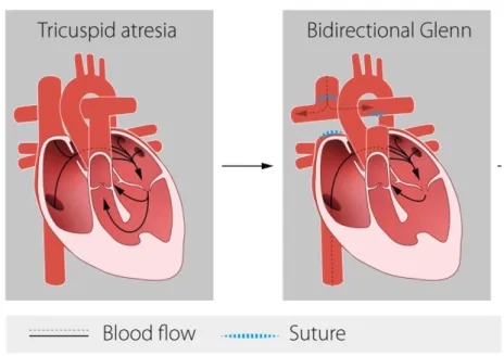
Bidirectional Glenn procedure (BDG)
Stage II of the Fontan procedure is the BDG or bidirectional cavopulmonary connection (BCPC), which involves transection of the SVC and, oversewing of the proximal end. The distal end is anastomosed end-to-side with the pulmonary arteries. This is typically performed between three to six months of age. At this point the pulmonary vascular resistance has declined and the pulmonary arteries have grown to an adequate size. During BDG procedure shunts or PA-bands are removed.
The Glenn shunt circulation is characterised by passive flow. Venous return passively drains from the SVC into the pulmonary arteries. Advantages of the Glenn circulation are that the pulmonary blood flow now is effective and adequate, and the single ventricle now has a reduced pressure and volume load.
However, since the IVC empties to the single ventricle that pumps the blood to the systemic circulation the child will still be cyanotic (Figure 2).
Figure 2. Bidirectional Glenn (BDG).
To the left: An example of univentricular heart (Tricuspid atresia and ventricular septum defect) before BDG. To the right: In the BDG procedure the superior vena cava (SVC) is connected to the pulmonary arteries.
Completion of Fontan circulation
In earlier days the atrio-pulmonary Fontan, was used. In this operation the right atrial appendage was directly anastomosed to the pulmonary artery, providing a pathway for blood from the IVC and SVC to the pulmonary circulation. The atrial septum was left intact, and there should be no residual shunting between the atria. This form of Fontan is associated with more late complications and may need conversion to cavopulmonary connection at a later date (24).
Total Cavopulmonary connection (TCPC)
will have decreased. After the TCPC the venous return flows passively into the pulmonary arteries, then the oxygenated blood returns to the single ventricle which pumps to the systemic circulation. This procedure results in the child being relieved of cyanosis. The deoxygenated coronary sinus is typically left to drain into the systemic circulation, consequently the patient does usually not have a 100 % blood oxygen saturation.
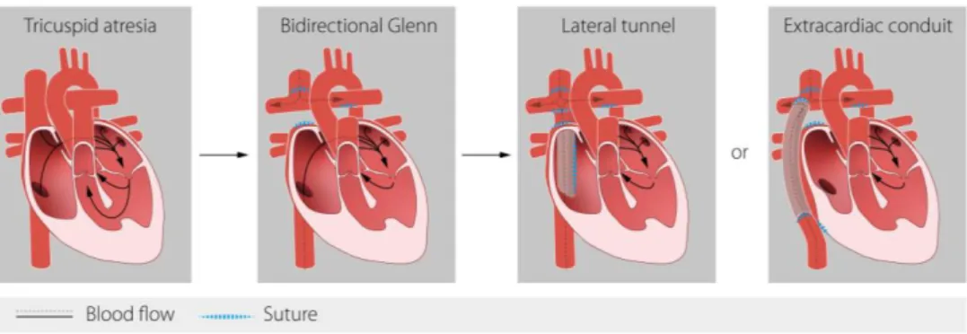
Lateral tunnel (LT) and extracardiac conduit (EC)
The surgical technique used for IVC connection in TCPC surgery, described in 1988 (25) has evolved from an intra-atrial lateral tunnel (LT) to the extracardiac conduit (EC) (Figure 3). The intra-atrial LT surgery is performed via cardiopulmonary bypass and cardioplegic arrest (or induced ventricular fibrillation). A GORE-TEX® baffle is constructed in order to direct the blood flow
from the IVC to the superior end of an atriopulmonary anastomosis. A small portion of atrium remains in the circuit to provide growth potential, and acts to minimise the risk of dilatation and arrhythmias compared to the older atrio-pulmonary Fontan method. Thus, there is still scarring in the right atrium after the LT operation. The EC was described in 1990 (26). The IVC is transected and the cardiac end is over sewn. A GORE-TEX® tube graft is sutured to the IVC and
the other end of the tube is connected end to side to the right PA.
Figure 3. Surgical stages of Fontan circulation.
To the left: example of univentricular heart (TA and VSD) before BDG. In the middle to the left: In the BDG procedure the superior vena cava (SVC) is connected to the pulmonary arteries. To the right: The two variants of the total cavopulmonary connection (TCPC); lateral tunnel (LT) or extracardiac conduit (EC).
The EC has several theoretical advantages, including flexibility in anatomically difficult situations (heterotaxy), the avoidance of sinus node manipulation, decreased suture lines and pressure in the right atrium (decreasing arrhythmogenic potential), and avoidance of cardioplegic arrest since no access to the right atrium is needed (27, 28). Potential drawbacks of the EC Fontan include the lack of growth potential and the risk of thrombosis in the prosthetic conduit. These theoretical advantages of EC versus LT have not been fully investigated, and controversy remains in this field (27, 29, 30).
In Sweden the Fontan type of surgery was sporadically used until the beginning of the 1990s before developing into routine management in patients with univentricular heart defects. A centralisation process was performed in 1992 and the pediatric hospitals in Gothenburg and Lund were assigned to perform all pediatric cardiac surgery. Beginning in 1998, the preferred surgical method in Sweden has switched from LT to EC.
Fenestration
A fenestration can be created during TCPC surgery. A fenestration is a small defect that allows shunting between the venous and systemic circulations. After Fontan surgery a fenestration offers a bypass, which reduces venous congestion, and increasing ventricular pre-load ultimately leading to increased cardiac output. With the increase in cardiac output comes a decrease in arterial saturation. Fenestration has been proven to minimise post-operative low cardiac output, pleural and pericardial effusions and ascites, as well as long-term complications such as diminished exercise performance, and protein-losing enteropathy (PLE) (31-33). The mechanism for this is that flow through the fenestration increases systemic ventricular preload, which in turn improves cardiac output. In addition, in LT TCPC, the fenestration limits the increase in systemic venous pressure and thereby might inhibit development of postoperative pleural effusion and ascites. Despite the potential benefits of fenestration, there are drawbacks including the risks of systemic embolization and systemic desaturation (34). In approximately 30-40% of the patients fenestrations close spontaneously; it can also be closed by a catheter intervention (34, 35). In Sweden fenestration is not routinely used.
Physiology of the Fontan circulation
Optimal cardiac output in the Fontan circulation requires attention to volume status (preload), vascular resistance (afterload), heart rate, rhythm, and myocardial contractility.
The normal heart has two ventricles that pump blood in synchrony to the pulmonary and systemic circulations. In the Fontan circulation, the blood-flow to, and through, the pulmonary circulation is driven passively by the remaining post capillary energy. Since there is no ventricle pumping to the pulmonary circulation, systemic venous pressure remain elevated compared to a normal biventricular circulation (36).Many of the common problems seen post-Fontan surgery are related to resistance in the pulmonary vascular bed. High resistance in the pulmonary vasculature creates a bottleneck, with congestion upstream and restricted flow downstream (36). In a Fontan circulation it is necessary that the systemic venous pressure, the resistance in the pulmonary vasculature and the ventricular filling pressures remains low. The contractility of the ventricle can increase the cardiac output to a certain degree, but the pulmonary vascular resistance has a much larger influence on cardiac output. The limit of the cardiac output is the preload, and well developed pulmonary arteries is extremely important (36). Thus, the Fontan patient do not tolerate hypovolemia or arrhythmias well (37).
The single ventricle, during the stages to complete Fontan circulation, is exposed to different volume loading conditions. During fetal life and after the initial palliation there is a large volume overload, approximately 250-350% of normal/ body surface area (BSA) (38). After BDG, the volume load is reduced to about 90% of normal /BSA (38). The Fontan operation will result in further reduction of volume loading to 50-80 % of load of the normal ventricular load (38). During exercise a person with Fontan circulation will experience limitations compared to persons with normal biventricular hearts. During exercise, in a normal circulation, pulmonary blood flow is increased though reduction of pulmonary vascular resistance due to vasodilatation and increased work of the right ventricle. A person with Fontan circulation has two major disadvantages during exercise; firstly, the reactivity of the pulmonary vasculature is limited and a right ventricle is absent (36). Regular exercise may have positive effects on the pulmonary vascular resistance by vessel recruitment and vasodilatation (39). Secondly, a regular atrial rhythm with AV synchrony is one of the most important prerequisites for the long-term effective functioning of this preload dependent circulation, especially during exercise.
In this abnormal type of circulation, even mild forms of arrhythmia may therefore be deleterious for the patient with Fontan circulation by hampering cardiac output.
Prognosis
Due to advances in surgical methods and pre-, peri- and post-operative medical care for these patients, life expectancy is steadily improving. Post-operative survival 20 years after Fontan procedure is approximately 85% (32, 40). In patients over 16 years, the incidence of sudden death is moderately elevated; 2.1/1000 patient years, compared with all patients with congenital heart disease with an event rate of 0.4 deaths/1000 patient years (41). Co-morbidities or complications are becoming more and more important. Early detection of Fontan associated disease is the key to reduce mortality.
Complications
Improvements of the diagnostics, cardiac surgery, and intensive care procedures have made it possible for children with different complex univentricular heart malformations to be palliated with excellent survival (28, 32, 40). Patients with Fontan circulation enjoy a nearly normal life, including mild-to-moderate physical activities (42). In a large study of Fontan survivors more than 90% were in New York Heart Association (NYHA) class I or II (43). As patients live longer, long term outcomes and late complications are now becoming more apparent. Follow-up of patients with Fontan circulation is challenging since there is a high risk for morbidity and mortality and also a large variability between patients with very good and very poor outcome (40). Many factors such as underlying anatomical diagnosis, elevated right atrial or central venous pressure, history of PLE contribute to mortality or heart transplantation (40).
PLE: is a rare and troublesome complication of the patients (44). The pathophysiology is not fully understood. A multifactorial proposed mechanism include; a response to the altered hemodynamics, especially low cardiac output with increased mesenteric vascular resistance, an inflammatory process and an altered function of the enterocytes (45). During follow-up symptoms of diarrhoea, abdominal pain, peripheral oedema, pleural and pericardial effusions, ascites and failure to thrive indicate PLE. Laboratory results show low serum albumin and increased alpha-1-antitrypsin in faeces (46). Treatment of PLE is individualised, with the aim of improving cardiac output. Underlying arrhythmia causing decrease in cardiac output should be ruled out. Pacemaker treatment may be indicated. PLE is considered an indication for heart transplantation.
Plastic bronchitis: Is a very serious rare complication. In plastic bronchitis bronchial casts obstruct the airways (47). Plastic bronchitis may require takedown of the Fontan circuit or cardiac transplantation (48).
Liver disease: Liver disease is an increasingly recognised complication post Fontan palliation. Hepatic fibrosis progressing to high grade cirrhosis, and even hepatocellular carcinoma, is a serious complication in adults with Fontan circulation (49).
Kidney disease: Patients with Fontan circulation are often at risk of developing reduced kidney function. The pathophysiological mechanisms proposed are end-organ dysfunction due to low cardiac output and venous congestion (50). Thrombosis: Patients with univentricular heart defects are at increased risk of thromboembolic events though out life. In the neonatal period, occlusion of the systemic-pulmonary shunt is a significant risk with devastating consequences (51). As patients get older thromboembolic events continues to be a common complication (52). Patients with atrial arrhythmia are at increased risk of thromboembolism (52).
Fontan failure
The Fontan circulation is initially well-tolerated by most patients. Early Fontan circuit failure is most commonly the result of underappreciated preoperative risk factors or intraoperative myocardial injury (53).
Several factors contribute to the ventricular dysfunction seen in patients with Fontan circulation. The heart defect can in some cases involve some grade of cardiomyopathy. A morphological right ventricle can adapt to the hemodynamic demands of a Fontan circulation, but over time, it is more likely that a morphological right ventricle than a morphological left ventricle fails (54). The accumulated effect of multiple surgeries, as well as periods of with a volume-overloading of the ventricle during neonatal and childhood palliation may negatively impact long-term ventricular function (53). Furthermore, in the Fontan circulation, the ventricle must drive cardiac output through two resistance beds, resulting in a chronic increase in pressure work (55).
Arrhythmia
Atrial arrhythmias and sinus node dysfunction are the most frequent complications in patients with Fontan circulation (42, 56-62). The development of any arrhythmia, including both tachyarrhythmias and bradyarrhythmias, has been shown to be independent predictors of late Fontan failure and of sudden death (60).
Tachyarrhythmia
Supraventricular tachycardia (SVT)
The most common type of supraventricular tachycardia in Fontan patients is intra-atrial reentrant tachycardia (63). Risk factors for development of atrial tachycardia include heterotaxia syndrome, SND, older age at Fontan procedure, early postoperative tachycardia, atrio-pulmonary type of Fontan, and duration of follow-up since Fontan procedure (64-67). In the atrio-pulmonary Fontan there is a “stretch” in the right atrium, this contributes to the high incidence of arrhythmia (60, 67). Other pathophysiological mechanisms are fibrosis, scarring and suture lines within the atrial tissue that act as a substrate for atrial reentrant circuits to develop (68, 69). With the development of the EC Fontan surgical technique there was a hope of reduction in the incidence of arrhythmia. Since the EC conduit minimises atrial incisions, late complications like SND and atrial tachycardia were expected to decline (60, 70). Concerning late tachyarrhythmia after TCPC surgery studies comparing LT and EC have shown mixed and conflicting results. Lower incidence of atrial tachycardia in the EC group compared to the LT group has been shown in some studies (27, 71, 72), but other studies have shown no difference in the incidence of late atrial tachycardia between LT and EC Fontans (73, 74).
In the patient with Fontan circulation, persistent atrial tachycardia is a serious condition, associated with morbidity and mortality (37, 75). When arrhythmia presents careful investigation concerning electrolyte and thyroid function but also ventricular function, AV-regurgitation, outflow obstruction, obstruction in the Fontan circuit is mandated. Immediate treatment options include electrical cardioversion, overdrive pacing manoeuvres or anti arrhythmic drugs. Long-term therapies include anti-arrhythmic drugs, intermittent cardioversions, catheter ablation or Fontan revision surgery combined with Maze procedure (63, 76, 77).
Ventricular tachycardia (VT)
Ventricular arrhythmia is not as common as atrial tachycardia, however VT and complex ventricular extra systoles occur among children and adolescents with Fontan circulation (62, 71). VT is assumed to be an important cause of sudden death in patients with Fontan circulation (40). However, the clinical relevance of asymptomatic ventricular arrhythmia is, found on Holter recordings in children with Fontan circulation, is uncertain; non-sustained VT was not associated with sudden cardiac events in patients with Fontan palliation in one study (78).
Bradyarrhythmia
Sinus node dysfunction (SND)
SND is found in 11-45% of patients with Fontan circulation (29, 58, 59, 71, 79-82). SND is broad array of abnormalities in sinus node and atrial impulse formation and propagation (83). A definition often used in pediatric cardiology research studies is one or more of the following: 1) minimal or mean heart rate 2 standard deviations (SD) below the mean value for age and gender (84), 2) a dominant junctional rhythm, 3) sinus pauses of three or more seconds on Holter recording and/or 4) peak heart rate during exercise lower than 80% of the predicted value for age and gender (56, 71).
SND in patients with Fontan circulation is likely to occur as a result of either damage to the sinus node during surgery or reduced blood supply to the sinus node that may result in fibrosis (58, 81, 85). In some cases the underlying anatomy, such as left atrial isomerism, may contribute to development of SND (18, 86). Late SND is more likely to occur in patients with SND in the early postoperative period (56). A lower incidence of SND in patients with EC when compared with LT has been shown in some studies (79, 81, 87). In contrast, others have found a higher incidence of SND among patients with EC when compared with patients with LT (29, 58).
Sinus node dysfunction is often associated with limited exercise capacity. With loss of AV-synchrony, there is a risk of aggravation of AV valve regurgitation, which in turn may contribute to development of atrial tachycardia through atrial remodeling (88, 89). Pacemaker treatment is indicated when the SND is associated with documented symptoms (83, 90). Symptoms compatible with cerebral hypoperfusion include syncope, pre-syncope dizziness and fatigue associated to bradycardia. Furthermore, an inadequate heart rate response to physical activity “chronotropic incompetence” is a symptom of SND (83, 90). Atrio-ventricular (AV) block
AV block may occur spontaneously, more commonly in certain univentricular heart defects (heterotaxy syndrome, AVSD, AV/VA discordance) or following surgical procedures or catheter interventions (90). Surgical AV block was significantly more common in patients with univentricular heart defects (3.29%) compared to patients after cardiac surgery for bi-ventricular repair (0.87%) {Marshall, 2016 #428}. The aetiology to surgical AV block is described as multifactorial; transection of the conduction system, as well as ischemia, oedema, and blunt trauma as isolated features or in combination, are all described as probable causes of AV block (91).
Pacemaker implantation
Previous studies have reported a 7-25% incidence of permanent pacemaker implantation in patients with Fontan circulation (59, 61, 92, 93). Two studies have shown left morphology of the single ventricle to be a risk factor for pacemaker implantation (92, 93), however, other studies could not find this correlation (28, 62). Stephenson et al. reported that Fontan survivors with L-looping registered in the North American Pediatric Heart Network Fontan Cross-sectional cohort had an increased need for pacemaker treatment (62). Another study on the same cohort showed that patients with pacemakers were taking a greater number of medications, and had undergone more cardiac procedures (92). Additionally, one study has found that the risk for permanent pacemaker implantation was higher after LT than after EC Fontan surgery (93). However, other studies did not find LT to be associated with a higher risk of pacemaker implantation than EC, after adjusting for time after Fontan surgery (59, 71, 74). Criteria for pacemaker implantation in Fontan patients include; SND with documented symptomatic bradycardia, chronotropic incompetence, tachy-brady syndrome, high degree of AV-block or any degree of AV-block associated with symptoms, ventricular dysfunction or ventricular arrhythmias presumed to be due to AV-block (83, 90).
SND is one of the major indications for pacemaker implantation among patients with Fontan circulation (94). Pacemaker treatment for SND has a favourable effect on hemodynamics, may relieve symptoms such as fatigue and permits effective treatment with anti-arrhythmic drugs (80). Pacemaker treatment may also protect against atrial reentrant tachycardias in patients with tachy-brady syndrome (95). In patients with Fontan circulation, pacemaker implantation has been shown to predict adverse events, such as death or heart transplantation (96). In the Pediatric Heart Network Fontan Cross-Sectional Study, patients with pacemakers were shown to have poorer functional status and mildly decreased systolic ventricular function compared to patients without pacemakers (92). Cardiac resynchronization therapy (CRT) is a well-recognized treatment in systolic heart failure in patients with biventricular hearts. There is limited evidence for CRT in Fontan circulation. Recommendations for CRT say that it may be considered in single ventricle patients with an ejection fraction (EF) ≤35%, NYHA function Class II-IV, or wide QRS complex ≥150 milliseconds (90). Epicardial pacing is the preferred method in patients with Fontan circulation (97). A transvenous approach can be used but it is technically challenging and there are risks of Fontan obstruction and thromboembolism (98).
ECG monitoring after Fontan surgery
Ambulatory EGC-Holter monitoring, often for 24-hours, is widely used to screen for arrhythmia (99). Periodic cardiovascular exercise testing, in order to exclude chronotropic incompetence, arrhythmia during exercise or desaturation is also recommended (100).
Intermittently occurring arrhythmia may be hard to detect on conventional Holter-recordings (101, 102). Another option for rhythm surveillance is a handheld patient activated ECG device. In paper III we used the Zenicor®-ECG
(Figure 4). This hand-held ECG devise allows multiple short (30 second) recordings of ECG over a longer period of time than possible with a Holter monitor. ECG trace bipolar extremity lead I is recorded when the patients press their thumbs against two sensors on the hand-held device. One of the first clinical applications for the hand-held ECG was for detection of recurrent atrial fibrillation (AF) (103). In adults, intermittent short ECG recording during a four week period proved more effective in detecting AF and paroxysmal supraventricular tachycardia in patients with ambiguous symptoms arousing suspicions of arrhythmia than 24-hour Holter ECG (104). In children hand-held ECG has been evaluated for detection of paroxysmal supraventricular tachycardia with 92% sensitivity (105). Ability to identify abnormal ECGs in the same study showed a 77% sensitivity and 92% specificity (105).
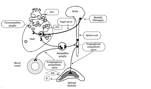
The autonomic control of the circulation
The autonomic nervous system (ANS) acts unconsciously and among other functions it regulates the circulation by the sympathetic and the parasympathetic limbs. The latter generally have opposite effects on the circulation, where one system activates a physiological response and the other one inhibits it. The sympathetic nervous system acts like a mobilizing system and the parasympathetic nervous system is a dampening system. By controlling the sinoatrial (SA) node, the sympathetic nervous system increases the heart rate and the contractility of the heart muscle, whereas the parasympathetic nervous system decreases heart rate. The parasympathetic nervous control originates in the vasomotor centre in the medulla oblongata and affects the heart via the vagal nerve (Figure 5). The pre-ganglionic neurons pass close to the aorta and superior vena cava and branch into epicardial subplexuses (106). Small nerve fibres form an extensive neural network of interconnecting nerve fibres. These nerves innervate the atria, SA and AV nodes, conducting tissue and ventricles (107). Histological studies have investigated the positioning and location of the cardiac ganglia. In the normal human heart the right atrium is innervated by two subplexuses, the left atrium by three, the right ventricle by one, and the left ventricle by three subplexuses (106). Each ganglionated plexus is composed of sympathetic, parasympathetic and mixed nerve fibres (108). In a study of the topography of the human heart 836 ± 76 ganglia were identified and by estimating the number of neurons within epicardiac ganglia, it was calculated that approximately 43000 intrinsic neurons might be present in adult hearts and 94000 neurons in young hearts (fetuses, neonates, and children) (106). The human epicardiac ganglia are formed and located in their definitive position already from 15 weeks after gestation (109). Large populations of cardiac ganglia are located adjacent to the nodal tissue of the heart. The SVC and IVC have moderate collections of cardiac ganglia on their posterior surfaces. In particular, the SVC, near the junction of the right atrium and inferior to the SA node, is moderately populated with ganglia (110).
The sympathetic nerves arise from the paravertebral ganglia, where the Stellate ganglion is particularly important (111) (Figure 5). The adrenal medulla is considered a modified sympathetic ganglion. Cells in the adrenal medulla are innervated by sympathetic preganglionic neurons and medulla release norepinephrine and epinephrine to the circulation. The sympathetic activity is mediated via norepinephrine which has a slower metabolism, this is why the sympathetic activity is more slowly mediated, but with longer lasting effects on the heart rate and heart rate variability compared with parasympathetic control (112).
Figure 5. The autonomic innervation of the heart.
The heart in a posterior view. Note dense populations of ganglia in relation to the SVC= superior vena cava, and the IVC= inferior vena cava.
Under normal conditions, there is little efferent sympathetic neural input to the sinoatrial node, however, there is substantial efferent parasympathetic input via the vagal nerve which slows the sinus node rate. Thus, resting heart rate is determined by both sympathetic and parasympathetic tone. Baroreceptors register blood pressure and send this information to the medulla oblongata. Medulla oblongata will respond through a change in the autonomic tone, in order to maintain pressure. Increased activity of the sympathetic nervous system is an important mechanism of the body to compensate altered hemodynamics due to heart disease. However, chronic sympathetic activation cause maladaptive and even detrimental effects to the cardiovascular system and the heart (113). Chronically increased sympathetic activity with elevated plasma catecholamines can be found in the setting of myocardial dysfunction (114). Elevated levels of catecholamines change electrophysiological properties of the myocardium and promote arrhythmia, through different mechanisms; i.e., enhanced automaticity, triggered activity, or re-entry (115). When there is sympathetic dominance the risk for arrhythmia and sudden death is increased (115-117). The parasympathetic nervous system may have important antiarrhythmic effects by reducing the heart
rate and counteracting the pro-arrhythmic effects of the sympathetic nervous system (118). On the other hand, excessive vagal tone is undesirable since excess vagal tone can result in SA-block and syncope.
Autonomous regulation, specifically of the cardiac ganglia, seems to be of importance for the protection from arrhythmic events. Cases of sudden cardiac death associated with arrhythmias in which no apparent pathology was present in the myocardium, coronaries, nodal tissue or conductive tissue of the heart, but where localised inflammation of cardiac ganglia was seen, have been described (119).
Surgical trauma to cardiac ganglia may also lead to withdrawal of cardio-protective vagal influences and predisposition toward arrhythmogenesis. When creating the bidirectional cavopulmonary connection (BDG), the SVC is transected, with the proximal end oversewn and the distal end anastomosed end-to-side to the pulmonary arteries. This might affect the ganglia located on the posterior surfaces of SVC near the junction of the right atrium. During cardiac surgery for the creation of the extracardiac conduit, the IVC is transected which could damage the ganglia located close to the IVC-atrial junction or at the medial and posterior surface of the IVC. In particular, the region on the SVC near the junction of the right atrium and inferior to the SA node is densely populated with ganglia (110).
Chronic changes appear in the cardiac autonomic control in order to compensate for hemodynamic alterations due to the single ventricle malformation itself, or to the different hemodynamic situations before and after BDG and after TCPC, or due to the surgery-related damage to the autonomic innervation of the heart. Assessment of cardiac autonomic function may provide insights to future disease progression. Increased sympathetic activity and decreased parasympathetic activity is strongly associated with myocardial dysfunction. A dominance in sympathetic activity plays a significant role in the progression of ventricle dysfunction and may contribute to long term sequelae, including fibrosis, after Fontan surgery (120).
Thus, to a certain extent the problem with the interpretation of low HRV is like
that of the chicken and the egg. During the interpretation of low HRV one must
keep in mind that increased sympathetic activity and decreased parasympathetic activity may not only be caused by autonomic dysfunction but also be caused by a physiological response to a decreased ventricular function, related to worse prognosis.
Heart rate variability (HRV)
A healthy heart does not perform as a metronome, i.e. it does not beat with a fixed rate. The oscillations in heart rate in a healthy heart are complex since the cardiovascular system constantly adjusts to physical inputs to keep homeostasis. Thus, the interval of heart beats is not constant but varies in certain patterns. HRV measures these beat-to-beat fluctuations in the RR intervals of the ECG. Analysis of HRV patterns allows the identification and measurement of underlying physiologic rhythms. One such rhythm is associated with breathing. Breathing gives rise to variations of thoracic blood pressure, which mainly affect the venous return to the heart. The baroreflex loop will then via the parasympathetic nervous system give rise to compensatory modulations of the heart rate. In 24 hour-ECG recordings, the predominant physiologic rhythm that accounts for the most HRV is the circadian rhythm, with relatively increased sympathetic activity associated with higher heart rates during the daytime and increased vagal activity associated with lower heart rates during the night (121). The strength of these rhythms is expressed by the magnitude of various frequency-domain and time-domain HRV measures. However it is important to observe that HRV, apart from reflecting the normal cardiac autonomic control (normal sinus rhythm), also reflects random variations due to underlining abnormalities in cardiac control (erratic rhythm).
How to measure HRV
HRV parameters normally are determined based on long-term recordings during daily activities over 24-hour periods, or as short-term (5 to 30 minutes) ECG recordings. A 24-hour registration better represent processes with slower fluctuations (e.g., circadian rhythms) and the cardiovascular system’s response to a wider range of environment stimuli, than short-term recordings (123). Short term ECG recordings are reliable under controlled conditions; lying supine or standing position, and restrictions of drugs including coffee some hours before testing (124). However, the use of very short HRV recordings (10 seconds) has also been suggested. These very short recordings do not test the ability to change heart rate, but can detect markedly reduced HRV; in particular, if performed on multiple occasions (125).
Before HRV analysis is performed, ECG recordings are evaluated using regular procedures for analysis of standard 24-hour ambulatory ECG recordings. Assessment of underlying rhythm, cardiac conduction disturbances and the presence and frequency of arrhythmic beats carried out. RR intervals are normally automatically detected in the ECG recordings. Therefore, even after careful manual editing, additional error correction is often necessary before HRV analysis, by using automatic computerised filters to remove arrhythmic beats,
noise and or artefacts that interfere with the analysis of HRV (122). In children, with sinus arrhythmia, the filtering process has to allow a wider range in RR intervals, since the choice of the threshold is a trade-off between the ability of removing ectopic beats and the risk for removing normal sinus beats in subjects with pronounced respiratory sinus arrhythmia (122).
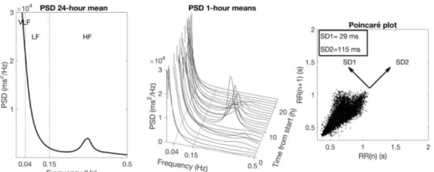
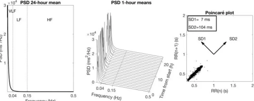
Spectral analysis
Overall HRV is normally characterized by the variance of the RR intervals over a given time interval, which is equivalent to the power of the signal. Spectral analysis works like a mathematical prism that divides the total power of HRV into components with different frequency and amplitude. The spectral analysis is graphically represented by a plot of the power spectral density (PSD), which shows the distribution of power into a large number of frequency components (123) (Figure 6). The definition of three HRV spectral components is based on the different underlying rhythms that the HRV signal is assumed to reflect. The area within each frequency region is then calculated representing the power of the spectral components. The PSD is expressed in milliseconds squared (ms2)
divided by cycles per second (ms2/Hz) (121). Thus, the spectral components are
expressed in ms2. In this thesis, the mathematical method used to calculate the
power spectral density (PSD) is Fast Fourier transformation(124). The following frequency domain indices are determined:
Total power (Ptot) in ms2 – Ptot is the total variance in HRV.
Very low frequency power (VLF, PVLF) in ms2 – VLF: (0.003 to 0.04 Hz).
VLF is assumed to reflect parasympathetic activity related to heat control and the renin-angiotensin system. The VLF band cannot be analyzed from shorter (< 5 min) ECG recordings since the periods of oscillation measured have very low frequencies. Twenty-four-hour ECG registrations are preferred (121). The most low-frequent fluctuations in the VLF region are strongly associated with increased mortality (125-127) in the adult populations with cardiac disease, however there is uncertainty regarding the physiological mechanisms responsible for activity within this band. The heart’s intrinsic nervous system appears to contribute to the VLF rhythm (121). The thermoregulation and the renin–angiotensin system may also influence VLF power (128). Moreover, parasympathetic activity may contribute to VLF power since parasympathetic blockade decreases VLF significantly (129).
Low-frequency power (LF, PLF) in ms2 – LF: (0.05 to 0.15 Hz). Represents
believed to arise because of modulations of the blood pressure via the baroreflex. The LF band is produced by both the parasympathetic and the sympathetic nervous systems in combination with regulation of blood pressure by baroreceptors (123).
High-frequency power (HF, PHF) in ms2 – HF: (0.15 to 0.50 Hz).
Represents the parasympathetic modulation of the heart rate. The variations are synchronous with the breathing. The HF band is produced by the parasympathetic nervous system alone (130). HF corresponds to the heart rate variations related to the respiratory cycle (129).
LF/HF ratio –Often used to estimate the "sympathovagal" balance. However, this term is something of a misnomer since lower frequency fluctuations may be related to both sympathetic and parasympathetic activity. The LF/HF ratio should be based on 24 hour ECG recordings. The assumptions underlying the LF/HF ratio is that LF power is
generated mainly by the sympathetic nervous system, while HF power is produced by the parasympathetic nervous system. A low LF/HF ratio reflects parasympathetic dominance and a high ratio reflects a sympathetic dominance (130).
Note that, if HRV is very irregular or even completely random (as in atrial fibrillation), then power is distributed over a wide frequency range, with nearly the same amplitude at all frequencies in the PSD; also referred to as white noise. Compare with white light with a uniform mixture of all colours.
The Poincaré diagram
The spectral analysis presumes that the HRV signal consists of different oscillating rhythms within different frequency regions. To describe more complex systems, non-linear methods, such as the Poincaré diagram can be used. In the Poincaré diagram each RR interval is plotted against the following RR interval. To quantify the variability an ellipse is fitted around the points in the plot a two-dimensional vector analysis of a Poincaré plot is used to measure separately the standard deviation in two perpendicular directions (131) (Figure 6):
SD1 representing the instantaneous beat-to-beat RR interval variability which is influenced by the parasympathetic mediated incautious action on the sinus node.
SD2 representing the continuous long-term variation in mean RR interval, which is influenced by sympathetic innervation.
SD1/SD2 the SD1/SD2 ratio is often increased during arrhythmia. It has also been considered as a marker of the autonomic balance during absence of arrhythmia, since it correlates with the LF/HF ratio (132).
This plot is useful to detect if arrhythmia is affecting the HRV signal (133). A Poincaré plot in a subject with normal HRV and not affected by arrhythmias, will look like an ellipse in 5-minutes recordings and as a comet in 24-hour recordings (Figure 6). If there are arrhythmias, the plot will have a V-shaped or a more complex pattern with clusters of points within particular areas on both sides of the diagonal of the diagram.
Low HRV (Figure 7) is associated with worse outcomes in numerous settings including; increased mortality and a greater risk of cardiac events in population studies (125, 134), increased mortality after myocardial infarction (127, 135), increased mortality in patients with heart failure (136) and increase mortality in patients with atrial fibrillation (137). Increased HRV is not always better since pathological conditions such as arrhythmia, and cardiac conduction abnormalities elevate HRV and elevated HRV has been found to be strongly linked to increased risk of mortality in adults (138). Patients with normal HRV measures, even if they have suffered an acute myocardial infarction, are at very low risk of mortality (117).
Figure 6. Examples of normal HRV
Examples of normal HRV; (left and middle) Power spectrum with a marked day-night variation in the respiratory-related peak near 0.3 Hz. The Poincaré plot (right) shows a “comet” pattern, where the variability increases with increasing R-R interval.
Figure 7. Examples of low HRV
Examples of low HRV; (left and middle) Power spectrum. The Poincaré plot (right) shows a “torpedo” pattern, with very little variability.
Age- and gender dependency of HRV
In childhood, HRV does not differ between females and males (139). During childhood, there is a positive correlation between age and HRV. HRV increases most rapidly during infancy and continues to increase, though at a slower rate, in early childhood and late childhood (140). After puberty HRV gradually decrease.
Clinical application of HRV
The most widely spread clinical use of HRV is in the monitoring of labour (cardiotocography, CTG). The variability of the fetal heart beats correlates with fetal viability. The postulated mechanism for this observation is that the fetal heart rate is modulated on a beat-to-beat basis by the parasympathetic and sympathetic nervous systems. Depression of the central nervous system secondary to anoxia leads to a loss of this fine beat-to-beat modulation of the heart rate and, hence, to a more metronome-like heartbeat (112). HRV is also used in clinical practice in patients with diabetes mellitus and familiar amyloidosis for detection and quantification of autonomic neuropathy (141).
HRV in congenital heart disease
Pre-operatively, patients with atrial septal defects have reduced HRV, but HRV normalizes post operatively (142-144). Also, in neonates with early repair of coarctation of the aorta, HRV was reduced compared to healthy babies. At five
years age, HRV in these children was at the same level as the healthy controls (145). Furthermore HRV studies in infants with transposition of the great arteries (TGA) demonstrated significantly lower PHF and PLF preoperatively, when
compared with healthy infants (146). In patients with tetralogy of Fallot, Butera et al found a significant reduction in HRV, particularly in patients with non-sustained ventricular tachycardia (147). In a large cohort of 258 patients with congenital heart disease of different types the association between the New York Heart Association (NYHA) functional classes I–IV and HRV was studied. HRV parameters were decreased compared to controls in NYHA class II-IV, but not in NYHA class I (148). Impaired autonomic nervous activity is associated with an increased risk of sudden cardiac death in patients with congenital heart disease (116).
HRV in univentricular heart defects and Fontan circulation
Already in fetal life, HLHS is associated with reduced HRV (149). Until now, there is only one published study that compares HRV during operations to complete Fontan circulation. HRV was analysed in 900-second ECG recordings. The study found a higher root mean square of successive RR interval difference (RMSSD) and a lower PLF in the BDG group (150). After Fontan surgery HRV is reduced
(151-153) and HRV continues to decrease over time after TCPC surgery (154). Bossers et al found no differences in HRV parameters between LT and EC except for a higher LF/HF ratio in EC group (71).
HRV and arrhythmia
Reduced HRV is also associated with a higher risk of arrhythmias such as supraventricular and ventricular arrhythmias (137, 155). In adults, reduced HRV implies a shift of the sympatho-vagal balance toward sympathetic dominance, and reduced vagal tone has been shown to precede onset of arrhythmia (156). There is also evidence that the HRV is reduced in children with idiopathic ventricular tachycardia (155), and that HRV correlates negatively to the extent of disease in patients with arrhythmogenic right ventricular cardiomyopathy (157). Our group has previously shown a change in HRV in Fontan patients who develop arrhythmias, compared with non-arrhythmic patients, even before the onset of arrhythmia (158).
Specifically, in studies by Bergfeldt et al and Sosnowski et al, HRV has been described to be significantly higher in adult patients with SND than in controls (159, 160). It was shown that patients with SND had an abnormal pattern using Poincaré plots for HRV-analysis (159). Furthermore, measures of HRV were highly predictive of high-degree atrioventricular (AV)-block after acute myocardial infarction in adults (161).
Overall Aim
The overall aim of this thesis was to study heart rate variability, arrhythmia and pacemaker treatment in children with Fontan circulation, with the purpose of contributing to the reduction of long term complications in this patient group.
Specific Aims
To investigate HRV in ambulatory 24-hour ECG recordings (Holter- ECG) in a large cohort of children with TCPC and to compare patients operated with LT with EC (Paper I).
To study HRV in between surgical stages to study the temporal profile of HRV changes in relation to the surgical steps (Paper II).
To investigate whether handheld ECG monitoring could be useful for the detection of silent arrhythmias and as a screening tool and for detection of changes in HRV in patients with Fontan circulation (Paper III).
To examine the prevalence of and risk factors for pacemaker treatment in a large national cohort of patients with Fontan circulation (Paper IV). To study if changes in HRV could be detected on 24-hour ECG recordings
Overview of included studies
Study Aims Study group
I To investigate HRV in ambulatory 24-hour ECG recordings in a large cohort of children with TCPC and to compare patients operated with LT with EC
Swedish patients with Fontan circulation, (n= 112)
Healthy controls (n=66)
II To investigate HRV in ambulatory 24-hour ECG recordings in a cohort of children with TCPC, focusing on the effect of HR and HRV in relation to surgical steps
Swedish patients with univentricular heart defects (n=89)
Healthy controls (n=38)
III To investigate whether handheld ECG monitoring in patients with Fontan circulation could be useful for two purposes:
Detection of silent arrhythmias and As a screening tool for detection of
cardiac autonomic dysfunction
Patients with Fontan circulation from the northern part of Sweden (n=27) Healthy controls (n=41)
IV To examine the prevalence of and risk factors for pacemaker treatment in a national consecutive national cohort of patients with Fontan circulation
All cases in Sweden with Fontan circulation operated on between 1982 and 2017 (n=599)
V To study if changes in HRV could be detected in 24-hour electrocardiogram (ECG) recordings in patients with Fontan circulation and SND
Patients with TCPC and pacemaker treatment due to severe SND (n=12) Patients with TCPC and SND without
pacemaker treatment (n=11) Healthy controls (n=66) TCPC controls (n=90)