THESIS
ACUTE BEET JUICE INGESTION IMPROVES ESTIMATES OF INSULIN SENSITIVITY IN OBESE ADULTS
Submitted by Joseph William Beals
Department of Health and Exercise Science
In partial fulfillment of the requirements For the Degree of Master of Science
Colorado State University Fort Collins, Colorado
Summer 2014
Master’s Committee:
Advisor: Christopher Bell Matthew Hickey
Copyright by Joseph William Beals 2014 All Rights Reserved
ii ABSTRACT
ACUTE BEET JUICE INGESTION IMPROVES ESTIMATES OF INSULIN SENSITIVITY IN OBESE ADULTS
Poor glucose regulation is strongly associated with low nitric oxide (NO)
bioavailability; a characteristic that may be improved with stimulation of NO generating pathways. For example, endothelial nitric oxide synthase null mice demonstrate improved glucose metabolism following sodium nitrate ingestion. Dietary nitrates are sequentially reduced in the oral and gastric cavities to NO, a process that is attenuated by rinsing with an antibacterial mouthwash. We hypothesized that acute dietary nitrate consumption will improve glucose tolerance. 9 sedentary, healthy, obese adults (2 male; body mass index: 33.7 ± 4.0 kg/m2: age: 45±7 years; mean ± SE) were studied. Using a randomized crossover design, four oral glucose tolerance tests were performed (equal carbohydrate load). To assess the influence of dietary nitrate, subjects
consumed either 500mL of beet juice + 25g glucose, or 500mL of water + 75g glucose, with and without prior antibacterial mouthwash use. Beet juice was selected because it is rich in nitrate. Venous blood samples were collected for the determination of glucose and insulin concentrations. Neither the circulating glucose nor insulin responses were influenced by beet juice and/or mouthwash (P>0.05). However, the Matsuda Index, an estimate of insulin sensitivity, was greater for beet juice compared with beet juice
preceded by mouthwash (104.6 ± 11.7 vs. 83.5 ± 11.1; P<0.05). These preliminary data suggest that acute dietary nitrate ingestion may promote insulin sensitivity in obese adults.
iii
TABLE OF CONTENTS
ABSTRACT ... ii
CHAPTER I: REVIEW OF LITERATURE ... 1
CHAPTER II: INTRODUCTION ... 15
CHAPTER III: METHODS AND MATERIALS ... 17
CHAPTER IV: RESULTS ... 20
CHAPTER V: DISCUSSION ... 22
REFERENCES ... 26
1 CHAPTER I
REVIEW OF LITERATURE
Type 2 Diabetes
25.8 million (8.3%) Americans currently have type 2 diabetes (1); this is roughly double the number of cases in 1970 (2). This trend is expected to continue, leading to 1 in 3 adults diagnosed by 2050 (1,2). In response, numerous treatment options have been developed for those affected. Oral anti-diabetic agents are being prescribed to over half of patients with type 2 diabetes (3). However, lifestyle modification through diet and exercise remains to be a commonly recommended treatment for diabetes (4).
Glucose Homeostasis and the Pancreatic Hormones
Regulation of whole body glucose homeostasis is central to human metabolism and is the fundamental insufficiency in diabetes. The rates of appearance (Ra) and
disappearance (Rd) ultimately determine the concentration of glucose in the blood. The
glucose Ra is primarily determined by intestinal absorption of glucose and hepatoportal
glucose uptake during the postprandial period, whereas in a fasted state, hepatic and renal glucose production and release are the primary determinants. The glucose Rd is
2
independent tissues. During the postprandial period, insulin-dependent glucose disposal is much more influential than during a fast.
The pancreas plays a pivotal role in glucose homeostasis by sensing the plasma glucose concentration and releasing hormones in response. The pancreatic Islets of Langerhans contain specialized cells called α and β-cells that produce glucagon and insulin, respectively. When plasma glucose concentrations fall, such as during a fast, the α cells respond by secreting glucagon. A primary target of glucagon is the
hepatocytes in the liver where it stimulates gluconeogenesis and glycogenolysis, which together act to supply glucose for the plasma. When plasma glucose rises such as during the postprandial period, the β-cells secrete insulin. Insulin has numerous actions, but with regard to glucose homeostasis its main actions are at the hepatocyte, adipose tissue and skeletal muscle. In hepatocytes, insulin opposes the actions of glucagon by stimulating glycogenesis and glycolysis. In skeletal muscle and adipose tissue, insulin stimulates glucose uptake and inhibits lipolysis. The absolute magnitude of glucose uptake into the skeletal muscle is larger than other organs due to the skeletal muscle's large mass and its ability to store glucose as glycogen. Although glycogen concentrations are highest in skeletal muscle when expressed as an absolutely
quantity, liver glycogen stores are a great deal larger on a per unit mass basis. When these processes are disrupted, homeostasis is lost and hyper- or hypoglycemia result.
3
Pancreatic Hormones
Insulin Secretion
After a meal, insulin is the most abundant hormone in circulation and facilitates proper storage of the nutrients absorbed from a meal. Insulin is synthesized and released from the β-cells in the Islets of Langerhans of the pancreas in response to elevated glucose (5). Insulin exocytosis is inherently linked to glucose metabolism in the β-cell (5). In the β-cells, glucose absorption occurs by facilitated diffusion, through the insulin-independent glucose transporter, GLUT-2 (6) and is subsequently
phosphorylated by glucokinase (7). Glucokinase is considered a "glucose sensing" enzyme and is the rate limiting step in glucose metabolism in pancreatic β cells (8). Glucokinase has a high Km of ~14.7mM (9), compared with a normal physiologic blood
glucose concentration range of 4-7mM (10). This relationship allows β-cell glucose metabolism to increase throughout the physiologic range and is critical to maintaining glucose homeostasis (11) and glucose-stimulated insulin secretion (GSIS) in the β-cells (12,13).
Insulin secretion is pulsatile (14) and biphasic (15). The size of each secretory burst is determined by the magnitude of the hyperglycemia (16). GSIS involves a series of intracellular events that are triggered by a rise in glucose phosphorylation by
glucokinase and leads to calcium influx, membrane depolarization and insulin granule exocytosis (17). Membrane depolarization is mediated by adenosine triphosphate (ATP) -sensitive potassium channels (Katp), which close in response to an increase in
the ATP:ADP ratio (18). Depolarization opens the L-type Calcium channels, located next to the insulin granules, allowing calcium to enter the cell (19). Mitochondrial
4
proteins such as the α ketoglutarate carrier (20) and reduced cytochrome c (21) are released into the cytosol during calcium influx and stimulate insulin exocytosis, but the exact mechanism of calcium-induced insulin release remains unclear. We do know, however, that extracellular calcium influx is required for granule exocytosis (22).
Glucagon Secretion
Glucagon is an important counter-regulatory hormone that responds to
decreasing blood glucose concentrations. As expected, glucagon release from the α-cells is inhibited by glucose in isolated α-cells (23), mouse pancreatic α-α-cells(24) or insulin in mouse pancreatic α-cells (24). In support, glucokinase is also present in the α-cells (25) and, similar to the β-cells, links glucose metabolism to Katp channel closure and
subsequent depolarization (26). Intriguingly, this mechanism has an inhibitory role on glucagon secretion (26,27) whereas it stimulates insulin release. Although currently an area of intense interest, much remains to be revealed on the mechanisms of glucagon secretion.
Major Glucoregulatory Tissues
Hepatocytes
The liver is responsible for maintaining blood glucose concentrations during a fast and storing glucose during the postprandial period. This role is central to whole-body glucose homeostasis. The liver is one of the few tissues that express glucose 6 phosphatase (G6P), which removes a phosphate from glucose 6 phosphate creating
5
free glucose (28); this allows glucose to be released into circulation during a fast.
Alternatively, skeletal muscle does not express this enzyme. Glycogen stored within the skeletal muscle must be metabolized in the myocyte and is not available for other
tissues. In addition, hepatocytes have first pass access to the glucose absorbed in the gut via the hepatoportal circulation. Similar to the pancreas, the GLUT-2 transporter (29) and glucokinase (2) are expressed in the liver. Due to the kinetic properties of these enzymes (High Km and High Vmax) the liver clears a significant portion of the
glucose from a meal while it is in portal circulation (28).
Postprandially, insulin is the primary circulating hormone. Because insulin is secreted directly into the portal circulation, a large portion of the insulin is also cleared by the hepatocytes (16). Although glucose transport into the hepatocyte is insulin-independent, insulin is important in many aspects of glucose metabolism in these cells. Circulating insulin interacts with the extracellular α-subunits of its receptor causing the intracellular β-subunits to dimerize and autophosphorylate on tyrosine residues (30). Phosphorylation of the β-subunits permits the docking of the insulin receptor substrates (IRS) 1 and 2 and activation of phosphoinositol 3 kinase PI3K (31). PI3K binds the IRS protein and phosphorylates Protein kinase B (PKB, also known as Akt) and protein phosphatase-1 (PP-1), in the hepatocytes (31). PKB regulates glycogen synthase (GS), the rate-limiting step in glycogen synthesis by phosphorylating and inactivating glycogen synthase kinase 3 while PP-1 activates GS by removing a phosphate (28). PKB also suppresses gluconeogenesis by phosphorylating the forkhead box protein O1 (FOXO1) which then migrates out of the nucleus reducing transcription of the key gluconeogenic enzymes G6P and phosphoenolpyruvate carboxykinase (PEPCK) (28). Coupled with
6
insulin's depression of glucagon release at the pancreas, insulin has a profound impact on the uptake of glucose for storage and the prevention of its release from the
hepatocyte. These actions greatly reduce glucose concentrations in the blood. Although glucose transport into the hepatocyte is insulin-independent, insulin is important in many aspects of glucose metabolism in these cells. Hepatic glucose metabolism is depicted in figure 1 (28).
Glucagon is elevated during a fast and opposes the actions of insulin to promote the release of glucose from hepatocytes. In the hepatocytes, glucagon binds its G-protein coupled receptor stimulating adenylate cyclase and the production of cyclic adenosine monophosphate (cAMP) (32). In response to rising cAMP concentrations, protein kinase A (PKA) phosphorylates and activates glycogen phosphorylase which is responsible for glycogenolysis and gluconeogenesis in the hepatocyte (32). Further, glucagon stimulated PKA the expression of G6P and PEPCK for greater abundance of the enzymes for gluconeogenesis (28). These processes stimulate glucose release into the blood to maintain normoglycemia.
Skeletal Muscle
Skeletal muscle is another important tissue in the regulation of glucose
metabolism. Skeletal muscle is a large reservoir for glucose due to its high capacity to synthesize glycogen and its absolute tissue mass (33). After a meal, insulin stimulates glucose transport and storage into skeletal muscle. Unlike the liver, the primary glucose transporter in the skeletal muscle is the insulin-dependent GLUT-4 (34). GLUT-4
7
response insulin (31,34). Insulin binding to its receptor leads to activation of PI-3K and subsequently PKB, which stimulates GLUT-4 translocation (35). Glucose is
phosphorylated by hexokinase II to form G6P (36), which prevents its escape from the myocyte and allosterically stimulates GS (37,38). Insulin increases the activity and protein content of hexokinase II (39) which further supplements glucose uptake. In addition, insulin stimulated PKB activation also leads to greater GS activity (40).
Collectively skeletal muscle is primed to store the glucose from the blood in response to a meal.
Adipose tissue
In adipocytes, opposing insulin's action, glucagon acts to stimulate lipolysis during a fast. Glucagon activates adenylate cyclase stimulating the formation of the second messenger cAMP, increased PKA activity, and phosphorylation of hormone-sensitive lipase (HSL) (10,32). HSL activity leads to the release of NEFAs into the circulation.
In adipocytes, insulin stimulates glucose uptake, triglyceride synthesis, and prevents lipolysis. Similar to skeletal muscle, insulin stimulates glucose transport through the translocation of GLUT-4 to the cell membrane (10). Glucose is stored in adipose primarily as glycerol on triglycerides (10) although not in large quantities. Perhaps the most important action Insulin's primary action on the adipocytes is to block lipolysis, primarily by inhibition of hormone-sensitive lipase (41); this occurs in part by activation of phosphodiesterase, the enzyme that converts cAMP to AMP (42). These actions prevent the release of NEFAs in the blood.
8
Impaired Glucose Metabolism
Insulin resistance
Insulin resistance is a hallmark of type 2 diabetes and is characterized by a failure of insulin to stimulate an appropriate reduction in blood glucose levels. Importantly, significant insulin resistance can exist without the presence of overt diabetes. A detailed discussion of the mechanisms of insulin resistance and
impairments to the insulin signaling pathway is not warranted here, but readers are directed to the following reviews (43–45). In adipose tissue, insulin resistance leads to poor regulation of lipolysis and high postprandial circulating NEFAs; high NEFAs cause insulin resistance in the hepatocytes and skeletal muscle (46). In hepatocytes, the primary defect is the inability of insulin to prevent the release of glucose into the blood (28). Continued hepatic glucose release coupled with a lack of uptake leads to a profound extension of the glucose excursion during the postprandial period. Additionally, in skeletal muscle, disruption of the insulin signal prevents GLUT-4 translocation and glycogen synthesis (44). Altogether, these impairments lead to
significantly longer glucose excursions and hyperglycemia. With hyperglycemia, insulin secretion becomes chronically elevated in the early stages of diabetes.
Hyperinsulinemia is a very strong predictor of the onset of diabetes and may play a pathogenic role in the progression of the disease (47). In early stages of insulin resistance, the β cells are capable of responding with adequate insulin, but as this worsens the β-cells decompensate and fail to keep up; this marks the development of overt diabetes (48).
9
Insulin Signaling in the Endothelium
An important component of insulin-mediated glucose uptake into skeletal muscle is insulin's ability to recruit microvascular blood flow to the skeletal muscle (49,50). Insulin recruitment of blood flow to the muscle is through PKB-dependent production of nitric oxide (51,52). Nitric oxide (NO) is an important regulator of blood flow to skeletal muscle (53). Nitric oxide is produced by nitric oxide synthase (NOS) of which there are three isoforms: Inducible NOS (iNOS), neuronal NOS (nNOS), and endothelial NOS (eNOS) (54). eNOS and nNOS are also referred to as constitutive or
calcium-dependent NOS (cNOS); these isoforms are constitutively expressed in mammalian tissues (54). NOS catalyzes the reaction that converts molecular oxygen and the amino acid l-arginine to NO and l-citrulline. In order for this reaction to proceed, the cofactor 6R-5,6,7,8-tetrahydrobiopterin (BH4) is required and is an important regulator of NO production (55).
Cyclic guanylyl monophosphate (cGMP) is the second messenger responsible for initiating the cascade of nitric oxide signaling (56). This second messenger is
formed from guanosine triphosphate (GTP) by the soluble guanylyl cyclase (sGC) (57). The primary effector of the physiological response to nitric oxide is the
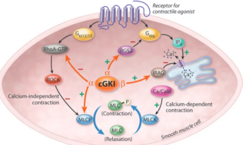
cGMP-dependent protein kinase (PKG) (58). In the smooth muscle of the vasculature, PKG activates myosin light chain phosphatase and reduces intracellular calcium
concentrations, which leads to smooth muscle relaxation and vasodilation (figure 2. from (53)) (for a detailed review see 54). Insulin mediated vasodilation recruits blood flow to skeletal muscle for storage. Additionally, in skeletal muscle, nitric oxide
10
uptake of glucose through an insulin-independent mechanism (59). Nitric oxide can also posttranslationally modify proteins in a process called nitrosylation.
S-Nitrosylation refers to the addition of a NO group to a cysteine residue on target proteins (60). For glucose homeostasis, nitrosylation of GLUT-4 vesicles facilitate greater
trafficking to the sarcomere in the skeletal muscle (61), and increases glucokinase activity in the β cells of the pancreas (62). In total, nitric oxide signaling facilitates glucose uptake through both insulin-dependent and insulin-independent mechanisms into skeletal muscle during the postprandial period. Inhibition of NOS diminishes insulin-mediated glucose uptake in rat skeletal muscle (63,64). Not surprisingly, poor NO bioavailability is a common feature in diabetes (49,65). In support, patients with type 2 diabetes insulin fails to stimulate skeletal muscle NO production unlike their healthy counterparts (66). BH4 and L-arginine have both been implicated in reduced NO bioavailability.
Bioavailability of L-arginine in some cases may limit NO synthesis. In endothelial cells, arginase 1 and 2 catalyze the formation of ornithine from arginine (67) effectively reducing arginine's availability for the NOS enzyme. This is supported by findings that arginase 1 activity is increased in human coronary arterioles (68) and in the plasma (66) of type 2 diabetics. Correspondingly, it has been shown that arginase 1 activity is
positively correlated with impaired vasodilation in diabetic mice (69,70), yet during a hyperinsulinemic euglycemic clamp, insulin reduced arginase activity in type 2 diabetics and healthy controls, but was unable to increase NO production in type 2 diabetics compared with an approximate doubling in normal controls (66). These findings
11
suggest L-arginine may not be limiting in human diabetes; this is supported by the indefinite results of arginine supplementation (49).
A loss of NO bioavailability can also occur when the cofactor BH4 is limiting. When BH4 concentrations are insufficient, NOS becomes uncoupled and leads to
superoxide generation rather than NO (71–73). Superoxide can further react with NO to form the highly reactive peroxynitrite leading to further degradation of the intracellular NO and BH4 pools (74). BH2 is the product of oxidized BH4 and can begin to
accumulate in cells with unusually high oxidative stress (74). In fact, the ratio of BH4 to 7,8 dihydrobiopterin (BH2) is an important mediator of NO synthesis due to competitive BH2 binding by the NOS enzyme (71). In rats with fructose-induced endothelial
dysfunction, BH4 administration restored NO-dependent vasodilation in a similar
manner to the NO donor sodium nitroprusside (75). Further, the pharmaceutical analog for BH4, sapropterin improves blood flow to human skin in an NO-dependent manner (73). In diabetic rodent models, BH4 supplementation suppresses NOS-dependent hepatic gluconeogenesis and reduced blood glucose during a glucose tolerance test (76). These findings suggest that supplementation with BH4 may be attractive therapeutic option for increasing NO bioavailability. In humans however, oral BH4 treatment may lack efficacy due to oxidation while in circulation (77).
Insulin secretion is also responsive to NO signaling. Insulin secretion is both inhibited and stimulated by NO in the β-cell in a concentration-dependent manner. At high concentrations insulin secretion is depressed, but when the concentration is below 50 nM insulin secretion is stimulated (78,79). The stimulatory action on the β-cells is mediated, in part, by PKG-dependent phosphorylation and inactivation of the Katp
12
channel (79). At high concentrations, NO appears to blunt glucose metabolism at phosphofructokinase and the mitochondria (80). Further, NOS co-localizes with the insulin granules of the β-cell and is activated by increased intracellular calcium
concentrations (81). Similarly, glucokinase also associates with insulin granules; when glucose concentrations rise glucokinase dissociates from the insulin granules and has greater enzymatic activity (82) facilitated by the post translational modification of
glucokinase by S-nitrosylation leading to its dissociation from the secretory granule (62).
Dietary Nitrate as a Nutriceutical
Recently, dietary nitrate has gained considerable interest as a means to improve NO bioavailability in symptomatic and healthy subjects independent of NOS. Dietary nitrate is available in most vegetables and is highly concentrated in beets, cured meats, and spinach (83). In the oral cavity, there are bacteria that possess the enzyme nitrate reductase which convert nitrate to nitrite (84–88). Nitrite produced in the oral cavity is then either further reduced to NO in the acidic environment of the stomach (88,89) or absorbed as nitrite (87). Once in circulation, the mechanisms of nitrite bioactivation are still relatively unclear, but several potential interactions have been proposed including interactions with hemoglobin, myoglobin, and carbonic anhydrase (90), and circulation to the salivary glands for excretion into the oral cavity for additional bacterial reduction (88). The importance of oral nitrate reduction is highlighted by the ability of antibacterial mouthwash to lower the plasma nitrite response to nitrate ingestion (87). Regardless, dietary nitrate increases the bioavailability of NO in a variety of models, including humans.
13
With respect to diabetes, numerous models have been used to study the effects of dietary nitrate on glucose homeostasis. Dietary sodium nitrate has been shown have positive effects in eNOS-deficient mice. In this model sodium nitrate ingestion
significantly reduced the glucose excursion after an intraperitoneal glucose tolerance test compared with water (91). The blood pressure in these mice was also reduced following dietary nitrate administration. Addition of the NOS inhibitor N (G)-nitro-L-arginine methyl ester (L-NAME) increased blood pressure in both groups, but the
dietary nitrate group resisted this increase for several hours indicating a substantial role for dietary nitrate in generating NO independent of the NOS pathway in these mice (91). In human participants with elevated risk of cardiovascular disease a 30-day dietary nitrate supplementation increased plasma nitrite and nitrate concentrations (92). Plasma nitrite is associated with improved glucose homeostasis in rodents (91) and increased cGMP concentrations and lower blood pressure in humans (93). Moreover, potassium nitrate reduced the area under the insulin and glucose curves during an oral glucose tolerance test in healthy subjects (94).
In opposition, beet juice ingestion has also been shown to have no effect on blood pressure or insulin sensitivity in patients with type 2 diabetes (95). In this study, subjects were given either 250mL of beet juice or placebo for two weeks. However, these subjects were tested after an overnight fast. These data suggest that short-term supplementation with dietary nitrate may be insufficient to elicit long-term adaptation. Alternatively, due to the rapid metabolism of the NO, the beneficial effects of dietary nitrate ingestion could be limited to the postprandial period. Indeed, a recent meta-analysis assessing both supplementation and acute treatments with dietary nitrate
14
found that inorganic nitrate/beet juice ingestion to be significantly associated with reduced blood pressure in humans (96). Despite this, the potential therapeutic benefit of nitrate ingestion on glucose homeostasis remains unclear. There is currently a great deal of evidence supporting a role of acute dietary nitrate metabolism improving the NO bioavailability in humans. Moreover, NO bioavailability has been shown to have a large role in the pathology of type 2 diabetes. Still, more studies are needed to clarify the potential for dietary nitrate to acutely improve glucose tolerance.
Hypothesis.
NO is an important physiological determinant of glucose tolerance.
Specific aim.
15 CHAPTER II
INTRODUCTION
Type 2 diabetes mellitus is associated with impaired macronutrient metabolism and vascular dysfunction. The primary metabolic defect is decreased insulin sensitivity resulting in elevated fasting blood glucose and insulin values (47). It has been shown in rodents that increased microvascular perfusion is an early event in insulin-mediated glucose disposal, occurring within minutes; an effect regulated by nitric oxide dependent processes (50). Indeed, nitric oxide (NO) -dependent vascular control has been shown to be impaired in type 2 diabetes mellitus (97). Moreover, in rodent skeletal muscle, inhibition of Nitric Oxide Synthase (NOS) completely suppressed the insulin-mediated recruitment of the microvasculature and dramatically lower glucose disposal in response to insulin (63,98). Dietary nitrate has recently gained interest as a means to improve NO bioavailability. Dietary nitrate is reduced by bacteria possessing the enzyme nitrate reductase in the oral cavity reducing salivary nitrate and nitrite to nitrite and NO,
respectively (88). Nitrite can also be reduced in the stomach to NO due to the low pH (89). In support, humans using antibacterial mouthwash prior to nitrate ingestion have significantly less salivary and plasma nitrite than when they consumed nitrate alone (87). Additionally, dietary nitrite leads to nitrosylation of cysteine residues on GLUT-4, leading to GLUT-4 incorporation in the membrane (61). Dietary nitrate also normalized glucose tolerance and fasting blood glucose in eNOS-deficient mice (91).
16
Improvements to NO bioavailability have promising therapeutic value for patients with impaired glucose tolerance. Specifically, dietary nitrate could provide an
accessible and affordable alternative to medications. Therefore, the purpose of this investigation is to determine the influence of dietary nitrate on glucose disposal during an oral glucose tolerance test (OGTT) in humans. We hypothesize that NO is an important physiological determinant of glucose tolerance.
17 CHAPTER III
METHODS AND MATERIALS
Research Participants.
Research participants were 10 (2 male, 7 female) sedentary, apparently healthy, overweight or obese adults. Study exclusion criteria consisted of fasting blood glucose concentration ≥ 100 mg/dL, pregnancy, regular use of tobacco products, and
medications that might confound the interpretation of data such as blood pressure lowering medications or antihyperglycemic agents. The experimental protocol conformed to the standards set by the Declaration of Helsinki of 1975, as revised in 1983, and was approved by the Institutional Review Board at Colorado State University. The nature, purpose and risks of the study were explained to each research participant before written informed consent was obtained.
Experimental Design.
On completion of health screening and baseline testing, research participants reported to the laboratory after an overnight fast. Participants refrained from dental hygiene for the previous 12 hours, including gum chewing and avoided foods known to contain nitrates such as leafy green vegetables, beets, and cured meats for 24 hours prior to their visit. Upon arrival to the laboratory, a Teflon catheter was placed in an antecubital vein for repeated blood sampling. Using a crossover design, four oral
18
glucose tolerance tests (OGTT) were performed with a 7-day washout period. To assess the influence of dietary nitrate, subjects consumed either 500mL of beet juice + 25g glucose, or 500mL of water + 75g glucose, with (Beet Juice, Water) and without prior antibacterial mouthwash use (Beet Juice + MW, Water + MW). Beet juice was selected due to its high concentration of nitrate. Mouthwash conditions are intended to blunt the reduction of nitrate to nitrite in the oral cavity
Oral Glucose Tolerance Testing
Glucose tolerance was determined in response to 500 mL of organic beet juice (Biotta; CAJ food products; Carmel, IN) sweetened with 25 grams of dextrose or 500 mL of water sweetened with 75 grams of dextrose. The carbohydrate load for each
condition was approximately 75 grams total. During mouthwash trials subjects first rinsed with 10 mL of a hydrogen peroxide mouthwash (1.5% H2O2 Peroxyl; Colgate Oral
Pharmaceuticals, Inc., NewYork, NY) for one minute followed by two, one-minute rinses with 10 mL of an antibacterial mouthwash (chlorhexidine digluconate; Corsodyl, BCM Ltd., Nottingham, UK). Subjects then consumed their carbohydrate beverage and remained supine for the duration of testing.
Blood Sampling.
Blood glucose was determined on arrival and at the following time-points 5, 10, 20, 30, 45, 60, 90, and 120 minutes using a blood glucose analyzer (2300 Stat Plus, Yellow Springs Instruments, Yellow Springs, Ohio). Blood Samples were collected
19
upon arrival and at time-points 10, 20, 30, and 120 minutes and placed into serum separator tubes for insulin analysis. Samples were centrifuged within 30 minutes and serum was immediately separated into 1mL aliquots. Serum was stored at -80° C for subsequent analysis. Insulin was determined using a commercially available ELISA kit (Alpco; Salem, NH).
Statistical Analyses.
This was a randomized crossover design with repeated measures, thus we examined the influence of dietary nitrate on blood glucose and serum insulin using a two-way repeated measures ANOVA (beet juice vs. water, mouthwash vs. no
mouthwash). The area under the curve (AUC) for glucose and insulin were determined using the trapezoidal method and then analyzed using a two-way repeated measures ANOVA. Additionally, insulin sensitivity was estimated using the Matsuda Index, which is highly correlated (r = 0.73) with the hyperinsulinemic euglycemic clamp (99), using the following equation:
10000 √ (FBG x FSI) x (mean G x mean I) Where:
FBG = fasting blood glucose FSI = fasting serum insulin Mean G = two hour glucose mean Mean I = two hour insulin mean
These estimations of insulin sensitivity were compared by two-way repeated measures ANOVA. The level of statistical significance was set at P < 0.05. Data within text and tables are expressed as mean ± SE.
20 CHAPTER IV
RESULTS
Ten subjects were enrolled in the experiment, but one participant's results were excluded from the analysis due to substantially high fasting blood glucose (>126 mg/dL). Nine subjects completed all four conditions of the study without any adverse events and are included in all analyses. The remaining participant's fasting blood glucose concentrations were in the healthy range, <100 mg/dL. Participants were assessed for body composition using dual-energy x-ray absorptiometry (DXA-IQ; Lunar Radiation corp., Madison, WI, software version 4.1). Selected physiological
characteristics are presented in Table 1.
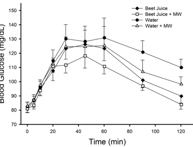
Acute Dietary Nitrate and Blood Glucose
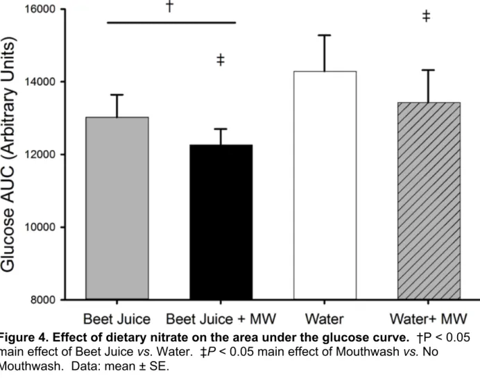
Circulating glucose responses are presented in figure 3. Acute dietary nitrate ingestion does not influence the circulating glucose concentrations during an oral glucose tolerance test (figure 3.). However, both Beet Juice conditions had a modestly lower glucose AUC, 9% (figure 4) and the final glucose concentration (figure 5) was 17% lower in the Beet Juice and Beet Juice + MW conditions as compared with the Water conditions (main effect of beet juice, P < 0.05). Intriguingly, there was also a main effect of mouthwash resulting in an approximate 6% lower glucose AUC and 9% lower circulating glucose at 120 minutes compared with the no mouthwash conditions,
21
figures 4 and 5, respectively (P < 0.05). The differences in peak glucose failed to attain significance at P = 0.08.
Acute Dietary Nitrate and Serum Insulin
Circulating insulin levels were not different between conditions (figure 6). Insulin AUC in both Beet Juice conditions was lower compared with the Water conditions (main effect of Beet Juice. P < 0.05). Peak insulin was 18% lower in Beet Juice compared with Water (P < 0.05).
Insulin Sensitivity
The results of the Matsuda Index calculations are presented in Figure 8. The Matsuda Index was approximately 25% greater in the Beet Juice condition than the Water condition (P < 0.05 vs. water) indicating greater insulin sensitivity. There were no other interactions or main effects for the Matsuda Index.
22 CHAPTER V
DISCUSSION
The primary findings of the current investigation are: 1) Estimated insulin
sensitivity is greater when 75g of carbohydrate is consumed in beet juice compared with 75g of carbohydrate in water. 2) Improved insulin sensitivity is evidenced by reduced area under both the glucose and insulin curves when 75g of carbohydrate is consumed in beet juice.
To our knowledge, we are the first to evaluate the influence of acute dietary nitrate intake on insulin sensitivity in an obese population. Central to the strength of our study design is the timing of the nitrate ingestion in a meal-like setting within a naturally occurring food source. This design accounts for the short half-life of NO in circulation by delivering the nitrate concurrently with the carbohydrate. We hypothesized that dietary nitrate is the primary bioactive component in beet juice that improves insulin sensitivity, however, beet juice is rich in many nutrients and phytochemicals. To
evaluate the effect of the nitrate in the beet juice, we used an antibacterial mouthwash to attenuate the reduction of nitrate to nitrite. Importantly, the nitrate load was the same in both Beet Juice conditions.
Plasma nitrite concentrations and time-to-peak concentrations increase dose-dependently for several hours following beet juice ingestion (100). Consumption of 280 mL of beet juice elicits an approximate four-fold increase in plasma nitrite after one hour, which rises to a seven-fold increase after two hours (100). Additionally, no further
23
lowering of blood pressure is detected when volumes of beet juice greater than 140 mL are consumed (100). Therefore, the volume of beet juice consumed in this study, 500 mL, is expected to be sufficient to elicit a physiological response.
We witnessed a lower postprandial glucose excursion with Beet Juice that persisted for two hours. In order to explain these observations we considered the specific carbohydrate composition of the carbohydrates in the beverages. The main effect of Beet Juice on the glucose AUC and 120 minute glucose values indicates improved glucose tolerance when 75g of carbohydrate is consumed in beet juice
compared with 75g of carbohydrate in water. However, approximately two-thirds of the carbohydrate in the Beet Juice trials was sucrose (101), which is catalyzed into glucose and fructose by sucrase-isomaltase located in the brush border of the intestinal mucosa (102). Fructose metabolism in humans is quite different than glucose in that fructose is primarily cleared by the liver and does not elicit a significant insulin response (103). This fact may explain a portion of our current findings, but there was not a main effect of Beet Juice on insulin sensitivity indicating other mechanisms. A main effect of Beet Juice on the final glucose concentration is clinically relevant in that this test is often used to diagnose diabetes. It has been suggested that peripheral insulin resistance is the primary cause for blunted responses during an OGTT (99). Our results indicate that beet juice likely influences the uptake of glucose into skeletal muscle.
In the current investigation Beet Juice, and potentially dietary nitrate, was
associated with a lower postprandial insulin excursion. With respect to insulin secretion, nitric oxide is stimulatory at lower concentrations, but inhibitory at greater
24
were sizeable enough to inhibit insulin secretion, an effect diminished in the mouthwash condition. This is supported by our findings that insulin secretion in the Beet Juice was significantly lower compared with Water, but the Beet Juice + MW condition was not. Inhibition of insulin secretion could be an important aspect of how dietary nitrate regulates glucose metabolism. The lower area under the glucose curve could also account for this observation due to reduced glucose stimulated insulin secretion in β-cells.
The Matsuda Index incorporates fasting and postprandial measures of glucose and insulin in order to more accurately depict insulin sensitivity (99). We found that estimated insulin sensitivity was significantly greater in the Beet Juice trial when compared with the Water trial. However, the Beet Juice + MW trial was not different from Water suggesting that dietary nitrate could have influenced insulin sensitivity, but the comparison within the Beet Juice conditions was not significant (P=0.16). To verify that mouthwash had no impact on insulin sensitivity, we included the water conditions. Unexpectedly, we found that mouthwash significantly lowered the glucose AUC and the final glucose concentration. The presence of carbohydrate sensors in the oral cavity (104) could explain this phenomenon, possibly through enhanced sweet taste sensitivity or greater access to the sensors, but this cannot be confirmed in the present study. However, the glucose response to mouthwash may be somewhat masking the effects of dietary nitrate on the estimates of insulin sensitivity.
We are not the first to investigate the influence of dietary nitrate on insulin
sensitivity. Recently, acute dietary nitrate was found to improve (94) and have no effect (105) on glucose tolerance. Acute potassium nitrate supplementation reduced the
25
glucose AUC and increased the insulin response to an OGTT in healthy males (94). However, in these experiments the nitrates were administered to participants in the healthy weight range compared with the obese subjects in our study. Alternatively, our results suggest that dietary nitrate blunts the insulin response.
Conclusion
Beet Juice improves estimates of insulin sensitivity, partially mediated by a
dramatic reduction of the insulin response to a carbohydrate challenge. Importantly, our data suggest that consuming nitrate within the context of a meal is an important timing consideration when attempting to influence glucose metabolism. While we selected beet juice for the present investigation, many vegetables are rich in nitrate. Vegetables are an important component of a nutritious diet and our findings may have revealed another important benefit of vegetables.
26
REFERENCES
1. CDC - Chronic Disease - Diabetes - At A Glance [Internet]. [cited 2014 Mar 1]. Available from:
http://www.cdc.gov/chronicdisease/resources/publications/aag/ddt.htm
2. CDC - Incidence and Age at Diagnosis - Data & Trends - Diabetes DDT [Internet]. [cited 2014 Jan 22]. Available from:
http://www.cdc.gov/diabetes/statistics/incidence_national.htm
3. Smyth S, Heron A. Diabetes and obesity: the twin epidemics. Nat Med. 2006 Jan;12(1):75–80.
4. Klein S, Sheard NF, Pi-Sunyer X, Daly A, Wylie-Rosett J, Kulkarni K, et al. Weight Management Through Lifestyle Modification for the Prevention and Management of Type 2 Diabetes: Rationale and Strategies A statement of the American Diabetes Association, the North American Association for the Study of Obesity, and the American Society for Clinical Nutrition. Diabetes Care. 2004 Aug 1;27(8):2067–73. 5. Straub SG, Sharp GWG. Glucose-stimulated signaling pathways in biphasic insulin
secretion. Diabetes Metab Res Rev. 2002 Nov;18(6):451–63.
6. Johnson JH, Newgard CB, Milburn JL, Lodish HF, Thorens B. The high Km glucose transporter of islets of Langerhans is functionally similar to the low affinity
transporter of liver and has an identical primary sequence. J Biol Chem. 1990 Apr 25;265(12):6548–51.
7. Meglasson MD, Burch PT, Berner DK, Najafi H, Vogin AP, Matschinsky FM.
Chromatographic resolution and kinetic characterization of glucokinase from islets of Langerhans. Proc Natl Acad Sci. 1983 Jan 1;80(1):85–9.
8. German MS. Glucose sensing in pancreatic islet beta cells: the key role of glucokinase and the glycolytic intermediates. Proc Natl Acad Sci. 1993 Mar 1;90(5):1781–5.
9. Pilkis SJ. Identification of human hepatic glucokinase and some properties of the enzyme. Proc Soc Exp Biol Med Soc Exp Biol Med N Y N. 1968 Dec;129(3):681–4. 10. Saltiel AR, Kahn CR. Insulin signalling and the regulation of glucose and lipid
metabolism. Nature. 2001 Dec 13;414(6865):799–806.
11. Grupe A, Hultgren B, Ryan A, Ma YH, Bauer M, Stewart TA. Transgenic knockouts reveal a critical requirement for pancreatic beta cell glucokinase in maintaining glucose homeostasis. Cell. 1995 Oct 6;83(1):69–78.
27
12. Efrat S, Leiser M, Wu YJ, Fusco-DeMane D, Emran OA, Surana M, et al. Ribozyme-mediated attenuation of pancreatic beta-cell glucokinase expression in transgenic mice results in impaired glucose-induced insulin secretion. Proc Natl Acad Sci. 1994 Mar 15;91(6):2051–5.
13. Terauchi Y, Sakura H, Yasuda K, Iwamoto K, Takahashi N, Ito K, et al. Pancreatic -Cell-specific Targeted Disruption of Glucokinase Gene DIABETES MELLITUS DUE TO DEFECTIVE INSULIN SECRETION TO GLUCOSE. J Biol Chem. 1995 Dec 22;270(51):30253–6.
14. Matthews DR, Lang DA, Burnett MA, Turner RC. Control of pulsatile insulin secretion in man. Diabetologia. 1983 Apr;24(4):231–7.
15. Cerasi E, Luft R. The Plasma Insulin Response to Glucose Infusion in Healthy Subjects and in Diabetes Mellitus. Acta Endocrinol (Copenh). 1967 Jun 1;55(2):278– 304.
16. Song SH, McIntyre SS, Shah H, Veldhuis JD, Hayes PC, Butler PC. Direct Measurement of Pulsatile Insulin Secretion from the Portal Vein in Human Subjects
1. J Clin Endocrinol Metab. 2000 Dec;85(12):4491–9.
17. Frankel BJ, Atwater I, Grodsky GM. Calcium affects insulin release and membrane potential in islet beta-cells. Am J Physiol. 1981 Jan;240(1):C64–72. 18. Cook DL, Hales CN. Intracellular ATP directly blocks K+ channels in pancreatic
B-cells. Nature. 1984 Sep 20;311(5983):271–3.
19. Bokvist K, Eliasson L, Ammala C, Renstrom E, Rorsman P. Co-localization of L-type Ca2+ channels and insulin-containing secretory granules and its significance for the initiation of exocytosis in mouse pancreatic B-cells. EMBO J. 1995 Jan 3;14(1):50–7.
20. Odegaard ML, Joseph JW, Jensen MV, Lu D, Ilkayeva O, Ronnebaum SM, et al. The Mitochondrial 2-Oxoglutarate Carrier Is Part of a Metabolic Pathway That Mediates Glucose- and Glutamine-stimulated Insulin Secretion. J Biol Chem. 2010 May 28;285(22):16530–7.
21. Jung S-R, Kuok ITD, Couron D, Rizzo N, Margineantu DH, Hockenbery DM, et al. Reduced Cytochrome c Is an Essential Regulator of Sustained Insulin Secretion by Pancreatic Islets. J Biol Chem. 2011 May 20;286(20):17422–34.
22. Henquin JC. Relative importance of extracellular and intracellular calcium for the two phases of glucose-stimulated insulin release: studies with theophylline.
Endocrinology. 1978 Mar;102(3):723–30.
23. Pipeleers DG, Schuit FC, Van Schravendijk CF, Van de Winkel M. Interplay of nutrients and hormones in the regulation of glucagon release. Endocrinology. 1985 Sep;117(3):817–23.
28
24. Ravier MA, Rutter GA. Glucose or Insulin, but not Zinc Ions, Inhibit Glucagon Secretion From Mouse Pancreatic α-Cells. Diabetes. 2005 Jun 1;54(6):1789–97. 25. Heimberg H, De Vos A, Moens K, Quartier E, Bouwens L, Pipeleers D, et al. The
glucose sensor protein glucokinase is expressed in glucagon-producing alpha-cells. Proc Natl Acad Sci U S A. 1996 Jul 9;93(14):7036–41.
26. Zhang Q, Ramracheya R, Lahmann C, Tarasov A, Bengtsson M, Braha O, et al. Role of KATP Channels in Glucose-Regulated Glucagon Secretion and Impaired Counterregulation in Type 2 Diabetes. Cell Metab. 2013 Dec 3;18(6):871–82. 27. MacDonald PE, Marinis YZD, Ramracheya R, Salehi A, Ma X, Johnson PRV, et
al. A KATP Channel-Dependent Pathway within α Cells Regulates Glucagon Release from Both Rodent and Human Islets of Langerhans. PLoS Biol. 2007 May 15;5(6):e143.
28. Klover PJ, Mooney RA. Hepatocytes: critical for glucose homeostasis. Int J Biochem Cell Biol. 2004 May;36(5):753–8.
29. Fukumoto H, Seino S, Imura H, Seino Y, Eddy RL, Fukushima Y, et al.
Sequence, tissue distribution, and chromosomal localization of mRNA encoding a human glucose transporter-like protein. Proc Natl Acad Sci. 1988 Aug
1;85(15):5434–8.
30. Obberghen EV, Rossi B, Kowalski A, Gazzano H, Ponzio G. Receptor-mediated phosphorylation of the hepatic insulin receptor: Evidence that the Mr 95,000
receptor subunit is its own kinase. Proc Natl Acad Sci. 1983 Feb 1;80(4):945–9. 31. Tsuchiya A, Kanno T, Nishizaki T. PI3 kinase directly phosphorylates Akt1/2 at
Ser473/474 in the insulin signal transduction pathway. J Endocrinol. 2014 Jan;220(1):49–59.
32. Habegger KM, Heppner KM, Geary N, Bartness TJ, DiMarchi R, Tschop MH. The metabolic actions of glucagon revisited. Nat Rev Endocrinol. 2010 Dec;6(12):689– 97.
33. Gollnick PD, Piehl K, Saubert CW, Armstrong RB, Saltin B. Diet, exercise, and glycogen changes in human muscle fibers. J Appl Physiol. 1972 Oct 1;33(4):421–5. 34. Klip A, Pâquet MR. Glucose Transport and Glucose Transporters in Muscle and
Their Metabolic Regulation. Diabetes Care. 1990 Mar 1;13(3):228–43.
35. Peck GR, Chavez JA, Roach WG, Budnik BA, Lane WS, Karlsson HKR, et al. Insulin-stimulated Phosphorylation of the Rab GTPase-activating Protein TBC1D1 Regulates GLUT4 Translocation. J Biol Chem. 2009 Oct 30;284(44):30016–23. 36. Deeb SS, Malkki M, Laakso M. Human hexokinase II: sequence and homology to
29
37. Bouskila M, Hunter RW, Ibrahim AFM, Delattre L, Peggie M, Diepen JA van, et al. Allosteric Regulation of Glycogen Synthase Controls Glycogen Synthesis in Muscle. Cell Metab. 2010 Nov 3;12(5):456–66.
38. Palm DC, Rohwer JM, Hofmeyr J-HS. Regulation of glycogen synthase from mammalian skeletal muscle – a unifying view of allosteric and covalent regulation. FEBS J. 2013;280(1):2–27.
39. Mandarino LJ, Printz RL, Cusi KA, Kinchington P, O’Doherty RM, Osawa H, et al. Regulation of hexokinase II and glycogen synthase mRNA, protein, and activity in human muscle. Am J Physiol - Endocrinol Metab. 1995 Oct 1;269(4):E701–E708. 40. Cross DA, Alessi DR, Cohen P, Andjelkovich M, Hemmings BA. Inhibition of
glycogen synthase kinase-3 by insulin mediated by protein kinase B. Nature. 1995 Dec 21;378(6559):785–9.
41. Kraemer FB, Shen W-J. Hormone-sensitive lipase control of intracellular tri-(di-)acylglycerol and cholesteryl ester hydrolysis. J Lipid Res. 2002 Oct 1;43(10):1585– 94.
42. Senft DG, Schultz G, Munske K, Hoffmann M. Influence of insulin on cyclic 3′,5′-AMP phosphodiesterase activity in liver, skeletal muscle, adipose tissue, and kidney. Diabetologia. 1968 Dec 1;4(6):322–9.
43. Xu E, Schwab M, Marette A. Role of protein tyrosine phosphatases in the modulation of insulin signaling and their implication in the pathogenesis of obesity-linked insulin resistance. Rev Endocr Metab Disord. 2014 Mar;15(1):79–97. 44. Makki K, Froguel P, Wolowczuk I. Adipose Tissue in Obesity-Related
Inflammation and Insulin Resistance: Cells, Cytokines, and Chemokines. ISRN Inflamm. 2013 Dec 22;2013:139239.
45. Dali-Youcef N, Mecili M, Ricci R, Andrès E. Metabolic inflammation: connecting obesity and insulin resistance. Ann Med. 2013 May;45(3):242–53.
46. Delarue J, Magnan C. Free fatty acids and insulin resistance. Curr Opin Clin Nutr Metab Care. 2007 Mar;10(2):142–8.
47. Weyer C, Hanson RL, Tataranni PA, Bogardus C, Pratley RE. A high fasting plasma insulin concentration predicts type 2 diabetes independent of insulin resistance: evidence for a pathogenic role of relative hyperinsulinemia. Diabetes. 2000 Dec;49(12):2094–101.
48. Weir GC, Laybutt DR, Kaneto H, Bonner-Weir S, Sharma A. Beta-cell adaptation and decompensation during the progression of diabetes. Diabetes. 2001 Feb;50 Suppl 1:S154–159.
30
49. Hoang HH, Padgham SV, Meininger CJ. L-arginine, tetrahydrobiopterin, nitric oxide and diabetes. Curr Opin Clin Nutr Metab Care. 2013 Jan;16(1):76–82. 50. Vincent MA, Clerk LH, Lindner JR, Klibanov AL, Clark MG, Rattigan S, et al.
Microvascular Recruitment Is an Early Insulin Effect That Regulates Skeletal Muscle Glucose Uptake In Vivo. Diabetes. 2004 Jun 1;53(6):1418–23.
51. Zhang Q-J, McMillin SL, Tanner JM, Palionyte M, Abel ED, Symons JD.
Endothelial nitric oxide synthase phosphorylation in treadmill-running mice: role of vascular signalling kinases. J Physiol. 2009 Aug 1;587(Pt 15):3911–20.
52. Dimmeler S, Fleming I, Fisslthaler B, Hermann C, Busse R, Zeiher AM. Activation of nitric oxide synthase in endothelial cells by Akt-dependent phosphorylation. Nature. 1999 Jun 10;399(6736):601–5.
53. Surks HK. cGMP-Dependent Protein Kinase I and Smooth Muscle Relaxation A Tale of Two Isoforms. Circ Res. 2007 Nov 26;101(11):1078–80.
54. Nathan C, Xie QW. Nitric oxide synthases: roles, tolls, and controls. Cell. 1994 Sep 23;78(6):915–8.
55. Tayeh MA, Marletta MA. Macrophage oxidation of L-arginine to nitric oxide, nitrite, and nitrate. Tetrahydrobiopterin is required as a cofactor. J Biol Chem. 1989 Nov 25;264(33):19654–8.
56. Moro MA, Russel RJ, Cellek S, Lizasoain I, Su Y, Darley-Usmar VM, et al. cGMP mediates the vascular and platelet actions of nitric oxide: confirmation using an inhibitor of the soluble guanylyl cyclase. Proc Natl Acad Sci. 1996 Feb
20;93(4):1480–5.
57. Liu Y, Ruoho AE, Rao VD, Hurley JH. Catalytic mechanism of the adenylyl and guanylyl cyclases: Modeling and mutational analysis. Proc Natl Acad Sci. 1997 Dec 9;94(25):13414–9.
58. Kuo JF. Guanosine 3’:5’-Monophosphate-Dependent Protein Kinases in Mammalian Tissues. Proc Natl Acad Sci U S A. 1974 Oct;71(10):4037–41. 59. Deshmukh AS, Long YC, de Castro Barbosa T, Karlsson HKR, Glund S,
Zavadoski WJ, et al. Nitric oxide increases cyclic GMP levels, AMP-activated protein kinase (AMPK)?1-specific activity and glucose transport in human skeletal muscle. Diabetologia. 2010 Jun;53(6):1142–50.
60. Broillet MC. S-nitrosylation of proteins. Cell Mol Life Sci CMLS. 1999 Jul;55(8-9):1036–42.
61. Jiang H, Torregrossa AC, Potts A, Pierini D, Aranke M, Garg HK, et al. Dietary Nitrite Improves Insulin Signaling Through GLUT4 Translocation. Free Radic Biol Med. 2013 Oct 21;
31
62. Rizzo MA, Piston DW. Regulation of β cell glucokinase by S-nitrosylation and association with nitric oxide synthase. J Cell Biol. 2003 Apr 28;161(2):243–8.
63. Vincent MA, Barrett EJ, Lindner JR, Clark MG, Rattigan S. Inhibiting NOS blocks microvascular recruitment and blunts muscle glucose uptake in response to insulin. Am J Physiol - Endocrinol Metab. 2003 Jul 1;285(1):E123–E129.
64. Bradley EA, Richards SM, Keske MA, Rattigan S. Local NOS inhibition impairs vascular and metabolic actions of insulin in rat hindleg muscle in vivo. Am J Physiol - Endocrinol Metab. 2013 Sep 15;305(6):E745–E750.
65. Honing MLH, Morrison PJ, Banga JD, Stroes ESG, Rabelink TJ. Nitric oxide availability in diabetes mellitus. Diabetes Metab Rev. 1998 Sep;14(3):241–9.
66. Kashyap SR, Lara A, Zhang R, Park YM, DeFronzo RA. Insulin Reduces Plasma Arginase Activity in Type 2 Diabetic Patients. Diabetes Care. 2008 Jan 1;31(1):134– 9.
67. Li H, Meininger CJ, Hawker JR, Haynes TE, Kepka-Lenhart D, Mistry SK, et al. Regulatory role of arginase I and II in nitric oxide, polyamine, and proline syntheses in endothelial cells. Am J Physiol - Endocrinol Metab. 2001 Jan 1;280(1):E75–E82. 68. Beleznai T, Feher A, Spielvogel D, Lansman SL, Bagi Z. Arginase 1 contributes
to diminished coronary arteriolar dilation in patients with diabetes. Am J Physiol - Heart Circ Physiol. 2011 Mar;300(3):H777–H783.
69. Romero MJ, Platt DH, Tawfik HE, Labazi M, El-Remessy AB, Bartoli M, et al. Diabetes-induced Coronary Vascular Dysfunction Involves Increased Arginase Activity. Circ Res. 2008 Jan 4;102(1):95–102.
70. Romero MJ, Iddings JA, Platt DH, Ali MI, Cederbaum SD, Stepp DW, et al. Diabetes-induced vascular dysfunction involves arginase I. Am J Physiol - Heart Circ Physiol. 2012 Jan;302(1):H159–H166.
71. Crabtree MJ, Smith CL, Lam G, Goligorsky MS, Gross SS. Ratio of 5,6,7,8-tetrahydrobiopterin to 7,8-dihydrobiopterin in endothelial cells determines glucose-elicited changes in NO vs. superoxide production by eNOS. Am J Physiol - Heart Circ Physiol. 2008 Apr 1;294(4):H1530–H1540.
72. Chen C-A, Druhan LJ, Varadharaj S, Chen Y-R, Zweier JL. Phosphorylation of Endothelial Nitric-oxide Synthase Regulates Superoxide Generation from the Enzyme. J Biol Chem. 2008 Oct 3;283(40):27038–47.
73. Stanhewicz AE, Alexander LM, Kenney WL. Oral sapropterin acutely augments reflex vasodilation in aged human skin through nitric oxide-dependent mechanisms. J Appl Physiol. 2013 Oct 1;115(7):972–8.
32
74. Alkaitis MS, Crabtree MJ. Recoupling the Cardiac Nitric Oxide Synthases: Tetrahydrobiopterin Synthesis and Recycling. Curr Heart Fail Rep. 2012 Sep;9(3):200–10.
75. Wang X, Hattori Y, Satoh H, Iwata C, Banba N, Monden T, et al.
Tetrahydrobiopterin prevents endothelial dysfunction and restores adiponectin levels in rats. Eur J Pharmacol. 2007 Jan 19;555(1):48–53.
76. Abudukadier A, Fujita Y, Obara A, Ohashi A, Fukushima T, Sato Y, et al. Tetrahydrobiopterin Has a Glucose-Lowering Effect by Suppressing Hepatic Gluconeogenesis in an Endothelial Nitric Oxide Synthase–Dependent Manner in Diabetic Mice. Diabetes. 2013 Sep 1;62(9):3033–43.
77. Cunnington C, Van Assche T, Shirodaria C, Kylintireas I, Lindsay AC, Lee JM, et al. Systemic and vascular oxidation limits the efficacy of oral tetrahydrobiopterin treatment in patients with coronary artery disease. Circulation. 2012 Mar
20;125(11):1356–66.
78. Kaneko Y, Ishikawa T, Amano S, Nakayama K. Dual effect of nitric oxide on cytosolic Ca2+concentration and insulin secretion in rat pancreatic β-cells. Am J Physiol - Cell Physiol. 2003 May 1;284(5):C1215–C1222.
79. Sunouchi T, Suzuki K, Nakayama K, Ishikawa T. Dual effect of nitric oxide on ATP-sensitive K+ channels in rat pancreatic β cells. Pflüg Arch - Eur J Physiol. 2008 Feb 1;456(3):573–9.
80. Kaneko YK, Ishikawa T. Dual Role of Nitric Oxide in Pancreatic β-Cells. J Pharmacol Sci. 2013;123(4):295–300.
81. Lajoix A-D, Reggio H, Chardès T, Péraldi-Roux S, Tribillac F, Roye M, et al. A Neuronal Isoform of Nitric Oxide Synthase Expressed in Pancreatic β-Cells Controls Insulin Secretion. Diabetes. 2001 Jun 1;50(6):1311–23.
82. Rizzo MA, Magnuson MA, Drain PF, Piston DW. A Functional Link between Glucokinase Binding to Insulin Granules and Conformational Alterations in
Response to Glucose and Insulin. J Biol Chem. 2002 Sep 13;277(37):34168–75. 83. White JW. Relative significance of dietary sources of nitrate and nitrite. J Agric
Food Chem. 1975 May 1;23(5):886–91.
84. Doel JJ, Benjamin N, Hector MP, Rogers M, Allaker RP. Evaluation of bacterial nitrate reduction in the human oral cavity. Eur J Oral Sci. 2005 Feb;113(1):14–9. 85. Kapil V, Haydar SMA, Pearl V, Lundberg JO, Weitzberg E, Ahluwalia A.
Physiological role for nitrate-reducing oral bacteria in blood pressure control. Free Radic Biol Med. 2013 Feb;55(C):93–100.
33
86. Van Maanen JM, van Geel AA, Kleinjans JC. Modulation of nitrate-nitrite conversion in the oral cavity. Cancer Detect Prev. 1996;20(6):590–6.
87. Govoni M, Jansson EA, Weitzberg E, Lundberg JO. The increase in plasma nitrite after a dietary nitrate load is markedly attenuated by an antibacterial
mouthwash. Nitric Oxide Biol Chem Off J Nitric Oxide Soc. 2008 Dec;19(4):333–7. 88. Duncan C, Dougall H, Johnston P, Green S, Brogan R, Leifert C, et al. Chemical
generation of nitric oxide in the mouth from the enterosalivary circulation of dietary nitrate. Nat Med. 1995 Jun 1;1(6):546–51.
89. McKnight GM, Smith LM, Drummond RS, Duncan CW, Golden M, Benjamin N. Chemical synthesis of nitric oxide in the stomach from dietary nitrate in humans. Gut. 1997 Feb;40(2):211–4.
90. Kim-Shapiro DB, Gladwin MT. Mechanisms of nitrite bioactivation. Nitric Oxide Biol Chem Off J Nitric Oxide Soc. 2013 Dec 6;
91. Carlström M, Larsen FJ, Nyström T, Hezel M, Borniquel S, Weitzberg E, et al. Dietary inorganic nitrate reverses features of metabolic syndrome in endothelial nitric oxide synthase-deficient mice. Proc Natl Acad Sci U S A. 2010 Oct
12;107(41):17716–20.
92. Zand J, Lanza F, Garg HK, Bryan NS. All-natural nitrite and nitrate containing dietary supplement promotes nitric oxide production and reduces triglycerides in humans. Nutr Res N Y N. 2011 Apr;31(4):262–9.
93. Kapil V, Milsom AB, Okorie M, Maleki-Toyserkani S, Akram F, Rehman F, et al. Inorganic nitrate supplementation lowers blood pressure in humans: role for nitrite-derived NO. Hypertension. 2010 Aug;56(2):274–81.
94. Lidder S, Hunt J, Omar S, Webb A. Acute effects of dietary nitrate on glucose handling and insulin levels during an oral glucose tolerance test in healthy subjects. Nitric Oxide. 2011 May;24, Supplement:S32.
95. Gilchrist M, Winyard PG, Aizawa K, Anning C, Shore A, Benjamin N. Effect of dietary nitrate on blood pressure, endothelial function, and insulin sensitivity in type 2 diabetes. Free Radic Biol Med. 2013 Jul;60:89–97.
96. Siervo M, Lara J, Ogbonmwan I, Mathers JC. Inorganic nitrate and beetroot juice supplementation reduces blood pressure in adults: a systematic review and meta-analysis. J Nutr. 2013 Jun;143(6):818–26.
97. Woodman RJ, Playford DA, Watts GF. Basal production of nitric oxide (NO) and non-NO vasodilators in the forearm microcirculation in Type 2 diabetes: associations with blood pressure and HDL cholesterol. Diabetes Res Clin Pract. 2006
34
98. Bradley EA, Richards SM, Keske MA, Rattigan S. Local NOS inhibition impairs vascular and metabolic actions of insulin in rat hindleg muscle in vivo. Am J Physiol - Endocrinol Metab. 2013 Sep 15;305(6):E745–E750.
99. Matsuda M, DeFronzo RA. Insulin sensitivity indices obtained from oral glucose tolerance testing: comparison with the euglycemic insulin clamp. Diabetes Care. 1999 Sep 1;22(9):1462–70.
100. Wylie LJ, Kelly J, Bailey SJ, Blackwell JR, Skiba PF, Winyard PG, et al. Beetroot juice and exercise: pharmacodynamic and dose-response relationships. J Appl Physiol. 2013 Aug 1;115(3):325–36.
101. Doll S, Rodier F, Willenbrink J. Accumulation of sucrose in vacuoles isolated from red beet tissue. Planta. 1979 Jan 1;144(5):407–11.
102. Van Beers EH, Büller HA, Grand RJ, Einerhand AWC, Dekker J. Intestinal Brush Border Glycohydrolases: Structure, Function, and Development. Crit Rev Biochem Mol Biol. 1995 Jan 1;30(3):197–262.
103. Cook GC. Absorption and metabolism of D(–) fructose in man. Am J Clin Nutr. 1971 Nov 1;24(11):1302–7.
104. Chambers ES, Bridge MW, Jones DA. Carbohydrate sensing in the human mouth: effects on exercise performance and brain activity. J Physiol. 2009 Feb 23;587(8):1779–94.
105. Larsen FJ, Schiffer TA, Ekblom B, Mattsson MP, Checa A, Wheelock CE, et al. Dietary nitrate reduces resting metabolic rate: a randomized, crossover study in humans. Am J Clin Nutr. 2014 Apr 1;99(4):843–50.
35 APPENDIX I
TABLES AND FIGURES
Table 1. Subject characteristics n=9 (2 males)
Variable Baseline
Age (years) 45 ± 4
Body mass (kg) 90.1 ± 10.9
Height (m) 1.65 ± 0.02
Body mass index (kg/m2) 33.7 ± 0.89
Percent body fat 43.2 ± 5.3
Fat free mass (kg) 51.4 ± 2.4
Fat mass (kg) 38.8 ± 1.6
36
Figure 1. ⊥ = Block/inhibit. ↓ = Stimulate/activate (28)
754 P.J. Klover, R.A. Mooney / The International Journal of Biochemistry & Cell Biology 36 (2004) 753–758 1. Introduction
Compounds absorbed in the gut pass through the liver where they can be absorbed and metabolized. To accomplish this, mature hepatocytes are arranged into irregular folded sheets surrounding the sinusoids where blood flows, separated only by a single layer of endothelial cells, interspersed Kupffer cells, and hep-atic stellate cells. Hepatocytes also maintain a connec-tion to the gut via the formaconnec-tion of canaliculi and larger ducts into which bile is secreted. While all hepatocytes are capable of carrying out the necessary metabolic and secretory tasks attributed to liver parenchymal cells, there are some differences that exist in subcellu-lar structure and function of hepatocytes with respect of localization within in the liver (Tosh, Alberti, & Agius, 1988). Afferent periportal hepatocytes report-edly have higher gluconeogenic activity, whereas the efferent perivenous hepatocytes have been shown
Fig. 1. Summary of glucose homeostatic pathways in the hepatocyte. In the fed state, the insulin-activated receptor signals primarily through tyrosine phosphorylation of its substrates, IRS-1/2. Tyrosine phosphorylated IRS-1/2 associate with and activate signaling intermediates, particularly phosphatidylinositol-3-kinase, which regulate downstream metabolic endpoints. The net effect promotes glucose utilization and inhibits glucose output via regulation of enzyme activity and gene induction. In the fasted state, glucagon signals through its G protein-coupled receptor to regulate metabolic endpoints which promote glucose output and suppress glucose utilization.
to possess higher activity of some glycolytic and li-pogenic enzymes. Together, hepatocytes play a criti-cal role in maintaining blood glucose levels within a narrow range while responding to the changing de-mands of the body. The focus of this review will be on hormonal regulation of mature hepatocytes to accom-plish glucose homeostasis, and hepatic-specific im-pairments in this process that are related to obesity, insulin resistance, and type 2 diabetes (Fig. 1).
2. Hepatocytes in the fasted state: net glucose production
During fasting, increased glucagon release by the ! cells residing in the pancreatic islets of Langerhans leads to a rise in plasma glucose levels. The binding of glucagon to its cognate receptor on hepatocytes activates the serine/threonine kinase PKA which
37
Figure 2. Myosin Light Chain phosphorylation determines smooth muscle contractility (53).
tested for inhibition of calcium release. Only the cGKI!-containing cells exhibited calcium regulation by cGMP.19 A potential limitation in this study is the possibility that the GFP moiety interfered with cGKI targeting. In a recent study by Christensen and Mendelsohn, the role of PKGI isoforms in thrombin receptor-mediated calcium mobilization was studied in both CHO cells stably expressing either cGKI isoform and human aortic smooth muscle cells expressing primarily cGKI! or cGKI". In CHO cells, cGKI! had a significantly greater calcium-lowering effect, whereas in cultured human aorta smooth muscle cells, only cGKI! lowered calcium in response to cGMP.20Thus neither of these cell culture-based studies that manipulated the expression of cGKI isoforms confirmed the expected isoform-specific functions based on cGKI targeting.
In the current study, Weber et al took the interesting approach of creating mouse lines that express either cGKI! or cGKI" on a cGKI null background. Expression of the cGKI genes was driven by the endogenous SM22! promoter, resulting in smooth muscle–specific expression. The cGKI isoforms were expressed at levels and activity 1.5- to 2.0-fold greater than control mice. The transgenic rescue mice had a life expectancy greater than the cGKI null mice, but less than the wild-type control mice. Interest-ingly, all of the tested smooth muscle functions in the cGKI transgenics were equivalent to those in the wild-type mice. These included intestinal transit of barium, jejunal, and aortic smooth muscle relaxation and inhibition of norepinephrine-induced calcium transients by cGMP. Although surprising, these data could still be explained by the known interactions between cGKI! and cGKI" with RGS2 and IRAG, respectively.
Weber et al performed a thorough examination of blood pressure in the null, wild-type, and the cGKI! and cGKI" rescue mice. The basal blood pressure was not different between wild-type and rescue mice, but was elevated as expected in the cGKI-null mice. Moreover, the hypotensive effect of nitrovaso-dilators, carbachol, and the ROCK inhibitor Y27632 were all preserved in the cGKI rescue mice. The latter manipulation is particularly interesting because cGKI! opposes the inhibitory effect of RhoA/ROCK on MLCP activity and inhibits RhoA directly by phosphorylation at Ser188.21One would therefore expect that the cGKI! rescue mice would have less RhoA/ ROCK activity and therefore less hypotension from Y27632.
The elegant approach by Weber et al avoids many of the pitfalls of the earlier studies of cGKI isoform-specific functions in the vasculature, including reliance on cell culture models and use of epitope-tagged cGKI isoforms. How then can we explain the apparently equivalent physiologic effect of cGKI! and cGKI" rescue? The authors provide two hypotheses. First, it is possible that the specificity for each isoform for their respective targets is less pronounced in vivo. This is possible because most of the experiments that characterized the cGKI isoform specific targets were performed in cell culture systems and with purified proteins. If there were more overlap between cGKI isoforms and their target interactions, then expression of individual cGKI isoforms might exhibit subtle, if any, differences from wild-type mice. Second, each cGKI isoform alone is sufficient to maintain circulatory physiology based on its known interactions. For example, cGKI! rescue can mediate cGMP-mediated vasodila-tion because it can lower calcium via its interacvasodila-tion with RGS2, and activate MLCP via its interaction with MYPT1. It is more difficult to reconcile how cGKI" rescue can fully reconstitute cGMP-mediated vasodilation without regulating MLCP.
There are several additional possibilities that may explain the apparent functional equivalence of cGKI! and cGKI" to rescue vascular function. The cGKI targets discussed here have well-described isoform-specific interactions, yet there are additional cGKI targets that do not bind in an isoform specific manner, whose role in vascular physiology is less clear, and there are likely more targets that are undiscovered.22,23 These cGKI targets may allow functional overlap between the cGKI iso-forms. Furthermore, although Weber et al observed similar expression of the cGKI isoform targets, IRAG, MYPT1, and RhoA, other critical proteins within these signaling pathways may be upregulated. Moreover, posttranslational modification (eg, phosphorylation) of these isoform-specific targets or splice variation may also contribute to altered activity of these signal-ing pathways without apparent differences in their protein expression levels. A third possibility represents a limitation to any transgenic approach when used to explore pathways regu-lated by differential targeting. Even modestly increased levels of protein overexpression may be adequate to obscure the specific-ity of protein targeting, particularly if the protein in question is present in excess of the targeting protein.
Figure. MLC phosphorylation determines
smooth muscle contractility. Contractile ago-nists lead to inositol 1,4,5 triphosphate (IP3) production or activation of RhoA (RhoA-GTP). IP3binding to its receptor in the sarcoplasmic reticulum leads to release of Ca2!. Ca2! /cal-modulin binds to and activates MLCK, which in turn phosphorylates MLC (calcium-dependent contraction). Activated RhoA binds to and acti-vates ROCK, leading to phosphorylation and inhibition of MLCP, inhibiting MLC dephosphor-ylation (calcium-independent contraction). cGKI mediates relaxation by inhibiting both calcium-dependent and -incalcium-dependent contraction. cGKI! activates MLCP by a direct interaction and by inhibition of RhoA, and activates RGS2 to inhibit G!qsignaling. cGKI" activates IRAG, which then inhibits Ca2!release by the IP
3 receptor.
Surks cGKI Isoforms and Smooth Muscle Relaxation 1079
by guest on January 24, 2014 http://circres.ahajournals.org/
38
Figure 3. Effect of dietary nitrate on circulating glucose concentrations. There
was no effect of dietary nitrate on the circulating glucose response to an OGTT. P > 0.05. Data: mean ± SE.
39
Figure 4. Effect of dietary nitrate on the area under the glucose curve. †P < 0.05
main effect of Beet Juice vs. Water. ‡P < 0.05 main effect of Mouthwash vs. No Mouthwash. Data: mean ± SE.
40
Figure 5. Effect of dietary nitrate on the final circulating glucose concentration.
†P < 0.05 main effect of Beet Juice vs. Water. ‡P < 0.05 main effect of Mouthwash vs. No Mouthwash. Data: mean ± SE.
41
Figure 6. Effect of dietary nitrate on circulating insulin concentrations. There was
no effect of dietary nitrate on the circulating insulin response to an OGTT. P > 0.05. Peak insulin concentrations were greater in Water compared with Beet Juice. *P < 0.05 Beet Juice vs. Water. Data: mean ± SE.
42
Figure 7. Effect of dietary nitrate on the area under the insulin curve. †P < 0.05
43
Figure 8. Effect of dietary nitrate on the Matsuda Index of insulin sensitivity.
There was no effect of dietary nitrate on the estimated insulin sensitivity. P = 0.15. Estimated insulin sensitivity was greater in Beet Juice as compared with Water. *P < 0.05 Beet Juice vs. Water. Data: mean ± SE.