Improved Functional MRI Activation Mapping in
White Matter Through Diffusion-Adapted Spatial
Filtering
David Abramian, Martin Larsson, Anders Eklund and Hamid Behjat
The self-archived postprint version of this journal article is available at Linköping
University Institutional Repository (DiVA):
http://urn.kb.se/resolve?urn=urn:nbn:se:liu:diva-165857
N.B.: When citing this work, cite the original publication.
Abramian, D., Larsson, M., Eklund, A., Behjat, H., (2020), Improved Functional MRI Activation Mapping in White Matter Through Diffusion-Adapted Spatial Filtering, ISBI 2020.
https://doi.org/10.1109/ISBI45749.2020.9098582
Original publication available at:
https://doi.org/10.1109/ISBI45749.2020.9098582
Copyright: Institute of Electrical and Electronics Engineers (IEEE)
http://www.ieee.org/index.html ©2018 IEEE.
Personal use of this material is permitted. However, permission to reprint/republish
this material for advertising or promotional purposes or for creating new collective
works for resale or redistribution to servers or lists, or to reuse any copyrighted
component of this work in other works must be obtained from the IEEE.
IMPROVED FUNCTIONAL MRI ACTIVATION MAPPING IN WHITE MATTER THROUGH
DIFFUSION-ADAPTED SPATIAL FILTERING
David Abramian
a,bMartin Larsson
cAnders Eklund
a,b,dHamid Behjat
ea
Department of Biomedical Engineering, Link¨oping University, Link¨oping, Sweden b
Center for Medical Image Science and Visualization, Link¨oping University, Link¨oping, Sweden c
Centre for Mathematical Sciences, Lund University, Lund, Sweden d
Department of Computer and Information Science, Link¨oping University, Link¨oping, Sweden
eDepartment of Biomedical Engineering, Lund University, Lund, Sweden
ABSTRACT
Brain activation mapping using functional MRI (fMRI) based on blood oxygenation level-dependent (BOLD) con-trast has been conventionally focused on probing gray mat-ter, the BOLD contrast in white matter having been gener-ally disregarded. Recent results have provided evidence of the functional significance of the white matter BOLD sig-nal, showing at the same time that its correlation structure is highly anisotropic, and related to the diffusion tensor in shape and orientation. This evidence suggests that conven-tional isotropic Gaussian filters are inadequate for denoising white matter fMRI data, since they are incapable of adapt-ing to the complex anisotropic domain of white matter axonal connections. In this paper we explore a graph-based descrip-tion of the white matter developed from diffusion MRI data, which is capable of encoding the anisotropy of the domain. Based on this representation we design localized spatial filters that adapt to white matter structure by leveraging graph signal processing principles. The performance of the proposed fil-tering technique is evaluated on semi-synthetic data, where it shows potential for greater sensitivity and specificity in white matter activation mapping, compared to isotropic filtering.
Index Terms— functional MRI, diffusion MRI, white matter, adaptive filtering
1. INTRODUCTION
Functional magnetic resonance imaging (fMRI) is a noninva-sive technique that allows investigation of the activity of the brain while performing a task or at rest. Most fMRI studies report activations in the gray matter, while reports of white matter activations are relatively sparse [1, 2, 3, 4, 5]. The sig-nificance of such reports is controversial [6], as the sources
This research has been supported by the Swedish Research Council un-der grants 2018-06689 and 2017- 04889, the Center for Industrial Informa-tion Technology (CENIIT) at Link¨oping University, and the Wallenberg AI, Autonomous Systems and Software Program (WASP) funded by the Knut and Alice Wallenberg Foundation. Correspondence should be addressed to H. Behjat, e-mail: hamid.behjat@bme.lth.se.
of the blood-oxygenation level dependent (BOLD) signal in white matter are not fully understood. Despite this, significant evidence directly linking the BOLD signal in white matter to neural activity has recently been presented [7].
The relative scarcity of reports of white matter activations can be partially explained by the anatomical and physiolog-ical differences between white and gray matter [6], which suggest the potential need for different experimental designs and analysis methods to optimize detection power in each tissue type. It has been shown, for example, that increased T2-weighting improves the sensitivity to callosal activations in an interhemispheric transfer task [8]. Therefore, we ex-pect the development of methods geared specifically towards white matter to be required in order to investigate the func-tional significance of the white matter BOLD signal.
An important distinguishing feature of the BOLD signal in white matter is that it exhibits a spatial correlation structure grossly consistent with the directions of water diffusion, as measured by diffusion tensor imaging (DTI) [9]. This corre-lation structure is present during rest and becomes even more pronounced under functional loading [10, 7]. As a conse-quence, isotropic Gaussian filtering, commonly used to in-crease the signal-to-noise ratio of the fMRI signal, may prove inadequate for use on the highly anisotropic white matter do-main.
The anisotropy of the white matter domain makes it suited for a graph-based description. Graph signal processing meth-ods have recently experienced significant development [11, 12], resulting in their successful application to the analysis of the BOLD signal in gray matter [13, 14]. In this work we propose a graph-based adaptive filtering approach for the white matter BOLD signal which incorporates information from diffusion MRI in order to adapt the filter design to the white matter mircostructure. We test our proposed filtering approach on semi-synthetic phantoms and show its increased sensitivity and specificity for activations in white matter.
2. METHODS 2.1. Preliminaries
We consider undirected, connected, weighted graphs G = (V, E , A), where V is a set of vertices with |V| = Nv, E
a set of edges connecting pairs (i, j) of vertices, and A is an adjacency matrix whose nonzero elements ai,j represent
the weight of edges (i, j) ∈ E. The diagonal degree ma-trix, denoted D, associated to A is defined with elements di,i=Pjai,j.
The normalized Laplacian matrix of G can then be ob-tained as L = I − D−1/2AD−1/2. As L is a real
symmet-ric matrix, it can be diagonalized, resulting in a set of Nv
real non-negative eigenvalues which define the graph spec-trum Λ, i.e., Λ = {0 = λ1 ≤ λ2. . . ≤ λNv
def
= λmax ≤ 2}.
The associated eigenvectors, denoted {χl}Nl=1v , form an
or-thonormal basis spanning the `2(G) space. Here `2(G)
repre-sents the Hilbert space of all square-integrable graph signals f : V → R defined on the vertex set V. A graph signal can therefore be seen as a vector f ∈ `2(G) whose n-th
com-ponent represent the signal value at the n-th vertex of G. A graph signal f can be filtered with spectral kernel K(λ) as
(Fkf ) [m] = Nv X l=1 ˆ k[l] ˆf [l]χl[m], (1)
where ˆk is the sampled version of K(λ) obtained as ˆk[l] = K(λl), l = 1, . . . , Nvand ˆf is the graph Fourier transform of
f (see [15] for an introduction to graph signal processing). 2.2. White matter graph construction
We define a white matter graph G(WM)as a graph whose ver-tex set V(WM)consists of all the white matter voxels in a brain volume (approximately 250k for the data used). The set of edges E(WM)is defined such that each voxel is connected to every voxel in a specified neighborhood, which we define as a 5 × 5 × 5 region, resulting in at most 124 neighbors for each white matter voxel.
We define the weight of the edge that joins a pair of voxels (i, j) in analogy to Iturria-Medina et al. [16] and Sotiropou-los et al. [17]. Let Oi(ω) denote the diffusion orientation
distribution function (ODF) associated to voxel vi, with its
coordinate center being the center of voxel vi. Let Ni
de-note the set of vertices in V(WM)that are adjacent to vertex
i; i.e., Ni : {k ∈ V(WM)|(i, k) ∈ E(WM)}. For any two
ver-tices i, j ∈ V(WM), let ~r
i,jdenote the vector pointing from the
center of vertex i to the center of vertex j, and define p(i, ~ri,j) =
Z
Ωi,j
Oni(ω)dω, (2)
where n ∈ Z+is a desired power factor to sharpen the ODFs and Ωi,jdenotes a solid angle of 4π/98 around ~ri,jsubtended
at the center of voxel vi. It is desirable to sharpen the ODFs
since they generally manifest only slight variations between directions of strong and weak diffusion. Using (2), the weight between vertices i and j, denoted wi,jis defined as
wi,j= p(i, ~ri,j) Ci +p(j, ~rj,i) Cj , (3)
where Ck = 2 maxl∈Nkp(k, ~rk,l). The normalization factor ensures having wi,j∈ [0, 1], ∀i ∈ V(WM), ∀j ∈ Ni.
Using diffusion MRI, Oi(ω) is estimated at a discrete set
of directions {~rk}Nk=1o from the center of an ODF, denoted
{Oi,k}Nk=1o , which can be used to approximate p(i, ~ri,j) as a
sum p(i, ~ri,j) ≈ 4π No X k∈Di,j Oni,k, (4)
where Di,j : {k | ~rk ∈ Ωi,j}. The resulting edge weights
amount to a measure of coherence in the directions of diffu-sion at neighboring voxels, with high weights associated with highly coherent diffusion and vice versa.
2.3. Graph filter definition
Conventional filters defined within the Euclidean domain, such as Gaussian filters, encompass a shift invariant impulse response. In contrast, vertex realizations of graph filters are shift-variant, leading to a unique spatial realization of the filter at each graph vertex. This property, combined with the weighting scheme in the proposed graph definition, results in graph filters adapted to the microstructure of white matter.
Due to the lack of shift-invariance, it is convenient, and conventional, to specify graph filters in the graph spectral do-main. In this work, we design and leverage smoothing filters associated with a heat kernel spectral profile, defined by
K(λ) = e−τ λ, ∀λ ∈ [0, λmax], (5)
where τ is a free parameter determining the spatial ex-tent of the filter. In particular, K(λ) ∈ L2(G), where L2(G) denotes the Hilbert space of all square-integrable K : [0, λmax] → R+. Although such filters are roughly
anal-ogous in shape to the Gaussian filters typically used for fMRI data analysis, there is no direct equivalence between them. 2.4. Graph filtering
Implementing graph filtering as in (1) requires calculation of all the eigenvectors of the Laplacian matrix {χl}Nl=1v, which is
practically infeasible for larger graphs such as those proposed here. Instead, we use a fast approximation algorithm [11]. Let P ∈ L2(G) be a polynomial approximation of kernel K(λ).
For a graph signal f , its filtering with kernel K can be found using P as ˜ cK= Nv X l=1 P(λl) ˆf [l]χl= P(L) Nv X l=1 ˆ f [l]χl= P(L)f , (6)
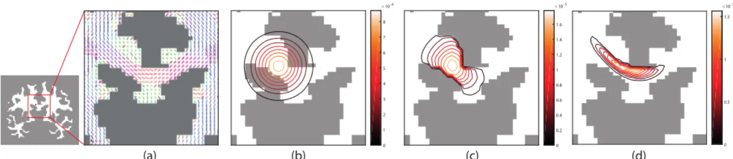
Fig. 1. (a) Main orientation of ODFs manifesting white matter fiber directions at a coronal slice in the vicinity of the corpus callosum. (b)-(d) Comparison of the contour profiles of three spatial filters, with different extents of adaptation to local anatomy, localized at a point within the corpus callosum marked with a red dot in (a). (b) An isotropic Gaussian filter adapts neither to local tissue morphology nor the underlying white matter structure. (c) A filter restricted to white matter adapts to the local tissue morphology, but not to the underlying white matter structure. (d) A filter defined on the proposed graph adapts both to the tissue morphology and the underlying white matter structure.
where ˜cK ∈ `2(G) with ˜cK[m] = (Fkf ) [m]; in the last
equality we exploit: Lχl= λlχl⇒ P(L)χl= P(λl)χl.
This approximation has the benefit that it does not require the explicit calculation of the eigenvectors. Instead, a poly-nomial of the Laplacian matrix is applied to the signal, which can be efficiently implemented with matrix-vector multipli-cation. Similarly to [11], we leverage a truncated Chebyshev polynomial expansion approximation of K(λ), as it has the benefit of approximating a minimax polynomial, minimizing an upper bound on the approximation error.
3. DATA
The MRI data used in this work was acquired from the Hu-man Connectome Project [18]. Structural images, parcella-tions, and diffusion data from the “100 Unrelated Subjects” subset of the 1200 subject release were used. Data from five of the subjects was excluded from the analysis due to incom-plete white matter coverage of the diffusion data. Addition-ally, fMRI data from a motor task from one subject was used. To evaluate the performance of the proposed graph-based filtering method we created a set of semi-synthetic phantoms, consisting of synthetic activation patterns generated on the ba-sis of tractography results from real diffusion data. Tractog-raphy was performed using generalized Q-sampling imaging [19] in DSI Studio [20]. Synthetic activation patterns were created from individual streamlines by diffusing an activation along the length of the streamline, starting from a random point.
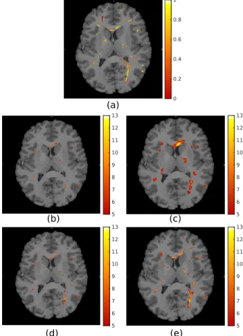
Activation patterns from 100 streamlines were combined to produce a single ground truth activation pattern for each subject. An example of such an activation pattern can be seen in Figure 2(a). Time-series phantoms for all subjects were then created by convolving the ground truth activation pattern with a regressor corresponding to a block fMRI paradigm. The resulting time-series were contaminated with additive white Gaussian noise of σ = 1.
4. RESULTS 4.1. Spatial adaptivity of graph filters
Figure 1 illustrates the differences in spatial adaptivity from various filter definitions. Isotropic Gaussian filters adapt nei-ther to the local tissue morphology nor the underlying white matter microstructure. They transcend tissue boundaries, integrating signal components associated to white matter as well as the adjacent gray matter/CSF. Moreover, the filters integrate signal components from multiple unrelated white matter tracts.
By restricting the analysis to the white matter tissue, the filters do not transcend tissue boundaries and thus only com-bine signal components from white matter. However, the fil-ters can not differentiate signal components from unrelated white matter tracts.
Filters defined on the proposed white matter graphs adapt both to the local tissue morphology and the under-lying microstructure. Their spatial profile closely matches the anisotropic diffusion orientation manifested by the ODFs. 4.2. Semi-synthetic phantom results
The 95 semi-synthetic time-series phantoms were filtered us-ing both isotropic Gaussian filterus-ing and the proposed graph filtering approach. The resulting filtered volumes were sub-jected to a standard GLM-based activation mapping using the SPM toolbox. Figure 2 shows a comparison of the results obtained by using the various filtering methods. For small fil-ter sizes, isotropic Gaussian filfil-tering is capable of detecting the subtle shapes of the activations, but with reduced sensi-tivity compared to graph filtering. More activations are de-tected with larger filter sizes, but at the cost of diminished specificity. In contrast, the shapes of the activations detected through graph filtering are consistent across filter sizes.
The t-maps from all phantoms were thresholded at multi-ple levels and compared with the available ground truth
acti-5 6 7 8 9 10 11 12 13 5 6 7 8 9 10 11 12 13 0 0.2 0.6 0.8 1 0.4 (a) (e) 5 6 7 8 9 10 11 12 13 5 6 7 8 9 10 11 12 13 (c) (b) (d)
Fig. 2. (a) Example synthetic activation pattern. Dot-shaped activations extend linearly in the plane orthogonal to the im-age. (b)-(e) t-maps obtained from analysis conducted using: (b)-(c) isotropic Gaussian filtering, FWHM = 2mm and 6mm respectively; (d)-(e) graph filtering, τ = 1.3 and 3.3 respec-tively. All t-maps thresholded at t = 5 and overlaid on the T1 image of the corresponding subject.
vation patterns in order to produce ROC curves, which were then averaged across subjects. Figure 3 shows the average ROC curves for a variety of filter sizes. The best performance was achieved with Gaussian filters of FWHM = 2mm and graph filters of τ = 1.4. The proposed filtering approach re-sults in increased sensitivity and specificity across all tested filter sizes. Importantly, the performance of Gaussian filters of FWHM > 2mm is worse than without the application of any filtering.
4.3. Task fMRI results
As a proof of concept, results from a single HCP subject performing a motor task are presented; see Figure 4. A sub-stantially greater extent of activations was detected in the corpus callosum from fMRI data filtered using the proposed approach than from data filtered with isotropic Gaussian smoothing. Similarly to the results obtained on phantoms, the
0 0.02 0.04 0.06 0.08 0.1 0.7 0.75 0.8 0.85 0.9 0.95
Fig. 3. Average ROC curves from 95 semi-synthetic phan-toms. The proposed graph-filtering approach shows increased sensitivity and specificity across filter sizes.
detection performance for isotropic Gaussian filtering deteri-orates with increasing filter sizes as a result of the increased ratio of extraneous signal mixed with the signal of interest. In contrast, the proposed filters show more homogeneous performance across filter sizes, as they consistently adapt to the shape of the underlying signal.
5. CONCLUSION
We have proposed a novel graph-based approach for filter-ing white matter fMRI data. The design enables construct-ing shift-variant spatial filters that adapt to the underlyconstruct-ing white matter structure, which enable revealing fine-grained, anisotropic activity patterns. Results on semi-synthetic data showed the potential of the proposed approach to enable im-proved specificity and sensitivity in white matter activity de-tection, compared to the use of isotropic Gaussian filtering. Our future work will focus on testing the proposed approach on a larger cohort of subjects, to probe white matter activa-tions across different functional loading tasks as well as to in-vestigate resting state BOLD signal fluctuations in white mat-ter.
(a) (d)
(b) (e)
(c) (f)
Fig. 4. Single subject activations in the corpus callosum from a left hand motor task using: (a)-(c) isotropic Gaussian fil-tering, FWHM = 2mm, 4mm and 6mm respectively. (d)-(e) graph filtering, τ = 1.4, 2.2 and 3.3 respectively. All t-maps thresholded at 5% FDR.
6. REFERENCES
[1] Erin L Mazerolle, Steven D Beyea, Jodie R Gawryluk, Kimberly D Brewer, Chris V Bowen, and Ryan CN D’Arcy, “Confirming white matter fMRI activation in the corpus callosum: co-localization with DTI tractog-raphy,” Neuroimage, vol. 50, no. 2, pp. 616–621, 2010. [2] Mara Fabri, Gabriele Polonara, Giulia Mascioli, Ugo
Salvolini, and Tullio Manzoni, “Topographical orga-nization of human corpus callosum: an fMRI mapping study,” Brain research, vol. 1370, pp. 99–111, 2011. [3] Jodie R Gawryluk, Erin L Mazerolle, Steven D Beyea,
and Ryan CN D’Arcy, “Functional MRI activation in white matter during the Symbol Digit Modalities Test,” Frontiers in human neuroscience, vol. 8, pp. 589, 2014. [4] Matthew J Courtemanche, Carolyn J Sparrey, Xiaowei Song, Alex MacKay, and Ryan CN D’Arcy, “Detecting white matter activity using conventional 3 Tesla fMRI: An evaluation of standard field strength and hemody-namic response function,” Neuroimage, vol. 169, pp. 145–150, 2018.
[5] Yali Huang, Stephen K Bailey, Peiguang Wang, Lau-rie E Cutting, John C Gore, and Zhaohua Ding, “Voxel-wise detection of functional networks in white matter,” Neuroimage, vol. 183, pp. 544–552, 2018.
[6] Jodie R Gawryluk, Erin L Mazerolle, and Ryan CN D’Arcy, “Does functional MRI detect activation in white matter? A review of emerging evidence, issues, and future directions,” Frontiers in neuroscience, vol. 8, pp. 239, 2014.
[7] Zhaohua Ding, Yali Huang, Stephen K Bailey, Yurui Gao, Laurie E Cutting, Baxter P Rogers, Allen T New-ton, and John C Gore, “Detection of synchronous brain activity in white matter tracts at rest and under func-tional loading,” Proceedings of the Nafunc-tional Academy of Sciences, vol. 115, no. 3, pp. 595–600, 2018. [8] Jodie R Gawryluk, Kimberly D Brewer, Steven D
Beyea, and Ryan CN D’Arcy, “Optimizing the detec-tion of white matter fMRI using asymmetric spin echo spiral,” Neuroimage, vol. 45, no. 1, pp. 83–88, 2009. [9] Zhaohua Ding, Allen T Newton, Ran Xu, Adam W
An-derson, Victoria L Morgan, and John C Gore, “Spatio-temporal correlation tensors reveal functional structure in human brain,” PloS one, vol. 8, no. 12, pp. e82107, 2013.
[10] Xi Wu, Zhipeng Yang, Stephen K Bailey, Jiliu Zhou, Laurie E Cutting, John C Gore, and Zhaohua Ding, “Functional connectivity and activity of white matter
in somatosensory pathways under tactile stimulations,” Neuroimage, vol. 152, pp. 371–380, 2017.
[11] David K Hammond, Pierre Vandergheynst, and R´emi Gribonval, “Wavelets on graphs via spectral graph the-ory,” Applied and Computational Harmonic Analysis, vol. 30, no. 2, pp. 129–150, 2011.
[12] David I Shuman, Benjamin Ricaud, and Pierre Van-dergheynst, “Vertex-frequency analysis on graphs,” Ap-plied and Computational Harmonic Analysis, vol. 40, no. 2, pp. 260–291, 2016.
[13] Hamid Behjat, Nora Leonardi, Leif S¨ornmo, and Dimitri Van De Ville, “Anatomically-adapted graph wavelets for improved group-level fMRI activation mapping,” NeuroImage, vol. 123, pp. 185–199, 2015.
[14] Hamid Behjat, Ulrike Richter, Dimitri Van De Ville, and Leif S¨ornmo, “Signal-adapted tight frames on graphs,” IEEE Transactions on Signal Processing, vol. 64, no. 22, pp. 6017–6029, 2016.
[15] Antonio Ortega, Pascal Frossard, Jelena Kovaˇcevi´c, Jos´e MF Moura, and Pierre Vandergheynst, “Graph sig-nal processing: Overview, challenges, and applications,” Proceedings of the IEEE, vol. 106, no. 5, pp. 808–828, 2018.
[16] Yasser Iturria-Medina, Erick Jorge Canales-Rodr´ıguez, Lester Melie-Garc´ıa, Pedro A Vald´es-Hern´andez, Ed-uardo Mart´ınez-Montes, Yasser Alem´an-G´omez, and JM S´anchez-Bornot, “Characterizing brain anatomical connections using diffusion weighted MRI and graph theory,” Neuroimage, vol. 36, no. 3, pp. 645–660, 2007. [17] Stamatios N Sotiropoulos, Li Bai, Paul S Morgan, Cris S Constantinescu, and Christopher R Tench, “Brain tractography using Q-ball imaging and graph theory: Improved connectivities through fibre crossings via a model-based approach,” Neuroimage, vol. 49, no. 3, pp. 2444–2456, 2010.
[18] David C Van Essen, Stephen M Smith, Deanna M Barch, Timothy EJ Behrens, Essa Yacoub, Kamil Ugurbil, Wu-Minn HCP Consortium, et al., “The WU-Wu-Minn human connectome project: an overview,” Neuroimage, vol. 80, pp. 62–79, 2013.
[19] Fang-Cheng Yeh, Van Jay Wedeen, and Wen-Yih Isaac Tseng, “Generalized q-sampling imaging,” IEEE trans-actions on medical imaging, vol. 29, no. 9, pp. 1626– 1635, 2010.
[20] Fang-Cheng Yeh, “DSI Studio,” http: //dsi-studio.labsolver.org/, [Online; accessed June 2, 2020].