1
Utveckling av analysmetoder för en tillämpning
inom beredskapsdiagnostik
Examensarbete 30p, Civilingenjör i bioteknik och läkemedelsdesign
Mälardalens Högskola, Institutionen för Biologi och Kemiteknik HT-07
2
Abstract
Bacillus anthracis is a risk class III organism and needs to be handled inside a biosafety level 3-laboratory. A major problem when working with airborne, spore-forming bacteria like
B. anthracis are the hazardous aerosols created when using an automated DNA-extraction method to prepare samples suspected to contain the organism.
This study has therefore evaluated the possibility of enclosing a DNA-extraction-robot inside an air tight container (glove box).
A prototype of a class III safety cabinet (also known as a glove box) was designed and built to enclose a BioRobot EZ1 from Qiagen. The purpose of this prototype was to evaluate the measurements needed to and also the feasibility of working with the robot inside the cabinet. During the manual DNA-extractions, there was some contamination found on the glove box gloves, probably due to the significantly lowered dexterity that was seen with the thick gloves. The enclosing of the robot revealed no obstacles as the machine was very easy to operate. In addition, protocols have been created for the operation of a transportable class III safety cabinet from Germfree available at SVA. The protocols include the different pressure tests that needed before every experiment take place and also decontamination steps before and after each run.
Bacillus cereus was used as a model organism for different DNA-extractions, i.e. automated and manual extractions. The extracted DNA was analysed by real-time polymerase chain reaction (PCR). DNA was also extracted and analysed from B. cereus-spores.
When using a manual DNA-extraction kit, B. cereus-DNA was detected at the femtogram level, i.e. 10-15 g DNA / PCR. When using the automated BioRobot EZ1, detection level was found to be at 10-16 g DNA / PCR. The PCR-efficiency for the manual kit was 89-90 % for all samples, whereas with the EZ1, efficiency was 99 %, showing the strengths of the magnetic bead separation used by the machine.
A novel PCR-machine, the AlphaHelix QuanTyper™, was evaluated and compared to an ABI 7500 with regards to efficiency, speed and consistency. The QuanTyper™ was found to be superior in ramping speeds, performing a 40-cycle real-time PCR-run with melting point analysis in only 14 minutes. The fastest run accomplished on the ABI 7500 took 1 h 40 min.
A ready made master mix for PCR was used for most tests (Platinum® SYBR® Green qPCR SuperMix-UDG), but faster and more robust enzymes are available and further studies need to be performed on the QuanTyper™ to fully evaluate the platform. Three target genes in Bacillus anthracis-DNA were analysed in only 38 minutes with efficiencies between 96-104 % for the virulence plasmids and detection at femtogram amount of DNA.
This master thesis has addressed rapid pathogen-detection with automated DNA-extraction and novel PCR-technology, coupled with a strong biosafety aspect.
The thesis will hopefully contribute to the surprisingly small area of biosafety and safety cabinet research.
3
List of abbreviations
BSL Biosafety Level
SVA The Swedish National Veterinary Institute
PCR Polymerase Chain Reaction
qPCR Quantitative Real-Time Polymerase Chain Reaction
ABI 7500 ABI 7500 Real-Time PCR System
QuanTyper™ Alpha Helix QuanTyper™
QIAamp® (-kit) QIAamp® DNA Mini Kit
EZ1 BioRobot EZ1
LAF Laminar Air Flow
HEPA High Efficiency Particulate Air
NBC Nuclear Biological Chemical
CDC Centers for Disease Control and Prevention
NIH National Institutes of Health
Pa Pascal
GB-3 Transportable Class III Glovebox with Airlock, Serial Number C-2170 and NBC Filtration System Serial Number C-2171
BHI Brain Heart Infusion
CFU Colony Forming Units
BSA Bovine Serum Albumin
Ct Threshold cycle
bp base pair
4
TABLE OF CONTENTS
LIST OF ABBREVIATIONS ... 3
1 INTRODUCTION ... 6
1.1INTRODUCTION TO MASTER THESIS ... 6
1.2BACILLUS ANTHRACIS ... 6
1.3BACILLUS CEREUS AS A MODEL ORGANISM ... 7
1.4PREPAREDNESS FOR THE DETECTION OF B. ANTHRACIS IN THE EVENT OF A DELIBERATE CONTAMINATION ... 7
1.4.1 Real-time PCR ... 8
1.4.2 ABI 7500 Real-Time PCR System ... 8
1.4.3 Alpha Helix QuanTyper™ ... 9
1.5SAMPLE PREPARATION ... 12
1.5.1 Manual DNA-extraction ... 12
1.5.2 Automated DNA-extraction ... 12
1.6BIOSAFETY ... 13
1.7SAFETY CABINETRY ... 14
1.7.1 Class I safety cabinet ... 14
1.7.2 Class II safety cabinet ... 14
1.7.3 Class III safety cabinet ... 15
1.7.4 Transportable class III glovebox from Germfree ... 16
1.8OBJECTIVES ... 17
1.8.1 Designing a class III safety cabinet prototype ... 17
1.8.2 Evaluating the prototype ... 18
1.8.3 Evaluating the class III cabinet from Germfree ... 18
1.8.4 DNA-preparation and quantitative PCR analysis ... 18
1.8.5 Evaluation of the Alpha Helix QuanTyper™ ... 19
2 MATERIALS AND METHODS... 19
2.1CONSTRUCTION OF GLOVEBOX-PROTOTYPE ... 19
2.2B. CEREUS STRAIN PREPARATION ... 19
2.3EVALUATION OF STERILIZATION PROTOCOL IN A CLASS III CABINET ... 19
2.4DNA EXTRACTION ... 20
2.4.1 Manual spin column kit on open bench using the QIAamp® DNA Mini Kit ... 20
2.4.2 Manual spin column kit inside class III safety cabinet by Germfree using the QIAamp® DNA Mini Kit ... 20
2.4.3 Manual spin column kit inside class III prototype using the QIAamp® DNA Mini Kit 21 2.4.4 Automated BioRobot EZ1 inside class III prototype ... 22
2.4.5 DNA concentration measurements ... 22
2.5DNA ANALYSIS BY REAL-TIME PCR ... 22
2.5.1 ABI 7500 ... 22
2.5.2 Alpha Helix QuanTyper™ ... 23
3 RESULTS ... 25
3.1DESIGNING A CLASS III SAFETY CABINET PROTOTYPE ... 25
5
3.2.1 B. cereus strain preparation ... 26
3.2.2 Feasibility of working with the prototype ... 26
3.3EVALUATING THE CLASS III SAFETY CABINET FROM GERMFREE ... 29
3.3.1 Evaluation of a decontamination protocol in a class III cabinet ... 29
3.4DNA-EXTRACTION AND QUANTITATIVE PCR-ANALYSIS... 29
3.4.1 Manual spin column kit on open bench using the QIAamp® DNA Mini Kit ... 29
3.4.2 Manual spin column kit inside class III safety cabinet by Germfree using the QIAamp® DNA Mini Kit ... 30
3.4.3 Manual spin column kit inside class III prototype ... 33
3.4.4 Automated BioRobot EZ1 inside class III prototype ... 34
3.5EVALUATION OF THE ALPHA HELIX QUANTYPER™ ... 34
3.6SUMMARY OF PCR-RESULTS ... 40
4 DISCUSSION ... 41
4.1 Biological safety cabinets ... 41
4.2 Enclosure of the EZ1 into a class III safety cabinet ... 42
4.3 The Alpha Helix platform ... 43
4.4 Future outlooks ... 44
5 ACKNOWLEDGEMENTS ... 45
6
1 Introduction
1.1 Introduction to Master Thesis
This master thesis at D-level (20 weeks) was performed at the National Veterinary Institute. In case of an outbreak of highly pathogenic microorganisms, rapid sampling, preparation and analyses are crucial for containment and treatment of the infected individuals. Therefore, the Swedish Emergency Management Agency (KBM) is funding this project performed in collaboration between the Swedish National Food Administration (SLV) and the National Veterinary Institute, producing standards for these tests. A safe, rapid and high-throughput analyses chain is the goal of the project.
Many different issues are to be, or have already been, addressed in this project.
Issues such as finding ways of handling different matrices containing hazardous pathogens, sample preparations and analyses methods, but also the safety aspects when working with these contagious pathogens.
The aim of this master thesis was to produce a prototype for a class III biological safety cabinet suitable for high throughput analyses, evaluate an already existing class III cabinet and also improve the analysis step of Bacillus anthracis.
Earlier work has produced standardized methods of sample preparation and DNA-analysis, but most of this work was performed in a class I/II safety cabinet inside a biological safety level-3 (BSL-3) laboratory. An objective is therefore to evaluate the usage of a class III safety cabinet when preparing and analyzing DNA.
As the B. anthracis is a BSL-3-organism the whole diagnostic chain must meet BSL-3 practice standards, but this master thesis mostly addresses the sample preparation-safety issues.
A class III safety cabinet prototype was designed to enclose a DNA-preparation robot. The prototype was then tested with a series of appropriate tests to evaluate its performance, with special attention to feasibility of working with the thick gloves and lowered dexterity.
1.2 Bacillus anthracis
The rod-shaped, gram-positive Bacillus anthracis is the etiologic agent of anthrax, a highly infectious and fatal disease that, since the terrorist act in USA in 2001, is one of the most feared biological threats. B. anthracis is normally found in livestock and grazing animals and when the bacteria is subjected to oxygen, it germinates to form spores that have a high resistance to heat, drying and other factors that the vegetative form would perish from1.
It can infect virtually all mammalian species, with a few exceptions: pigs and horses are moderately susceptible, carnivores are relatively resistant and birds are almost totally resistant. Their high body temperature is believed to be the cause of this resistance2.
7 Anthrax is a zoonotic disease, which means it can infect humans via animals and the most
common cause of human infection is handling of wool, tissue from the animal, fodder or soil that contains spores1.
There are three routes of infection; gastrointestinal, cutaneous, and inhalational, the latter being the most serious with a mortality rate of nearly 100% if not aggressively treated with antibiotics in an early stage. After 3 days, almost all inhalational cases of anthrax progress to shock and death unless treated early. The gastrointestinal infection gives rise to the vomiting of blood and severe diarrhea. The mortality of gastrointestinal anthrax is also believed to be almost 100% 3.
1.3 Bacillus cereus as a model organism
Bacillus cereus is a rod-shaped, gram-positive, facultative aerobic bacterium with spore-forming capacity.4 It can cause food borne illness with vomiting, nausea and diarrhea, and this is often due to survival of bacterial spores in improperly cooked food5.
B. cereus is beta-hemolytic and motile which B. anthracis is not. It also lacks the two virulence plasmids pXO1 and pXO2, which makes its sibling so much more dangerous5.
Although there are differences, they are physiologically and in some cases also genetically very similar, which make B. cereus the perfect model organism for B. anthracis5.
1.4 Preparedness for the detection of B. anthracis in the event of a
deliberate contamination
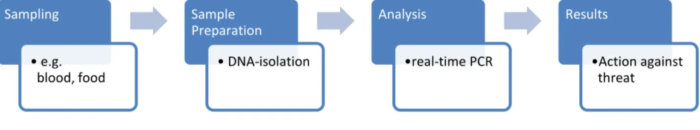
If B. anthracis spores were to be released into the air, in animal fodder, in food or by other routes, a fast and unambiguous detection method is needed to prevent the disease. In such a scenario a high number of samples would be expected and a high throughput method is therefore necessary. See Figure 1 for the full analysis chain when, for instance, B. anthracis spores were to be
released in public.
Figure 1. General workflow in the analysis of a bioterrorism microorganism.
Sampling • e.g. blood, food Sample Preparation • DNA-isolation Analysis •real-time PCR Results •Action against threat
8 In the past, positive confirmation of B. anthracis was performed by growth on a selective media or by using antibody strips against B. anthracis, but theses methods are not sufficiently fast and unambiguous, respectively. In 1992, Makino et al6 published a simple and effective Polymerase Chain Reaction (PCR)-analysis of B. anthracis in animals that was able to detect one single spore-forming unit per PCR assay, and in later years PCR has become one of the most effective ways of detection. This technique has been further improved with the onset of real-time
quantitative PCR analysis (qPCR) that is a quantitative analysis of the bacterial load. Also, the real-time PCR can utilize ready-made master mixes containing all reagents except the primers. The reduced labor time, and the use of automated DNA-extraction robots, allow for a high-throughput and automated analysis7, which is a criterion in the case of an emergency.
1.4.1 Real-time PCR
A standard PCR is one of the most widespread techniques in biological research8. Still, in the past, when requiring a quantitative result, the PCR-amplified DNA had to be analysed with dyes on a gel and this requires a lot of manual labour and is not very accurate8. This all changed with the advent of real-time PCR9 and since then, this technique is one of the most sensitive and accurate used for quantification methods of nucleic acids (DNA/RNA)8.
Real-time PCR uses basically the same reagents as with the conventional PCR, but with the addition of a fluorescent dye, probe or beacon. When the designated primers bind to a sequence and the reaction is underway, the dye (or probe/beacon) is activated, chelated or released and this increases the amount of fluorescent light emitted which the machine detects. This is repeated with each thermal cycle and a standard curve can be created from the data acquired. Then the sample‟s emission data is compared with the standard curve and a relatively exact concentration of DNA can be acquired.
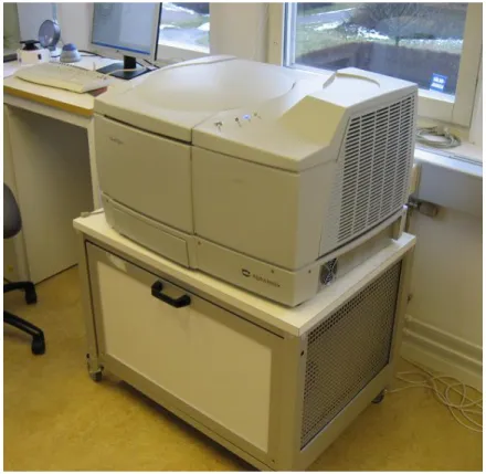
1.4.2 ABI 7500 Real-Time PCR System
The Applied Biosystems 7500 Real-Time PCR System (Applied Biosystems, Foster City, CA, USA) is a standard 96-well thermal cycler with real-time fluorescence detection. It is compatible with the most commonly used assays (gene expression analysis, SNP etc.) and dyes/probes (SYBR-Green I, TaqMan etc.)10. See Figure 2 for picture of the ABI7500.
9
Figure 2. The ABI 7500 real time PCR system 1.4.3 Alpha Helix QuanTyper™
The QuanTyper™ is a thermal cycler prototype that utilises SuperConvection™, developed by Alpha Helix (Uppsala, Sweden). This is a technique with powerful infrared heating capacity and also a very fast cooling system. The machine centrifuges (up to 8 000 x g) the samples during thermal cycling thus enabling a fast and homogenous temperature equilibration. A fan is mounted on top of the sample holder which cooled air is flowed through as the fan spins with the centrifugal rotor, and the cooling procedure is therefore done in seconds11. See Figure 3 for a picture of the rotor and fan of the QuanTyper™.
10
Figure 3. The QuanTyper™ with the lid open, showing the sample holder with the fan on top of it. Notice the air duct in the lid through which cold air is flowed down to the fan which cools the samples.
It has a capacity of 48 individual 0.2 mL PCR-tubes and several different kinds of probes or dyes can be used for real-time detection11. See Figure 4 for a picture of the sample holder.
Figure 4. The sample holder of the QuanTyper™. The red ring shows the temperature probe which contains the same amount of liquid as the samples, thus providing a very accurate temperature measurement.
11 The tubes are specifically designed for the machine and moulded by MicroPlast (Skara, Sweden). Standard PCR-tubes are not compatible with the QuanTyper™ as they lack the special profile that locks the tubes in place in the machine.
Also, the standard tubes are, in contrast to the Alpha Helix tubes, moulded from top to bottom, meaning that the plastic is injected into the mould from the bottom. Hence, the weakest point of the tube is in the bottom and that area is put to extreme stress when centrifuging the samples. Compared to the ABI 7500, which has a sample ramp rate of 1.6 C/sec10, the QuanTyper™ can heat or cool the samples at 8 C/sec (mean in the range 50 to 95 C)11.
The centrifugation also provides a kind of stirring in the tubes, which may increase reaction kinetics and decrease reaction times.
All of this enables the machine to complete a real-time PCR with quantitative data in 10 minutes11. Therefore, incorporating the QuanTyper™ into the diagnostic chain of B. anthracis could dramatically increase the speed for a rapid detection. See Figure 5 for a picture of the QuanTyper™.
12
1.5 Sample preparation
Before a qPCR-analysis is performed, a DNA preparation step is usually performed to achieve higher sensitivity. High DNA purity is a prerequisite for good detection since the matrix (fodder, feaces, food etc) may contain PCR inhibitors. Therefore a sufficient DNA-preparation and usage of appropriate buffers are crucial12.
1.5.1Manual DNA-extraction
A DNA-preparation can be performed manually with different kits and often a centrifuge.
Another method includes extraction using magnetic beads. The downside of using a manual kit is that it is time- and labor-demanding, often requiring over an hour of work.
A commonly used kit is the QIAamp® DNA Mini Kit (Qiagen, Hilden, Germany) that utilizes a spin column filter extraction. It can be used on different kinds of samples, such as buffy coat, whole blood, plasma, cultured cells, tissue etc.
The cells are first put through a lysis step, and then the DNA is bound to a membrane inside the spin column. The column fits inside a 2 ml collection tube and the tube is then centrifuged in a microcentrifuge between steps. The sample is then washed and eluted with purified DNA as the product. The downside of using this manual spin kit is the “hands-on” labor time, and also the increased risk of contamination compared to automated solutions.
A study by Timothy Barkham showed that for a viral DNA extraction, the “hands-on” time was 1 h for the QIAamp® kit13.
1.5.2 Automated DNA-extraction
Automated DNA-extraction robots can dramatically increase the processing capabilities of the extraction step, being able to perform a vast amount of samples at the same time.
It can also be a faster alternative and also produce more consistent results.
A downside of using a robot is that it will produce aerosols during operation which can be very hazardous when working with spore-forming bacteria like B. anthracis that can be spread by the aerosols and cause infection.
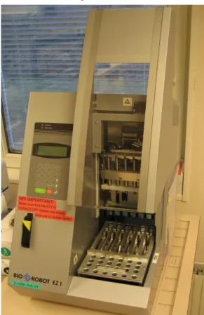
The BioRobot EZ1 workstation (QIAgen) is an automated DNA-purification robot. It uses a magnetic particle technology where the DNA is bound to the silica surface of the magnetic beads during the preparation procedures.
The DNA/beads are removed from the sample and then washed to remove impurities and the DNA is then eluted into 1.5 ml tubes. The machine can be used in clinical and forensic analysis14, 15.
The reagents needed for the purification are supplied in pre-filled cartridges, which greatly reduce the risk of contamination and also lower hands-on work time.
13 A study by Timothy Barkham showed that for a viral DNA extraction, the “hands-on” time with the robot was 10 min compared to 1 h for the QIAamp® kit13.
The machine uses pre-programmed EZ1 cards, which contains the protocols for the purification of choice. Six samples can be extracted simultaneously.
A negative aspect of the robot is that it produces aerosols when performing the DNA-purification and this is extremely hazardous when working with B. anthracis unless used in a proper biosafety way.
Barkham was also aware of this fact and tried to enclose the EZ1 into a safety cabinet, but the robot was too big for the standard cabinet13. See Figure 6 for a picture of the EZ1.
Figure 6. The QIAgen BioRobot EZ1.
1.6 Biosafety
The next aspect to address is that of the safety issues. When dealing with highly hazardous bacteria such as B. anthracis, personnel safety becomes an issue.
In Sweden, The Swedish Work Environment Authority classifies B. anthracis as a risk class 3-organism16.
Risk class 3 includes infectious substances with a risk of serious consequences at exposure16, and some of the organisms in class 3 have very dangerous aerosols. The spores of
B. anthracis can form an aerosol and be inhaled causing pulmonary anthrax and this is the main worry when working with the bacteria.
14 In USA, The Centers for Disease Control and Prevention (CDC) and The National Institutes of Health (NIH) have published the Biosafety in Microbiological and Biomedical Laboratories (B.M.B.L.) 4th Edition in which B. anthracis isclassified as a Biosafety Level 3 organism (BSL-3)17. Although BSL-2 is adequate when working with small quantities17.
1.7 Safety cabinetry
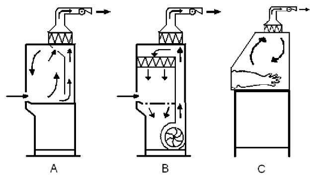
1.7.1 Class I safety cabinetThe class I safety cabinet has a front aperture through which the operator can conduct work. The cabinet uses a blower to achieve negative pressure and thus an inward air stream is created. Exhaust air is then filtered and often led through a duct into the laboratory ventilation. This solution protects the worker from bacterial aerosols and such, but it does not protect the product. Although it must be noted that studies has showed that the class I cabinet can indeed protect the product from cross-contamination18, but the class I is mainly developed and used for scenarios where personnel protection is preferred to product protection.
Could the class I cabinet then be used to prepare B. anthracis spores with sufficient biosafety? The answer is no, although the cabinet probably could contain the aerosols created, there are still big risks to be considered.
Studies have showed that a door slam or laboratory personnel moving around in the laboratory severely disrupts the airflow for a short period of time18. The optimal laboratory does not have people running around slamming doors, but in the event of a deliberate contamination incident, stress levels and work load may increase dramatically thus creating a stressful environment. Also, in the event of a power failure or a fuse melting, the inward airflow would stop and putting everyone in the lab at risk, or at least contaminate the laboratory. See Figure 7 for a basic
schematic of airflow and construction.
1.7.2 Class II safety cabinet
To protect laboratory personnel working with class 3 organisms, the workspace and small machinery is often positioned in a class II safety cabinet (often referred to as a Laminar Airflow bench (LAF)).
The class II cabinet operates under negative pressure airflow into the cabinet to protect the personnel. To protect the product/bacteria, the work surface inside the cabinet is constantly flowed-through by High Efficiency Particulate Air (HEPA)-filtered air19.
This provides a good safety for the product, but gives only little protection for the personnel. The safety of this design depends on the airflow being absolute at all times, so when some sort of machinery, e.g. a DNA-preparation robot, is inserted into the cabinet, the airflow could probably be disrupted and the cabinet no longer fills its purpose. Macher et al.20 showed that a simple
15 movement of the cabinet operator‟s hand affected the laminar flow, so it is reasonable to assume that when inserting a machine that obstructs at least half of the work area, safety is compromised. And, as with the class I cabinet, it is sensitive to cross drafts created from air conditioning and heating21 and also to mechanical failure of the fan.
At SVA, a BioRobot EZ1 was enclosed in a class II safety cabinet and a potassium iodide discus test was done to evaluate the airflow and safety of the configuration. The airflow became
turbulent with the robot inside and the safety cabinet did not pass the safety regulations.
As stated earlier, the purpose of this study was to enclose a robot in a safety cabinet so the class II cabinet is not the optimal solution for this problem. See Figure 7 for a basic schematic of airflow and construction of a class II safety cabinet.
Using a totally sealed container is probably the best approach to solve this problem. This container will protect the personnel from the aerosols created by the robot and dramatically reducing the risk of contamination.
1.7.3 Class III safety cabinet
A higher class of safety cabinet is available, the class III safety cabinet (often referred to as a glovebox). It represents the highest standard of personnel and environmental protection from aerosols and microbiological contaminants. It is suitable for work on both class III and class IV organisms. It is also used in organic chemistry when handling oxygen sensitive chemicals, as the glovebox allows for a different atmosphere inside.
The boxes are available in different configurations, e.g. fitted with carbon-filters, HEPA-filters, refrigerators and other laboratory equipment22. One or more sides of the container are usually transparent and fitted with gloves to allow management of elements inside the box.
There is also a possibility to lower the pressure in the box, so if there is a leak, air would flow into the box rather than out in the laboratory23.
It can also be configured for positive pressure operation, which is preferred when product protection is preferred over personnel protection.
See Figure 7 for a basic schematic of airflow and construction.
The cabinet can be connected to a chemical dunk tank or an autoclave to allow for disinfection of the material entering and exiting the cabinet. Instead of an autoclave, the cabinet can be
connected to a class II safety cabinet via an air lock.
As oppose of the class II cabinet, the class III cabinet is not dependent on laminar air flow, hence, it is not sensitive to interior modifications. Placing a DNA-preparation robot in the class III cabinet poses merely spatial problems, as the robot is not a small piece of machinery. Placing machinery into a safety cabinet has been attempted before; in 1979, Chatigny et al. enclosed an ultracentrifuge into a class III safety cabinet for evaluation24. The problem with enclosing the
16 BioRobot EZ1 is the measurements of the robot. Custom-made safety cabinets can be ordered but, the measurements must first be evaluated.
An issue when working with the thick gloves is the loss of dexterity. Sawyer et al. showed that there was a statistically significant detrimental effect on dexterity when working with gloves compared to working with bare hands and that this may increase the risk of accidents25.
Figure 7. Side view-schematic of airflow and basic construction of the three different safety cabinet classes. A = class I, B = class II, C = class III. The areas with a triangular pattern are HEPA-filters. Picture is modified from original26.
1.7.4 Transportable class III glovebox from Germfree
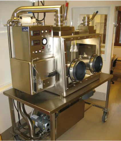
Transportable Class III Glovebox with Airlock, Serial Number C-2170 and NBC Filtration System Serial Number C-2171 (Germfree, Ormond Beach, FL, USA) is available at SVA (see figure 8). The glove box will be referred to as GB-3 in this report.
The box is designed to be used as a mobile platform and is mounted on top of a movable bench. It is fitted with a nuclear, biological, chemical (NBC) filter unit, which filters the air from
biological threats with HEPA-filters and from chemical threats with carbon filters It meets all the safety requirements specified in the CDC-NIH B.M.B.L., 4th Edition17 .
It is constructed of all welded stainless steel and a viewing window made of polycarbonate. All the intake air is filtered through a HEPA-filter, the outgoing air is first filtered through two HEPA-filters and two carbon-filters too fully clean the air from contaminants and chemicals. The NBC filtration system maintains a flow of 1.7 m3 / min with greater than -149 Pascal negative pressure in the glovebox.
17 According to Germfree, the recommended flow rate into the box when a glove has failed is about 0.5 m/s. This flow rate is achieved when the box is operated at a negative pressure of -250 Pascal. It is therefore of highest importance that the pressure is kept lower than -250 Pa when working with B. anthracis and other spore-forming bacteria.
The gloves are made from butyl, which is one of the easiest glove materials to work with, being very flexible and sensitive27.
Figure 8. The transportable class III biological safety cabinet from Germfree
1.8 Objectives
The aim of this master thesis was to produce a prototype for a class III biological safety cabinet, evaluate a class III cabinet and also improve the PCR-analysis step for B. anthracis.
1.8.1 Designing a class III safety cabinet prototype
The first objective of this study was to do a literary study on biological safety cabinets and biological safety issues surrounding this area. Then, a prototype of a class III safety cabinet was designed and eventually built by the technical staff at SVA. The main purpose of this prototype was to enclose the BioRobot EZ1.
18 A lot of aspects were addressed, what type of material the gloves needs to made of, if there is to be air filtration, measurements of the box to facilitate work etc.
The prototype was made from simple materials and in no way resembling the final product in terms of biosafety, but the form and function was basically the same.
1.8.2 Evaluating the prototype
The prototype was also evaluated in terms of dexterity and performance when working with the BioRobot EZ1.
The BioRobot utilises pre-filled reagent cartridges that is very easy to load into the machine14, and this should reduce risks of accidents when working with the thick gloves.
The necessary kits, chemicals and samples was inserted into the box through the lock. A series of tests was conducted on the feasibility of operating the BioRobot EZ1 and preparing the DNA from the samples. In these tests, cultures of B. cereus were used as a model organism because of the hazards of working with B. anthracis.
1.8.3 Evaluating the class III cabinet from Germfree
Also, as a side project, tests were performed on an existing class III glovebox, model GB-3 (Germfree), to evaluate its performance and ergonomics.
This box was not big enough to enclose the EZ1; therefore, the DNA-preparation was performed using a manual spin kit from QIAgen. A decontamination protocol was also tested. Other tests may later be added, and hopefully this will uncover difficulties and problems during glovebox work that is important to remedy before the glovebox is actually used in a real emergency.
1.8.4 DNA-preparation and quantitative PCR analysis
The time it took to purify a bacterial sample was noted and compared, both from the EZ1 inside the prototype and from the manual spin kit (both inside the GB-3 (Germfree) and inside the prototype).
The time it took the DNA to be analysed after preparation was also noted. The PCR machine commonly used in the laboratory is the ABI 7500 Real-Time System (Applied Biosystems) and it was used as a reference.
Spores from B. cereus were lysed in lysis buffer with and without lysozyme and the DNA-yields were compared and analysed with PCR.
B. cereus spores and culture went through the whole analysis chain and were analysed on the ABI 7500.
19
1.8.5 Evaluation of the Alpha Helix QuanTyper™
The QuanTyper™ (Alpha Helix) was compared with the ABI 7500 (Applied Biosystems) in terms of performance and reliability.
The aim was to evaluate the possibilities to incorporate the QuanTyper™ into the analysis chain of pathogenic microorganisms.
First, a compatible master-mix was found that showed stable results on the platform and then different samples and primer pairs was tested. DNA from both B. anthracis and B. cereus was analysed. The DNA analysed was prepared from cultures and from spores.
2 Materials and Methods
2.1 Construction of glovebox-prototype
Measurements of the BioRobot were gathered and the space needed to safely conduct work with the robot was estimated. Different requests, such as glove ports on three sides of the box and a railing system, were evaluated and a basic design of the box was produced. The design was delivered to the technical services at SVA for production.
Gloves were ordered from North (North Safety Products, Cranston, RI, USA). The gloves were 10 inches wide with the product number 10LA1832A/10H and were made of nitrile.
2.2 B. cereus strain preparation
The B. cereus strain KBM1 was taken from the -70°C freezer and inoculated in brain heart infusion (BHI), (SVA, Uppsala, Sweden) and incubated overnight at 37°C in a Termaks KB8400 incubator (Termaks AS, Bergen, Norway). The culture was serially diluted in 0.86 - 0.9 % NaCl solution (SVA). The dilutions was plated on horse blood agar (SVA) and incubated at 37°C overnight, and the colonies was counted to achieve a colony forming units per millilitre number (CFU/ml) on the culture.
The culture was divided into 20 aliquots in 1.5 mL tubes and put in a -20°C freezer.
2.3 Evaluation of sterilization protocol in a class III cabinet
A decontamination protocol has been developed for B. anthracis and was here tested for compatibility on B. cereus inside a class III safety cabinet by Germfree. All of the following work under this headline has been performed inside the glovebox.
Two hundred µL aliquots of the B. cereus strain KBM1 in BHI was put into three 1.5 mL tubes, and to each of these tubes, 670 µL of Buffer MTL (Qiagen) was added. Buffer MTL contains guanidium thiocyanate.
The tubes were vortexed and 800 µL of the mixtures was put into new 1.5 mL tubes with screw caps, to get rid off the bubbles/foam on the wall of the tubes. The tubes were incubated at 80°C for 20 min and plated on horse blood agar plates (SVA) and incubated at 37°C overnight. The
20 fingertips of the butyl gloves inside the glovebox were also tested for contamination by touching a plate with horse blood agar which was then incubated at 37°C overnight in a KB8400 incubator (Termaks).
2.4 DNA extraction
2.4.1 Manual spin column kit on open bench using the QIAamp® DNA Mini Kit
All solutions used are included in the QIAamp® DNA Mini Kit if not otherwise stated. A tube containing 1 mL of B. cereus-culture was taken from the –20°C freezer and thawed. The tube was centrifuged for 10 min at 5000 x g to create a pellet, which was suspended in 180 µL lysis buffer (20 mM Tris-HCl, pH 8.0; 2 mM EDTA; 1.2% Triton®), (SVA).
Twenty µL Proteinase K and 200 µL Buffer AL was added and mixed by vortexing. The tube was incubated at 56°C for 30 min and then for a further 15 min at 95°C followed by a short centrifugation.
Then the Tissue Protocol (QIAamp® DNA Mini Kit) was followed and elution was made in 2 x 100 µL AE buffer. The eluates were then stored in a -20°C freezer.
2.4.2 Manual spin column kit inside class III safety cabinet by Germfree using the QIAamp® DNA Mini Kit
The glovebox was started and all the start-up tests were performed to ensure the performance of the box.
Two tubes of frozen B. cereus culture (1.0 mL) were thawed and inserted into the glovebox along with the necessary material stated in the kit manual. An Eppendorf 5418 aerosol-safe micro centrifuge (Eppendorf, Hamburg, Germany) and a heating block were also inserted.
Two horse blood agar plates (SVA) were positioned on the sides of the workspace and the centrifuge as a control for aerosol formation.
The gloves were decontaminated using a sodium hypochlorite-solution before the experiment. All solutions used are included in the QIAamp® DNA Mini Kit if not otherwise stated. A clean tube without any culture was used as a negative control.
The tubes were centrifuged for 10 min at 5000 x g to create a pellet, which was suspended in 180 µL lysis buffer (20 mM Tris-HCl, pH 8.0; 2 mM EDTA; 1.2% Triton®), (SVA).
Twenty µL Proteinase K and 200 µL Buffer AL was added to each tube and mixed by vortexing. The tubes were incubated at 56°C for 30 min and then for a further 15 min at 95°C followed by a short centrifugation.
Then the Tissue Protocol (QIAamp DNA Mini Kit) was followed and elution was made in 2 x 100 µL AE buffer per sample.
21 The DNA concentration of the eluates were analysed in a NanoDrop ND-1000
Spectrophotometer (NanoDrop Technologies, Inc.). The eluates were then stored in a -20°C freezer.
The gloves were checked for contamination using an eSwab (Copan innovation, Brescia, Italy) which is a swab made for bacterial samples but was here used for DNA.
The gloves were thoroughly swabbed and the swab was inserted into the accompanying sample liquid. After a short vortex, the swab was discarded.
Two µg transfer-RNA (Ribonucleic acid, transfer from baker‟s yeast 10 mg/ml, Sigma Aldrich, St.Louis, MO, USA) was added to 400 µL of eSwab liquid to enhance the chance of extracting the small amounts of DNA in the liquid28. The 400 µL was extracted with the BioRobot EZ1 and eluted in 75 µL. 2.5 µL of the eluate was directly added to 22.5 µL PCR-master mix and analysed on the ABI 7500.
The horse blood agar plates were incubated at 37°C overnight in a KB8400 incubator (Termaks). The QIAamp-procedure above was repeated but with a solution of B. cereus-spores. In that experiment, 25 µL of spore-solution (Raven labs, Omaha, NE, USA) containing 107 spores/ml was extracted. The effect of lysozyme (From chicken egg white, ~70 000 units/mg solid, Sigma Aldrich), was evaluated. Lysis buffer (175µL) containing lysozyme (20 mg/mL Lysozyme, 20 mM Tris-HCl, pH 8.0; 2 mM EDTA; 1.2% Triton®) was added to one of the two samples, and the same lysis buffer, but without lysozyme, was added to the other sample. Other than that, they were put through the exact same method. A blank sample was also used to rule out false
positives. The extracted DNA was analysed on the ABI 7500.
2.4.3 Manual spin column kit inside class III prototype using the QIAamp® DNA Mini Kit
Two tubes of frozen B. cereus culture (1.0 mL) were thawed and inserted into the glovebox along with the necessary material stated in the kit manual. An Eppendorf 5418 aerosol-safe micro centrifuge (Eppendorf) and a heating block were also inserted.
The gloves were decontaminated using a sodium hypochlorite-solution before the experiment. All solutions used are included in the QIAamp® DNA Mini Kit if not otherwise stated. A clean tube without any culture was used as a negative control.
The tubes were centrifuged for 10 min at 5000 x g to create a pellet, which was suspended in 180 µL lysis buffer (20 mM Tris-HCl, pH 8.0; 2 mM EDTA; 1.2% Triton®), (SVA).
Twenty µL Proteinase K and 200 µL Buffer AL was added to each tube and mixed by vortexing. The tubes were incubated at 56°C for 30 min and then for a further 15 min at 95°C followed by a short centrifugation.
Then the Tissue Protocol (QIAamp DNA Mini Kit) was followed and elution was made in 2 x 100 µL AE buffer per sample.
22 The DNA concentration of the eluates were analysed in a NanoDrop ND-1000
Spectrophotometer (NanoDrop Technologies, Inc.). The eluates were then stored in a -20°C freezer.
2.4.4 Automated BioRobot EZ1 inside class III prototype
All consumables used under this headline were included in the EZ1 DNA Tissue Kit (Qiagen), if not stated otherwise. The EZ1 DNA Tissue Card (Qiagen) was inserted into the EZ1 (Qiagen), which contained the program for the extraction.
Two 1.5 mL aliquots of B. cereus-culture were thawed. Two hundred µL from each aliquot and were loaded into sample tubes and inserted into the EZ1 (Qiagen) along with the elution-tubes, tip holders with filer tips and the reagent cartridges. Two hundred µL sterile water was also extracted and used a negative control. Elution volume was set to 75 µL and the protocol was started.
2.4.5 DNA concentration measurements
The eluates from all the DNA-extractions performed were analysed in a NanoDrop ND-1000 Spectrophotometer (NanoDrop Technologies, Inc., Wilmington, DE, USA). It utilizes full-spectrum UV-Vis absorbance to analyse the amount of DNA in the sample. It requires no consumables and only 1 µL of sample.
The machine can give an estimate of the amount of DNA and also the purity of the sample. It can measure in the range of 2-3700 ng/ul of dsDNA.
2.5 DNA analysis by real-time PCR
2.5.1 ABI 7500A Power SYBR Green PCR Master Mix (Applied Biosystems) assay was used for all DNA samples as a reference. The Master Mix contains deoxyuridine triphosphates (dUTP) and is therefore compatible with uracil-N-glycosylase (UNG). 1 U/µl AmpErase UNG (Applied Biosystems) was used to rule out carryover contaminations.
The addition of Bovine Serum Albumin (BSA), (Sigma Aldrich) helps with minor inhibitions due to matrices12.
The prepared DNA from the different preparations was serially ten-fold diluted to a relative concentration of 1 x 10⁻⁸. See table 1 for PCR reagents recipe.
23
Table 1. Recipe for PCR-master mix. Volume is per reaction.
Reagent Volume of reagent per PCR (µL)
Final concentrations of reagents
Power SYBR Green PCR Master Mix
12.5 1X
ddH2O 7.5 -
BSA 2 % (w/v) 0.25 0.02 % (w/v)
UNG 1 U/µL 0.25 0.25 U/sample
Primer forward (10µM) 1.0 400 nM
Primer reverse (10 µM) 1.0 400 nM
Total amount 22.5
The primers (Operon, Huntsville, AL, USA) for B. cereus targeted the rpoB gene29 with the sequences
(5‟-TTGCTTGAAATTTATGAGCGTCTAC-3‟, 5‟- ATTGTTCCTTCTGCCGCTAAAA-3‟)29
The master-mix from Table 1 was prepared (volumes multiplied by number of reactions), and distributed on a 96-well micro plate. 2.5 µL of the different template dilutions was added to the different wells, also a negative and a positive control was added. The final concentration of the individual primers was 400 nM, with a BSA concentration of 0.02 % (w/v).
The PCR was started (50°C, 2 min; 95°C, 10 min; (95°C, 15 s; 60°C, 1 min) x 40; 95°C, 15 s; 60°C, 1 min; 95°C, 15 s).
The first step at 50°C is required by the UNG, and when the thermal cycling and amplification is done, a dissociation step is used to verify the amplicon.
2.5.2 Alpha Helix QuanTyper™
A Platinum® SYBR® Green qPCR SuperMix-UDG (Invitrogen, Carlsbad, CA, USA) assay was used for all DNA samples. The Master Mix contains dUTP and also UNG. The prepared DNA from the different preparations was serially ten-fold diluted to a dilution factor of 10-8. Different samples were tested such as DNA from vegetative B. cereus and also from spores. DNA from B. anthracis was also tested targeting three different sequences (one chromosomal- and two different plasmid-targets).
See table 2 for PCR reagents recipe.
Table 2. Recipe for PCR-master mix. Volume is per reaction.
Reagent Volume of reagent per PCR (µL)
Final concentrations of reagents
Platinum® SYBR® Green qPCR SuperMix-UDG 10.0 1X ddH2O 6.4 - Primer forward (10 µM) 0.8 400 nM Primer reverse (10 µM) 0.8 400 nM Total amount 18
24 The QuanTyper™ was configured for 20 µL reactions.
The primers (Operon) for B. cereus targeted the rpoB gene29 with the sequences
(5‟-TTGCTTGAAATTTATGAGCGTCTAC-3‟ and 5‟- ATTGTTCCTTCTGCCGCTAAAA-3‟)29
. To the 18 µL prepared master mix, 2 µL of template was added.
The PCR-program was optimized (50°C, 2 min; 95°C, 90 s; (95°C, 10 s; 60°C, 25 s) x 40). After all runs, a dissociation step was added by the use of a gradient going from 60°C to 93°C with a ramp rate of 0.5°C/s.
Another enzyme tested was the Phusion™ Flash (Finnzymes, Espoo, Finland), having most impressive performance as it has an extremely high processing speed coupled with a low error-rate30.
With the Phusion™ Flash High-Fidelity PCR Master Mix, a three step cycle was set
up (98°C, 10 sec; (98°C, 0 sec; 58°C, 3 sec; 72°C, 3 sec) x 40), the template was the human gene for corticotropin releasing hormone receptor 2 which yields an amplicon of 115 bp, and the assay was a SYBR Green assay. The reagents and template were supplied by Alpha Helix.
25
3 Results
3.1 Designing a class III safety cabinet prototype
After all the measurements had been done and all the input was gathered a design for a simple class III cabinet was made. For a schematic of the design, see Figure 9, 10 and 11.
Figure 9. Schematic of the class III safety cabinet prototype, showing the back of the cabinet to the left and the front of the cabinet at the right. The measurements are in centimetres (cm).
Figure 10. Schematic of the class III safety cabinet prototype, showing the left side of the cabinet to the left and the right side of the cabinet at the right. The diameter of the glove port on the left image is 25.4 cm. The measurements are in centimetres (cm).
26
Fig 11. Schematic of the class III safety cabinet prototype, showing two different possible designs from a top view. In the design to the right, the BioRobot EZ1 is positioned on top of a platform on rails which enables moving of the machine.
3.2 Evaluating the prototype
3.2.1 B. cereus strain preparationAfter counting the colonies on the plates the CFU/ml was calculated to be 6.25 x 10⁸.
This culture was used for all the DNA-preparations which were used for the PCR-runs if not otherwise stated.
3.2.2 Feasibility of working with the prototype
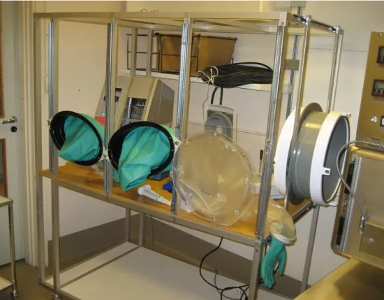
The prototype built was a very simplified version, basically only consisting of the front side with the glove ports. This was enough to evaluate the feasibility of working with the EZ1 and also doing a manual spin column kit DNA-preparation, with nitrile gloves fitted to the prototype. Also, an evaluation of the measurements needed to work with the robot was carried out. The railing system shown in Figure 11 was not used due to lack of time.
Due to some technical issues, the schematic was not followed exactly and the measurements of the built prototype are as follows.
Length: 127 cm Depth: 74 cm Height: 82 cm
Three gloves were attached to the prototype which could be adjusted in position and height to find the optimum positions. Two gloves are meant to be used for the majority of the work and the third glove is for more specific manoeuvres, such as extracting or inserting material through the airlock, or to operate the EZ1. See Figure 12 for a picture of the prototype.
27
Figure 12. Picture of the glovebox prototype with gloves attached and the EZ1 positioned inside
See figure 13 for a picture of the workspace when performing a manual spin column kit DNA-extraction.
28 It was found that this third glove was best suited to operate the EZ1, and using the paired oval glove ports for all other preparative work.
Since the EZ1 is easily operated and not much hand-on time is needed, the robot was positioned in the left upper corner of the prototype to free up as much space as possible. The optimum position of the EZ1 and the gloves is shown in figure 14.
Figure 14. The optimal positioning of the gloves and machinery. The EZ1 is to the left, the microcentrifuge to the right.
The glove to the far left was found to be too long as the EZ1 is occupying the area in front of it. A shorter glove is needed to operate the EZ1 (See Figure 14). The machine could not be turned on using the gloves as the button is located on the back of the machine. In the final glove box, the power to the machine would have to be operated from the outside.
If working only with the EZ1 robot in the glove box, a width of 90 cm is sufficient. The 127 cm of this prototype was only necessary when performing a manual extraction using for instance a manual spin kit beside the robot. In this case the extra width was needed by the centrifuge and heating block etc. (See Figure 13).There were some difficulties in reaching and decontaminating the whole work area since the gloves were too short for this purpose.
29 This could be remedied by the use of a glove on the back-side of the box, which could also be used by another person when performing tasks with a heavy workload or when moving machinery inside the box.
3.3 Evaluating the class III safety cabinet from Germfree
Manuals for operating the cabinet from Germfree were created as well as decontaminating and cleaning procedures of the different parts and surfaces of the box. A short run protocol was written that must be followed and properly filled in during every experiment to be able to follow the filter quality and to see pressure changes over time. The electrical outlets inside the box were also changed to Swedish standard.
3.3.1 Evaluation of a decontamination protocol in a class III cabinet
The protocol was followed successfully and no spillage occurred. There were two B. cereus colonies on one of the horse blood agar plates containing the killed culture. There was also growth on the plate that had contact with the fingers on the butyl gloves, but only at one finger imprint
3.4 DNA-extraction and quantitative PCR-analysis
3.4.1 Manual spin column kit on open bench using the QIAamp® DNA Mini Kit
The DNA-extraction procedure took 1h 40 min. The DNA concentration of the first eluate was 14.5 ng/µL (260/280 = 1.75; 260/230 = 0.92) measured with NanoDrop. The 260/230-ratio of 0.92 indicate that the sample is unclean and contains contaminating proteins. The second eluate had a concentration of 22.2 ng/µL (260/280 = 1.95; 260/230 = 2.47), and the ratios indicating that the sample purity was high. This eluate was used for further analyses. A DNA-extraction
performed on an open bench was used as a reference run to which all other preparations were compared. The PCR-analysis results are shown in Figure 15.
The run showed good sensitivity with an intercept of around 37, but with an efficiency of only 90 %. The limit of detection (LOD) was about 22.2 ×10⁻⁵ ng DNA/µL sample, i.e. 2.22 ×10⁻¹⁵ g of total DNA / µL sample.
30
Figure 15. Results from PCR-analysis of DNA-extraction on open bench on B. cereus culture. PCR was
performed on the ABI 7500 real time system. The cycle when sample fluorescence exceeds chosen threshold (Ct) is shown. R2 is a correlation coefficient of the trend line, Eff is the efficiency of the run.
3.4.2 Manual spin column kit inside class III safety cabinet by Germfree using the QIAamp® DNA Mini Kit
The DNA-extraction was completed in 2h 6 min, not taking the removal of material from the glovebox into account. For DNA-concentration measurements, see Table 3.
Table 3. Measurements performed on the NanoDrop spectrophotometer, showing
DNA-concentrations and absorbance ratios of different wavelengths to indicate purity of sample. 260/280-ratio should be ~1.8, 260/230-ratio should be ~1.8-2.2 (not to low).
The negative control was a tube without culture that was submitted to the same treatment as the other samples.
Sample Eluate Concentration (ng/µL) 260/280 260/230
1 a 57.8 2.20 2.09 b 94.1 2.23 2.68 2 a 50.6 2.17 1.84 b 93.1 2.29 2.54 Negative control a -0.1 -1.98 -0.14 b 0.0 -0.22 0.01
QIAamp DNA Mini Kit y = -3.581x + 37.9 R² = 0.997 Eff. = 90 % 10 15 20 25 30 35 40 0 1 2 3 4 5 6 7 Ct
31 The horse blood agar plates showed no growth even after three days incubation, showing that no aerosols formed around the centrifuge.
The 1a-eluate (see Table 3) was analysed with PCR.
For results from the PCR analysis on the DNA that was prepared inside the GB-3 glovebox (Germfree), see Figure 16. The calculated efficiency was 0.9157 with a correlation coefficient of 0.991.
The LOD was about 57.8 ×10⁻⁵ ng DNA/µL sample, i.e. 5.78 ×10⁻¹⁵ g of total DNA / µL sample.
Figure 16. Results from PCR-analysis of DNA-extraction performed inside a class III safety cabinet, PCR was performed on ABI 7500 real time system. Ct is the cycle when sample fluorescence exceeds chosen threshold. R2 is a correlation coefficient of the trend line, Eff is the efficiency of the run.
Both samples from the eSwab (Copan) liquid were positive. The concentration of DNA in the eSwab (Copan) liquid was calculated to 0.1 ng/µL, assuming a 100 % extraction from the EZ1. That is equal to some 50 ng DNA collected with the swab. A negative control eSwab (Copan) was also analysed but showed no amplification.
The DNA extracted from the spore-solutions was directly analyzed on the ABI 7500 without diluting the samples. As seen in Figure 17, there was a good yield of DNA when using the QIAamp® spin column kit on spores. The first eluates of the samples produce very similar results, and the same goes for the second eluates. The negative controls are all negative. As seen, there is no difference in amount extracted DNA when using lysozyme compared to without the enzyme. y = -3.542x + 37.40 R² = 0.991 Eff. = 92 % 10 15 20 25 30 35 40 0 1 2 3 4 5 6 7 Ct
32
Figure 17. Results from PCR-analysis of DNA-extraction performed inside a class III safety cabinet on B. cereus spores. Sample 1 is extracted with the use of lysozyme, sample 2 is extracted without lysozyme. The letters ‘a’ and ‘b’ indicates first and second eluate. PCR was performed on the ABI 7500 real time system. Both the cycle when sample fluorescence exceeds chosen threshold (Ct), and the melting point of the amplicons(Tm) is presented.
1a 1a 2a 2a 1b 1b 2b 2b 68 70 72 74 76 78 80 15 17 19 21 23 25 27 29 31 33 35 Tm (°C) Ct
Sample (nr) / eluate (letter)
Ct Tm
33
3.4.3 Manual spin column kit inside class III prototype
The DNA-extraction was completed in 2h 15 min, not taking the removal of material from the glovebox into account. For DNA-concentration measurements, see Table 4.
Table 4. Measurements performed on the NanoDrop spectrophotometer, showing
DNA-concentrations and absorbance ratios of different wavelengths to indicate purity of sample. 260/280-ratio should be ~1.8, 260/230-ratio should be ~1.8-2.2 (not to low).
The negative control was a tube without culture that was submitted to the same treatment as the other samples.
Sample Eluate Concentration (ng/µL) 260/280 260/230
1 a 28.1 1.89 1.63 b 9.4 1.70 1.94 2 a 16.1 1.89 1.18 b 7.2 1.88 1.46 Negative control a 1.3 0.59 0.55 b 0.7 0.36 0.32
The 1b-eluate (see Table 4) was analysed by PCR, see Figure 18 for results. The calculated efficiency was 0.889 with a correlation coefficient of 0.998
The run showed a lowered sensitivity with an intercept of around 39, and with an efficiency of only 89 %. The LOD was about 9.4 ×10⁻⁵ ng DNA/µL sample,
i.e. 0.94 ×10⁻¹⁵ g of total DNA / µL sample.
Figure 18. Results from PCR-analysis of DNA-extraction performed inside the prototype using a manual spin kit. PCR was performed on the ABI 7500 real time system. Ct is the cycle when sample fluorescence exceeds chosen threshold. R2 is a correlation coefficient of the trend line, Eff is the efficiency of the run.
QIAamp DNA Mini Kit y = -3.618x + 38.97 R² = 0.998 Eff. = 89 % 10 15 20 25 30 35 40 0 1 2 3 4 5 6 7 Ct
34
3.4.4 Automated BioRobot EZ1 inside class III prototype
The DNA-extraction was completed in 30 min the DNA concentration of the first sample was 60.3 ng/µL (260/280 = 1.85; 260/230 = 1.75) measured by NanoDrop. The second sample had a concentration of 44.0 ng/µL (260/280 = 1.90; 260/230 = 1.30).
The first sample was used in PCR-analysis and the results are shown in Figure 19. The run showed good sensitivity with an intercept under 37, and with an efficiency of 99 %. The LOD was about 60.3 ×10⁻⁶ ng DNA/µL sample, i.e. 6.03 ×10⁻¹⁶ g of total DNA / µL sample.
Figure 19. Results from PCR-analysis of DNA-extraction performed inside the prototype using the BioRobot EZ1. PCR was performed on the ABI 7500 real time system. Ct is the cycle when sample fluorescence exceeds chosen threshold. R2 is a correlation coefficient of the trend line, Eff is the efficiency of the run.
3.5 Evaluation of the Alpha Helix QuanTyper™
The Power SYBR Green PCR Master Mix (Applied Biosystems) was tested with the same reagent concentrations as when used on the ABI 7500, but no amplification was seen with any template concentration. Further tests with different reagent concentrations and temperature profiles showed little or no improvement.
A different enzyme was then tested; the Platinum Taq (Invitrogen) which showed good results from the first run. A ready-made master mix based on this enzyme (Platinum® SYBR® Green qPCR SuperMix-UDG) was then evaluated with regards to different temperature profiles and also the level of detection.
The master mix from Invitrogen successfully produced a 40-cycle quantitative PCR-run with a dissociation analysis in some 30 minutes.
BioRobot EZ1 y = -3.339x + 36.76 R² = 0.990 Eff.= 99 % 10 15 20 25 30 35 40 0 1 2 3 4 5 6 7 8 Ct
35 The greatly reduced reaction times did not affect the level of detection in any way, which was the same as with the ABI 7500 with the ABI Power SYBR Green master mix.
A benefit with this master mix is also the cross-platform capabilities, showing positive results on both the QuanTyper™ and the ABI 7500.
Although, some optimization was needed as the comparative run showed big differences in efficiency between the platforms, see Figure 20.
When using the separate PCR-reagents from Invitrogen, the standard curve was improved compared to when using the ready-made master mix (Figure 20).
Figure 20. Results from comparative run of the ABI 7500 and the QuanTyper™. The same master mix and template was used on both machines. The SYBR Green Master Mix from Invitrogen was used for a direct comparison, and then the separate reagents from Invitrogen is also shown as they produce better results with the QuanTyper™. R2 is a correlation coefficient of the trend line, Eff is the efficiency of the run and Tm is an average of the melting temperatures of all samples.
After an optimizing study, a two-step temperature profile (95°C, 10 sec denaturation, 60°C, 25 sec elongation) was found that yielded excellent efficiencies and melting point curves. These settings were used to analyse B. anthracis.
The criteria set to positively test for B. anthracis-DNA is the confirmation of three targets i.e. target genes on the two virulence plasmids and the chromosome itself. The total reaction time was 38 min for 40 cycles. Three reactions was set up, one for each of the two plasmids and one for the chromosome.
The DNA analysed had been prepared from two different avirulent B. anthracis-strains, one that contained only the pX01-plasmid (sterne strain, 7702) and one that contained only the
pX02-36 plasmid (Pasteur vaccine strain, 4229). Both samples were ten-fold serially diluted from a
solution containing ~5.7 ng DNA /µL. For the chromosomal reaction, both strains were serially diluted and analysed with the same target primers.
The master mix used was the Platinum® SYBR® Green qPCR SuperMix-UDG (Invitrogen). B. anthracis DNA prepared from the extremely pathogenic vollum strain was also available and analysed at the same time as the other two strains with the same master mix. The samples were not diluted and had a concentration of 5.7 ng DNA/µL.
The result from the B. anthracis assay that was analysed with the QuanTyper™ targeting the virulence plasmids is shown in Figure 21. The LOD of the two virulence plasmids was around the fifth ten-fold serial dilution of whole DNA, i.e. a concentration of ~5.7×10⁻⁵ ng DNA/µL.
Figure 21. Results from two PCR-runs of B. anthracis DNA specifically targeting the two virulence plasmids. R2 is a correlation coefficient of the trend line, Eff is the efficiency of the run and Tm is an average of the melting
temperatures of all samples.
Results from the B. anthracis-chromosomal analysis are shown in figure 22. Here, the LOD was around the fourth ten-fold serial dilution, i.e. a concentration of ~5.7×10⁻⁴ ng DNA/µL.
Plasmid pX01 y = -3.225x + 38.06 R² = 0.995 Eff. = 104 % Tm = 74.28 ± 0.105 Plasmid pX02 y = -3.433x + 35.97 R² = 0.995 Eff. = 96 % Tm = 74.50 ± 0.107 15 20 25 30 35 40 0 1 2 3 4 5 6 Ct
Relative log concentration of DNA
Plasmid pX01, sterne strain, 7702 Plasmid pX02, Pasteur vaccine strain, 4229
37
Figure 22. Results from the PCR analysis of B. anthracis DNA, targeting the chromosome. Both the sterne and the pasteur strains, the 7702 and the 4229, were analysed. R2 is a correlation coefficient of the trend line, Eff is the efficiency of the run and Tm is an average of the melting temperatures of all samples.
The vollum strain was tested to compare with the results from the sterne- and Pasteur-strains. The results from the vollum DNA PCR analysis are shown in Table 5. The vollum DNA produced good results with small or no deviations in Ct-values. Also, the melting point temperatures matched very well to the earlier analysed strains.
Table 5. Results from PCR with vollum-DNA as template, showing both the Ct- and the Tm-values.
The Ct-values of the references(sterne + Pasteur) strain are not shown as the template concentrations are not the same. The melting point temperature for the references is a calculated average of several reactions.
The melting points of the vollum-DNA are clearly matching the reference indicating that correct product formation occurred. Target Ct Tm (°C) vollum pX01 15.55 74.40 sterne pX01 74.28 vollum pX02 15.6 74.25 Pasteur pX02 74.50 vollum chromosome 15.6 77.10 sterne chromosome 77.14 Pasteur chromosome 76.99 Strain 7702 y = -3,55x + 36.32 R² = 0.995 Eff. = 91 % Tm = 77.14 ± 0.18 Strain 4229 y = -3.530x + 34.28 R² = 0.992 Eff. = 92 % Tm = 76.99 ± 0.11 15 20 25 30 35 40 0 1 2 3 4 5 Ct
Relative log concentration of DNA
B.anthracis, sterne strain, 7702
38 When using the Phusion™ Flash, after about 10 minutes, all the dilutions were clearly positive, and the cycling was completed in about 12 minutes and then a dissociation step was added. As seen in Figure 23, the Phusion™ Flash master mix yields excellent results on the
QuanTyper™ with an efficiency of 99 % coupled with a very accurate product formation as seen in the gel-analysis (Figure 21). The ABI 7500 on the other hand, produced a whole range of products and primer-dimers according to the gel and melting point analysis (Figure 24).
The analysis time was also lowered to a total of 14 minutes on the QuanTyper™ compared to 1h 40 minutes on the ABI 7500 which had a minimum elongation time of 32 seconds per cycle.
Figure 23. Results from the cross-platform comparison of the PCR-master mix Phusion™ Flash on the ABI 7500 and QuanTyper™. R2 is a correlation coefficient of the trend line, Eff is the efficiency of the run and Tm is an average of the melting temperatures of all samples.
QuanTyper y = -3.352x + 39.22 R² = 0.989 Eff = 99 % Tm = 85.47 ± 0.46 ABI 7500 y = -3.695x + 41.71 R² = 0.894 Eff = 86 % Tm = 85.07 ± 4.43 15 20 25 30 35 40 0 1 2 3 4 5 Ct
Relative log concentration of DNA
QuanTyper ABI 7500
39
Figure 24. Comparison between the ABI 7500 and the QuanTyper™ using the same Phusion™ Flash mastermix and template. The amplicons have here been analyzed on an E-gel (Invitrogen) agarose-gel. Well 1-2, 5-6, 9-10 is different dilutions run on the QuanTyper™. Well 3-4, 7-8, 11-12 is the same dilutions run on the ABI 7500. Courtesy of Alpha Helix
40
3.6 Summary of PCR-results
All the important results from the different PCR-assays tested are shown in Table 6. Results are from both PCR-platforms tested with different master-mixes and from different templates.
Table 6. Summary of the different SYBR Green-PCR–assays tested. No amplification was present if no melting point is presented. Some melting points are highlighted in different colors to indicate comparable data. Detection level is presented in the highest ten-fold serial dilution still showing detection and also the mass of whole DNA per PCR. Reaction volume was 20 µL for all QuanTyper-experiments, and 25 µL for the ABI7500-experiments.
Species, strain Platform Master Mix Primers Efficiency, Melt. Point, Det. Level Time for
target % °C (mass DNA) run, min
B.cereus, KBM1
rpoB-gene ABI 7500 Applied Biosystems OG136/OG137 89 77.96 ± 0.27 5 (5.6 fg) 135
rpoB-gene QuanTyper™ Applied Biosystems OG136/OG137 -- -- -- 50
rpoB-gene ABI 7500 Invitrogen OG136/OG137 90 79.78 ± 0.19 5 (5.6 fg) 125
rpoB-gene QuanTyper™ Invitrogen OG136/OG137 71 78.11 ± 0.36 5 (4.4 fg) 32
B.cereus spores
rpoB-gene ABI 7500 Applied Biosystems OG136/OG137 -- 78.00 ± 0.30 -- 135
B.anthracis, sterne 7702
plasmid pX01 QuanTyper™ Invitrogen iQBa3F/iQBa3R 104 74.28 ± 0.11 5 (11.4 fg) 38 chromosome QuanTyper™ Invitrogen iQBa4F/iQBa4R 91 77.14 ± 0.18 4 (114 fg) 38
B.anthracis, pasteur 4229
plasmid pX02 QuanTyper™ Invitrogen iQBa2F/iQBa2R 96 74.50 ± 0.11 5 (11.4 fg) 38 chromosome QuanTyper™ Invitrogen iQBa4F/iQBa4R 92 76.99 ± 0.11 4 (114 fg) 38
B.anthracis, vollum
plasmid pX01 QuanTyper™ Invitrogen iQBa3F/iQBa3R -- 74.40 ± 0.14 -- 38
plasmid pX02 QuanTyper™ Invitrogen iQBa2F/iQBa2R -- 74.30 ± 0.07 -- 38
41
4 Discussion
4.1 Biological safety cabinets
When working in the GB-3 (Germfree) cabinet contamination was detected on the gloves, clearly indicating that the lowered dexterity when using gloves is a problem, both when using the eSwab (Copan) after a DNA-extraction, and when evaluating the decontamination protocol.
Sufficient care must therefore be taken when handling pathogenic organisms inside the box. Decontamination of the gloves with isopropyl alcohol or sodium hypochlorite should be
performed between different steps. The choice of decontamination solution used must be decided carefully upon which organism/product is handled, and also according to the chemical resistance of the gloves.
Both with the GB-3 (Germfree) and the prototype, a severe detrimental effect on dexterity and sensitivity was seen as expected25. This was only a problem when using small consumables, such as sample tubes (1.5 mL), and not when using pipettes, centrifuges and heating blocks. Sample tubes (Eppendorf) with screw caps attached to the tube were found to be much easier to work with than the standard tubes.
The correct placement of small machinery and the height of the chair, when working sitting down, is crucial to minimize the discomfort of working with the box.
The pressure in the box should not be lower than -250 Pa as this makes the gloves very stiff and very tiresome to work with.
It took 1h 40 min to perform a DNA-extraction without the use of lysozyme using the QIAamp® DNA Mini Kit (Qiagen) on an open laboratory bench, compared to 2h 6 min inside the GB-3 (Germfree) cabinet. The insertion and removal of consumables and small machinery in and out of the box is not included in that time. The high safety level needed when handling hazardous pathogens in a glove box clearly makes the laboratory work tedious. The time difference is higher for manual work compared to automated DNA-extraction inside the box.
The use of lysozyme, an enzyme that can break the cell wall on especially gram-positives bacteria can be useful on spore-forming bacteria such as B. cereus,but as seen in Figure 17, no difference was seen between the samples that were treated with lysozyme compared to the ones treated with just heat.
It is of high importance that the safety measures don‟t interfere with the laboratory results, and the PCR-analysis performed on the different manual DNA-extractions showed no significant differences in sensitivity, efficiency or detection level. Although, there were differences between the automated EZ1 and the manual spin kit as the EZ1 produced much better results.