共Received 1 June 2010; accepted 23 June 2010; published online 11 August 2010兲
2-aminopyridine dimer has frequently been used as a model system for studying photochemistry of DNA base pairs. We examine here the relevance of 2-aminopyridine dimer for a Watson–Crick adenine-thymine base pair by studying UV-light induced photodynamics along two main hydrogen bridges after the excitation to the localized 1ⴱ excited-state. The respective two-dimensional potential-energy surfaces have been determined by time-dependent density functional theory with Coulomb-attenuated hybrid exchange-correlation functional共CAM-B3LYP兲. Different mechanistic aspects of the deactivation pathway have been analyzed and compared in detail for both systems, while the related reaction rates have also be obtained from Monte Carlo kinetic simulations. The limitations of the 2-aminopyridine dimer as a model system for the adenine-thymine base pair are discussed. © 2010 American Institute of Physics.关doi:10.1063/1.3464485兴
I. INTRODUCTION
The photostability under the ultraviolet共UV兲 irradiation is one of the most remarkable properties of DNA, which has played an important evolutionary role in protecting the ge-netic information against photodamages and in avoiding se-rious problems in the transcription and other biological pro-cesses. However, solar ultraviolet light can still create excited electronic states in DNA that lead to mutagenic pho-toproducts. It has been shown that many DNA photolesions, including bipyrimidine photodimer, are formed from singlet excited-states.1–4 Understanding the structure and properties of the initial excited singlet electronic state is essential in elucidating the photostable mechanism in DNA. Because of extremely short lifetimes of the singlet excited-states and complex combinations of several tautomeric forms of the base pairs, experimental studies on real DNA base pair are very difficult.
Various model systems to mimic DNA base pairs have thus been suggested over the years. Among them, 2-aminopyridine共AP兲 dimer, see Scheme 1, is probably the most widely used model for investigating basic photochemi-cal reaction mechanisms.5–9This dimer consists of the N–H donor and the aromatic N acceptor group of the single base and is also of doubly hydrogen-bonded structure. Earlier the-oretical studies on one-dimensional potential-energy surfaces of excited-states in the 2-AP dimer7have shown that a coni-cal intersection between a loconi-cal excited-state 共LE兲 and a charge transfer共CT兲 state along a proton-transfer coordinate was responsible for the deactivation of the 2-AP dimer in vacuum. This electron-driven proton-transfer process was considered to be the reason behind experimentally observed
short lifetime of the excited-state 共65⫾10 picoseconds兲 of the 2-AP dimer.5 Later theoretical studies on one-dimensional potentials of Watson–Crick adenine-thymine10 共A-T兲 and guanine-cytosine11 共G-C兲 base pairs revealed the
same deactivation pathways via excited-state proton-transfer, which seem to suggest that the 2-AP dimer is a reasonable model for the real DNA base pair.
However, there are limitations in the earlier models. For instance, previous theoretical calculations only considered one N–H bond as the reaction coordinate. In a doubly hydro-gen bond system, a quantitative study of the hydrohydro-gen or proton transfer process needs the knowledge of at least a reliable two-dimensional potential-energy surface 共2D PES兲 involving two hydrogen bond or proton coordinates. More-over, in DNA oligomers, the base pairs are covalently bonded to the sugar-phosphate backbone which restricts the intermolecular distance of the base pairs. This structural con-strain that cannot be enforced in a 2-AP dimer might have significant impacts on the electronic structure of the DNA base pair.
In the present work, we have examined the relevance of the 2-aminopyridine dimer as a model for the A-T base pair 共Scheme 1兲 by comparing 2D-PESs of their excited-states obtained from the same computational methods, which al-lows to directly demonstrating the similarities and differ-ences between these two systems. We focus on the ground state, low-lying local excited-state, and charge transfer states along the two hydrogen/proton transfer reaction coordinates. Time-dependent density functional theory 共TDDFT兲 has been employed with Coulomb-attenuated hybrid exchange-correlation functional 共CAM-B3LYP兲.12 CAM-B3LYP is specially designed for treating the long-range CT transitions, and it has been successfully used in recent CT transition study of the A-T base pair.13Based on the calculated 2D-PES
a兲Electronic mail: luo@kth.se. b兲Electronic mail: fangwh@bnu.edu.cn.
of the two systems, we have also carried out Monte Carlo simulations to examine the kinetics of the electron-driven proton-transfer process involved.
II. COMPUTATIONAL METHODS
A. Equilibrium structures and potential-energy surfaces
Equilibrium structures of the 2-AP dimer and the A-T base pair in the ground 共S0兲 state are optimized at B3LYP/ 6–31G共d,p兲 level without any geometrical constraints. For the construction of the 2D PES, the reaction path for proton or hydrogen-transfer has adopted the coordinate-driven minimum-energy path, i.e., for a given NH bond length or fixed N¯N distance, all remaining coordinates were opti-mized. Based on the optimized geometries of the ground state, the potential-energy surfaces for the excited-states of interest along the reaction path have been calculated by TDDFT method with CAM-B3LYP functional.12Since accu-rate high level calculations such as CASPT2 and CASSCF employing large active spaces are tedious and time-consuming, TDDFT is gaining increasing popularity as an efficient tool to calculate electronic excitations in biochem-istry systems because of their simplicity and moderate com-putational cost. For the DNA base pair, there were also some excellent TDDFT works before which confirmed the avail-ability for the A-T base pair.14,15It is noted that B3LYP func-tional has also been used to calculate the excitation energies and the results, in particular, for CT states, are not particu-larly satisfied. In this work, TDDFT calculations with CAM-B3LYP have been performed with DALTON 2.016 program, while geometry optimizations of the ground states are done withGAUSSIAN 03software packages.17
B. Microcanonical Monte Carlo rate constant The microcanonical rate constant k is given as
k =1
2
N共E兲
h共E兲, 共1兲
where共E兲 is the density of reactant states per unit energy and N共E兲 is the number of states. The factor 共1/2兲 comes from the fact that only one-way crossing can contribute to the rate constant. In the microcanonical ensemble, the den-sity of states is given by
共E兲 = 1
h3NN!
冕
␦关E − H共pN,qN兲兴dpNdqN. 共2兲 The integral over momentum can be solved analytically by separating the variables, when the density is written in the configuration space, 共E兲 = 1 ⌫
冉
3N 2冊
N!冉
冑
2m h冊
3N冕
关E − V共qN兲兴共3N−2兲/2 h ⫻关E − V共qN兲兴dqN . 共3兲So the configuration probability distribution for the microca-nonical ensemble is given by
f共E兲 ⬀ 关E − V共qN兲兴共3N−2兲/2h关E − V共qN兲兴. 共4兲
Then the rate constant can be written in the form of summa-tion of separated-state numbers by using the Monte Carlo method. In our studies, a nonadiabatic transition from the reactant 共LE state兲 to the product 共CT state兲 may occur at a configuration lying in the region of the crossing seam. A schematic PES model for our Monte Carlo simulation is given in Scheme 2. The sampled configurations are gener-ated on the LE state, while the PES of the CT state is only used for determining the crossing region. It looks like there is a small window in the LE state, the configurations located inside the window共the blue solid line兲 can jump to the prod-uct region共CT state兲 with 100% probability.
Finally, the nonadiabatic rate can be obtained by count-ing states lycount-ing in the crosscount-ing seam␦共S兲, where S is defined as the potential-energy difference of the reactant and product,
S = V1− V2, k共E兲 =1 2 兺i M h关E − V1共i兲兴␦共Si兲 h共E兲 . 共5兲
The density of states共E兲 is the derivative of state numbers at the total energy E, which could be obtained by numerical
SCHEME 1. 2-AP dimmer; A-T base pair.
calculations. Practically the Monte Carlo summation in Eq. 共5兲 is obtained by using the important sampling technique.18,19
III. RESULTS AND DISCUSSION A. Ground state geometry
The optimized structures for the 2-AP dimer and the A-T base pair in the S0state are depicted in Fig.1. It can be seen that the 2-AP dimer is formed through intermolecular hydrogen-bond bridges. The rN–H in the hydrogen bond bridge of the 2-AP dimer is 1.026 Å, which is 0.019 Å longer than other nonhydrogen bonded N–H bond. For the two main hydrogen bridges in the A-T base pair: N共A兲–H¯O共T兲 and N共T兲–H¯N共A兲, the lengths of the connected N–H bond are 1.022 and 1.049 Å, respectively in the S0 state. The rN–N distance between adenine and thymine molecules is 2.846 Å, which is shorter than that in the 2-AP dimer共3.030 Å兲. The two hydrogen-transfer products of these two molecules in the ground state are represented as their “tautomers” shown in Fig.1. The rN–Hin the tautomer of the 2-AP dimer is 1.734 Å, while the corresponding N–H bonds in hydrogen-transfer tautomer of the A-T are 1.442 and 1.701 Å, respectively. B. Potential-energy surface of the ground state
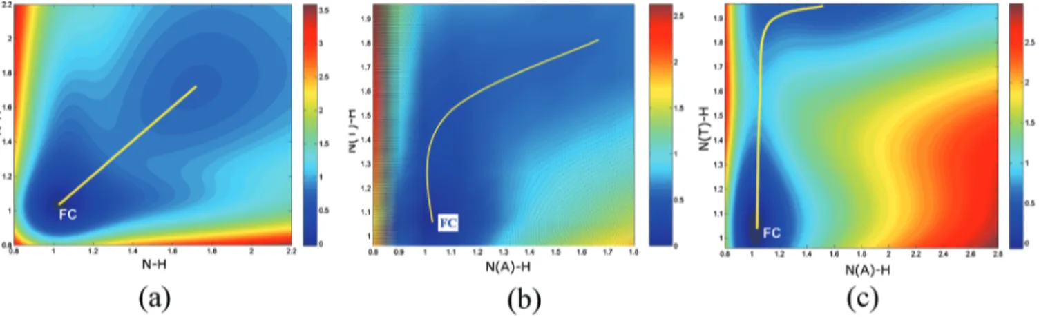
For the 2-AP dimer, based on the stationary structure in the ground state, we have optimized 225 geometries with a frozen NH distance 共rN–H兲 between two adjacent 2-aminopyridine molecules, which takes value from 0.8 to 2.2 Å with a step of 0.1 Å. In other words, there are 15ⴱ15 grid points for constructing 2D PES of the S0 state. The numerical method of 2D cubic spline interpolation has then been applied to obtain smooth potentials, see Fig.2共a兲. The two reaction coordinates are related to hydrogen positions in the two N–H hydrogen bridges of the 2-AP dimer. There are two obvious minima in the ground state PES, corresponding
to the 2-AP dimer and its tautomer. The tautomer is 17.2 kcal/mol higher in energy than the 2-AP dimer. The 2D PES of the ground state of the 2-AP dimer has a symmetrical characteristic along the diagonal line due to its symmetric geometry.
Different from the 2-AP dimer, in DNA oligomers, the base pair is bonded to the sugar-phosphate backbone via co-valent bonding with a defined intermolecular distance. Re-cently, Samoylova et al.20 reported that the electron/proton-transfer barrier is sensitive to the N¯N distance in the 2-AP dimer. Considering this external restriction, we have studied the A-T base pair in two different ways, that is, with free and with fixed intermolecular N¯N distance following the coordinate-driven minimum-energy path as mentioned above. In this case, for a given value of N共A兲–H¯O共T兲 共rN共A兲–H兲 or N共T兲–H¯N共A兲 共rN共T兲–H兲, or N¯N distance all remaining coordinates were optimized.
The calculated 2D PES for the case of free N¯N dis-tance is given in Fig.2共b兲. There are two minima with a very flat central transition region. The hydrogen-transfer path is not along the diagonal line. The corresponding N–H bonds in the hydrogen-transfer tautomer are 1.442 and 1.701 Å, re-spectively. The energy of the tautomer is found to be 13.2 kcal/mol higher than that of the initial A-T base pair. We have also found a transition state for the ground state hydrogen-transfer process. The hydrogen bonds, rN共A兲–H and rN共T兲–H, of the transition state along the N共A兲–H¯O共T兲 and N共T兲–H¯N共A兲 hydrogen bridges are 1.42 and 1.69 Å, re-spectively. The transition state in the S0state has almost the same energy as its tautomer.
Figure 2共c兲 illustrates the projected two-dimensional ground state PES of the A-T base pair with a fixed intermo-lecular distance rN共A兲. . .N共T兲共rN–N兲=3.0 Å. In this case, the PES of the ground state is much steep. It is also possible to locate the transition state between these two minima at the coordinate near rN共A兲–H= 1.1 Å, rN共T兲–H= 1.6 Å. The barrier
of this hydrogen-transfer pathway is 23.5 kcal/mol, which is almost 10 kcal/mol higher than those with a free N¯N dis-tance. One can at least conclude that for the A-T base pair, the reaction barrier in the ground state is very sensitive to the intermolecular N¯N bond distance.
For both 2-AP dimer and A-T base pair, many studies have been devoted to the hydrogen- and proton-transfer mechanisms in the ground and excited-states.15However, no consensus has been reached on whether it should involve a single or double hydrogen 共or proton兲 transfer mechanism. As can be seen in Fig. 2共a兲, the hydrogen-transfer exhibits concerted mechanism in the ground state due to the sym-metrical structure of the 2-AP dimer. Whereas from Figs. 2共b兲 and 2共c兲, we can conclude that the ground state hydrogen-transfer in the A-T base pair is a stepwise process. In this case, the hydrogen of N共T兲–H moves first with the other hydrogen bond N共A兲–H almost unchanged. Then the hydrogen of N共A兲–H moves and reaches the equilibrium po-sition of the tautomer structure. These two-step mechanism agrees with the results of Villani.21 It is thus important to note that in terms of ground state dynamics, the 2-AP dimer and the A-T base pair have very different hydrogen-transfer mechanisms. One could conclude that the 2-AP dimer is at least not a good model for studying the ground state dynam-ics of the A-T base pair.
C. Potential-energy surfaces of excited-states
Based on the optimized ground state geometries of the 2-AP dimer and the A-T base pair, by scanning the coordi-nates along the two main hydrogen bonds, we have obtained two-dimensional potential-energy surfaces of excited-states from TDDFT calculations. It is worth to note that to the best of our knowledge, these are the first complete two-dimensional PES of excited-states for these essential biologi-cal systems. Earlier studies provided mostly some particular minima and barrier points or one dimensional PES. 2D-PESs of the important singlet electronic excitations of the 2-AP dimer are shown in Fig.3共a兲. In addition to the ground state
S0, a local 1ⴱ excited-state on the monomer is marked as LE state in Fig.3共a兲. Sobolewski et al.7predicted a low-lying 1ⴱCT state, where one electron from theorbital of one molecule is promoted to theⴱorbital of the other molecule. This state is labeled as CT1 state in Fig.3共a兲. Symmetrically, the opposite electron transfer 1ⴱ CT state of the 2-AP dimer is set as CT1
⬘
. Recently, Samolylova et al.20identified the next higher1ⴱCTⴱstate which was about 1 eV higher than the low-lying CT state by CASPT2. It was found that this second CT state may also attribute to the ultrafast elec-tronic relaxation with ⱕ50 fs of the 2-AP dimer.From our TDDFT calculations, we have also reproduced this CT state 共labeled as CT2兲 which is also about 1 eV
FIG. 2. Two-dimensional potential-energy surfaces of the ground state for共a兲 2-AP dimer and for the A-T base pair 共b兲 with free and 共c兲 fixed N...N distance. The picture is obtained fromMATLAB 7.1.
FIG. 3. Potential-energy surfaces of the ground共S0兲, CT, and localized 共LE兲 states of 共a兲 the 2-AP dimer; and the A-T base pair with 共b兲 free and 共c兲 fixed
higher than the CT1 as shown in Fig.3共a兲. The good agree-ment between our CAM-B3LYP and previous CASPT2 results20highlights the usefulness of this functional for such systems. It can be seen that the energy of the 1ⴱ 共LE兲 character increases along the N–H reaction coordinate, while the CT states are essentially repulsive. There are crossing seams between the potential-energy of CT1 and CT2 states and the LE state at N–H distances longer than 1.2 and 1.5 Å, respectively.
Similarly, we have performed TDDFT calculations for potential-energy surfaces of excited-states of the A-T base pair. The results for the A-T base pair with free and frozen N¯N distance are presented in Figs.3共b兲 and3共c兲, respec-tively. It is known from previous CIS and CC2/cc-pVDZ calculations that the character of the lowest1ⴱ 共LE兲 state is of A→Aⴱtype.10However, from our TDDFT calculations with CAM-B3LYP, we have found two nearly degenerated LE states at 5.40 and 5.48 eV, of T→Tⴱ type and A→Aⴱ type, respectively. These two LE states are found to be al-ways close in energy with very similar potential shapes. To simplify the discussion, we only present the LE state of A
→Aⴱcharacter in Figs.3共b兲and3共c兲. We have also given the results of two important charge transfer states, CT1 共A→T兲 and CT2 共A→T兲, which are both 1ⴱ charge transfer from the base A to the base T, as well as the opposite charge transfer state from base T to base A, marked as CT共T→A兲. The energies of the LE and CT 共T→A兲 states increase along the N共A兲–H reaction coordinate. The strongly repulsive potentials of CT1共A→T兲 and CT2共A→T兲 states cross over with the LE state after N共A兲–H distance beyond 1.1 and 1.4 Å, respectively. Moreover, the CT共T→A兲 state crosses over the LE state when N 共T兲–H distance is longer than 1.6 Å.
The PESs of the A-T base pair with a fixed N¯N dis-tance differ from those with a free N¯N distance, as clearly demonstrated in Fig. 3共c兲. Basically their potential surfaces of the ground and excited states are steeper with higher re-action barriers. The constraint from the DNA backbone seems to have significant effects on the potential-energy sur-faces, hence the related ultrafast deactivation processes. It is noted that such a structural constrain cannot be enforced in the 2-AP dimer.
The difference in potential-energy surfaces of excited states of the 2-AP dimer and the A-T base pair can be clearly distinguished from Figs.3共b兲 and3共c兲. To highlight the dif-ference, we have listed the vertical excitation energies of
several relevant excited states in both 2-AP dimer and the A-T base pair in Table I. It can be seen that the vertical excitation energy of the LE state in the 2-AP dimer is 4.81 eV, which is about 0.6 eV lower than that in the A-T base pair. The CT1 and its symmetric charge transfer state CT1
⬘
have the same high energy共6.21 eV兲 due to the symmetry of the 2-AP dimer. The vertical energy共7.00 eV兲 of the second charge transfer state CT2 is about 0.8 eV higher in energy that CT1. In comparison with the 2-AP dimer, the CT1 共A→T兲 and CT 共T→A兲 of the A-T base pair are different in
vertical energy by as much as 1.7 eV. This energy difference can induce different reaction barriers which will be discussed later.
D. Deactivation mechanism in the 2-AP dimer and the A-T base pair
The given potential-energy surfaces for both 2-AP dimer and the A-T base pair provide a good opportunity to discuss and to compare deactivation mechanism involved in these systems. It is seen that upon the UV excitation at 274 nm, the 2-AP dimer is mainly excited to the lowest singlet local ex-cited state1ⴱ 共LE兲. Based on the PES in Fig.3共a兲, it can be anticipated that the excited electron on the LE state can overcome various barriers and reach the crossing seams be-tween the CT1 共or the CT2兲 and the LE states. It can be further relaxed to the ground state through the fast internal conversion 共IC兲 process at large N–H distance. We set this pathway as the path 1. Meanwhile, the crossing between the charge transfer state CT1
⬘
of opposite direction and the LE state can be another efficient deactivation pathway—named as the path 2. Both paths are quite competitive because of the involvement of same barriers caused by symmetric charac-teristics of the CT1 and the CT1⬘
states. With even shorter pump wavelengths, the electron can reach the surface of the CT2 that can directly relax along the N–H stretch pathway.With excitation wavelengths of 250–272 nm, the A-T base pair is excited to the 1ⴱ excited states which lies approximately 5 eV above the ground state.22After excita-tion to the LE states, there are also two possible relaxaexcita-tion paths involved. In contrast to the case of the 2-AP dimer, the notable difference in the relaxation process in the A-T base pair is that the barrier on the path 2 is much higher than that on the path 1. As concluded by Domcke et al.,10the CT state which is of the A→Tⴱtype corresponds to the formation of the A+-T− ion pair structure, which can drive the proton
CT1 6.21 0.00 CT1 5.85 0.01
CT2 7.00 0.00 CT2 7.35 0.00
transfer from the base A to the base T to compensate the charge separation. However, the ionization potential of the base T is higher than that of the base A. This makes the deactivation path 2 less efficient than the path 1. It means that the CT state of A→Tⴱtype is energetically more favor-able than the opposite T→Aⴱ type of CT state. The latter state has a higher electron/proton transfer barrier as shown in Fig.3共c兲. It is worth to notice that if the excitation energy is higher enough to reach the PES of the CT共T→A兲 state, the CT state of T→Aⴱ type can then become a possible deacti-vation channel along the N共T兲–H coordinate.
It is clear that the electron-driven proton charge transfer state always plays an important role in the relaxation pro-cesses in the singlet excited 1ⴱ 共LE兲. The height of the barrier which arises as a consequence of the LE/CT crossing along the proton-transfer coordinate is a decisive factor in this rate-determining step in the ultrafast deactivation pro-cess.
E. LE\ CT internal conversion rate
The PESs of electronic states in the 2-AP dimer and the A-T base pair are quite different in terms of energy position, energy separation, and potential shapes. It would be interest-ing to see how these differences affect the actual decay rates. For this purpose, we have carried out microcanonical Monte Carlo simulations to calculate the rate constant of the IC process via the intersection seam between the local excited 共LE兲 state and the charge transfer 共CT1兲 state. The transition from the LE to the CT1 state is obvious a nonadiabatic pro-cess in nature. The Landau–Zener nonadiabatic probability is often regarded as unit for an internal conversion process via the conical intersection.23As indicated by Scheme2, the size of the crossing region, as the ellipse zone marked in Scheme 2, will influence the number of selected configurations lo-cated in the crossing seam region. In other words, the nona-diabatic coupling matrix element V12is an important param-eter for the microcanonical Monte Carlo rate constant. Unfortunately, the calculations of the nonadiabatic coupling matrix element for 2-AP dimer and AT molecule are far too complicated and beyond the capacity of the used programs.
Although accurate coupling matrix elements are not
available, we can still discuss the effect of the coupling by a set of parameters, i.e., to highlight the relative changes. Our calculations indicate that the IC lifetime from the LE to the CT1 states is directly proportional to the nonadiabatic cou-pling between the two states. The IC lifetime of the 2-AP dimer and the A-T base pair as a function of driving energy are shown in Fig.4, when 2V12共labeled as ims in Fig.4兲 is chosen as 0.002 and 0.001 eV, respectively. The driving en-ergy is defined as the excess enen-ergy of the system after over-coming the barrier of the minimum-energy crossing point. It is noted that the present Monte Carlo simulation is based on the classical concept without considering the quantum tun-neling effect, which implies that the IC lifetime is infinity when the driving energy is less than zero. During the total 5⫻106 random moves, the minimum crossing point is lo-cated at共1.35 Å, 1.07 Å, 5.012 eV兲 for the 2-AP dimer, and 共1.14 Å, 1.00 Å, 5.588 eV兲 for the A-T molecule. From Fig. 4, we can see that the IC lifetime is very sensitive to the driving energy. For clarity, the IC lifetime is presented into two regimes, at the low driving energy 关Fig. 4共a兲兴 and the high driving energy 关Fig. 4共b兲兴. For both systems, the LE
→CT1 IC lifetime decays very rapidly with the increase of
the energy, as soon as the energy is enough to overcome the “barrier” of the crossing seam.
For the 2-AP dimer, the nonadiabatic transition from the LE to CT1 states is expected to occur in the time scale of dozens of picoseconds even when the total energy is only 0.001 eV higher than the barrier. Moreover, the IC lifetime will be shortened to the scale of dozens of femtoseconds in case the driving energy is higher than 0.01 eV. The lifetime of the excited state of the 2-AP dimer has been measured to be 65⫾10 ps by the femtosecond time-resolved mass spec-troscopy experiment with 274 nm excitation.5,20Our simula-tions might lead to two possible mechanistic interpretasimula-tions for this experimental result: 共a兲 the driving energy is suffi-ciently small in the experiment, which can result in long decay time in the scale of picoseconds; 共b兲 there might be another conical intersection due to the slow structural relax-ation. Further study is thus needed to resolve this puzzle. Obviously, the limitations of the Monte Carlo simulations used in this study could also be the possible source for the difference between the calculated and experimental lifetimes.
that the difference in the shape of the PES can also lead to different decay rates in two systems.
IV. CONCLUSION
In summary, we have provided a visual and more com-plete picture on the relevance of the 2-AP dimer as a model to mimic A-T base pair. Our calculations have shown that the 2-AP dimer model has limited value for understanding pho-tochemistry of real A-T base pair since it lacks some impor-tant structural information about the A-T base pair. For in-stance, the hydrogen-transfer mechanism in the ground state of the 2-AP dimer is completely different from the A-T base pair. Moreover, the sugar-phosphate backbone in DNA double strands forces the base pair to be at a reasonable intermolecular distance while this structural constrain cannot be enforced in the 2-AP dimer. The fixed intermolecular dis-tance can actually lower the reaction barrier and modifies the potential-energy surfaces. However, the specific difference in the PESs of these two systems seems to have very little ef-fects on the kinetics of the IC process as demonstrated by Monte Carlo kinetic studies. For this particular purpose, the 2-AP dimer could be a reasonable model for the A-T base pair.
ACKNOWLEDGMENTS
The work was supported by the Swedish National Infra-structure for Computing 共SNIC兲, the NSFC 共Grant Nos.
V. I. Danilov, O. N. Slyusarchuk, J. L. Alderfer, J. J. P. Stewart, and P. R. Callis,Photochem. Photobiol. 59, 125共1994兲.
5T. Schultz, E. Samoylova, W. Radloff, I. V. Hertel, A. L. Sobolewski, and
W. Domcke,Science 306, 1765共2004兲.
6E. Samoylova, V. R. Smith, H. H. Ritze, W. Radloff, M. Kabelac, and T.
Schultz,J. Am. Chem. Soc. 128, 15652共2006兲.
7A. L. Sobolewski and W. Domcke,Chem. Phys. 294, 73共2003兲. 8J. R. Roscioli and D. W. Pratt,Proc. Natl. Acad. Sci. U.S.A. 100, 13752
共2003兲.
9R. Wu and B. Brutschy,J. Phys. Chem. A 108, 9715共2004兲.
10S. Perun, A. L. Sobolewski, and W. Domcke,J. Phys. Chem. A 110,
9031共2006兲.
11A. L. Sobolewski, W. Domcke, and C. Hättig,Proc. Natl. Acad. Sci.
U.S.A. 102, 17903共2005兲.
12T. Yanai, D. P. Tew, and N. C. Handy,Chem. Phys. Lett. 393, 51共2004兲;
Y. Tawada, T. Tsuneda, S. Yanagisawa, T. Yanai, and K. Hirao,J. Chem. Phys. 120, 8425共2004兲.
13F. Santoro, V. Barone, and R. Improta,J. Am. Chem. Soc. 131, 15232
共2009兲.
14A. Tsolakidis and E. Kaxiras,J. Phys. Chem. A 109, 2373共2005兲. 15D. Varsano, R. Di Felice, M. A. L. Marques, and A. Rubio,J. Phys.
Chem. B 110, 7129共2006兲.
16
DALTON, release 2.0, a molecular electronic structure program 共2005兲, http://www.kjemi.uio.no/software/dalton/dalton.html.
17M. J. Frisch, G. W. Trucks, H. B. Schlegel et al.,
GAUSSIAN 03, Revision B.02, Gaussian, Inc., Pittsburgh, PA, 2003.
18A. J. Marks and D. L. Thompson,J. Chem. Phys. 96, 1911共1992兲. 19W. K. Hastings,Biometrika 57, 97共1970兲.
20E. Samoylova, W. Radloff, H. H. Ritze, and T. Schultz,J. Phys. Chem. A
113, 8195共2009兲.
21G. Villani,Chem. Phys. 316, 1共2005兲.
22M. Marazzi, U. Sancho, O. Castaño, W. Domcke, and M. Frutos,J. Phys.
Chem. Lett. 1, 425共2010兲.
23M. Olivucci, Theoretical and Computational Photochemistry, Vol. 16