Faculty of Veterinary Medicine and Animal Science Department of Animal Nutrition and Management
The effect of probiotic administration on
piglets performance and intestinal
microbiota
Alma Eriksson
Master´s thesis • 30 credits
The effect of probiotic administration on piglets perfor-
mance and intestinal microbiota
Probiotika som fodertillskott till smågrisar och dess effekt på tillväxt och tarmens bakterieflora
Alma Eriksson
Supervisor: Johan Dicksved, Swedish University of Agricultural Sciences, Department of Animal Nutrition and Management
Assistant supervisor: Lidija Arapovic, Swedish University of Agricultural Sciences, Department of Animal Nutrition and Management
Examiner: Torbjörn Lundh, Swedish University of Agricultural Sciences, Department of Animal Nutrition and Management
Credits: 30 credits
Level: Second cycle, A2E
Course title: Självständigt arbete i husdjursvetenskap
Course code: EX0872
Course coordinating department: Department of Animal Nutrition and Management
Place of publication: Uppsala
Year of publication: 2019
Cover picture: Alma Eriksson
Online publication: https://stud.epsilon.slu.se
Keywords: Lactobacillus plantarum, Lactobacillus reuteri, weaning, microflora, intestinal development
Swedish University of Agricultural Sciences
Faculty of Veterinary Medicine and Animal Science Department of Animal Nutrition and Management
Piglets are born with an undeveloped immune system and without a developed mi-crobiota in their gastro intestinal tract. Early establishment of the mimi-crobiota is im-portant for a good intestinal function but also immunologic maturation. In addition, it is of great importance with a well-developed microbiota early in life to prevent health issues and increase performance.
In this study, a feed supplement with two probiotic strains (Lactobacillus reuteri and Lactobacillus plantarum) was supplemented to piglets three times a week from the age of 3 days until weaning at 34 days. The supplement contained 8x10⁷ ± 3x10⁷ cfu L. reuteri and 2x10⁹ ± 5x10⁷ cfu L. plantarum at every occasion and was used to evaluate if it could provide any effects on performance, faecal score and intestinal microbiota. Thirty piglets from three litters were selected where five piglets received the probiotic supplement and five piglets received a placebo treatment (control group) in each litter. The piglets were held in intact litters with the mother sow until weaning. Supplementation with probiotics showed no significant effect on performance re-garding average daily weight gain or faecal score. The bacterial count for lactic acid bacteria isolated from fresh faecal samples was somewhat higher in piglets fed pro-biotics during the whole experimental period, and with significant greater counts at 42 days of age (P=0.041). Bacterial count for Enterobacteriaceae showed no signif-icant difference between treatment groups. The distribution of lactic acid bacteria in fresh faeces analysed with Maldi-Tof and the composition in collected rectal swab-samples analysed with the molecular fingerprinting method Terminal-restriction fragment length polymorphism (T-RFLP), did show relatively similar results from both treatment groups.
In conclusion, no clear effect could be seen on piglets performance or microbiota with probiotic supplement. However, the data set in this study was quite small, which can explain the difficulty to see a significant effect between treatment groups.
Keywords: Lactobacillus plantarum, Lactobacillus reuteri, weaning, microbiota,
in-testinal development, gastro inin-testinal tract
Abstract
Grisar föds utan ett färdigutvecklat immunsystem och utan en utvecklad bakterieflora i mag- och tarmkanalen. Det är därmed viktigt att tidigt i livet etablera en god bakte-rieflora, då det är avgörande för en god tarmfunktion och immunologisk mognad. Dessutom är en väl fungerande bakterieflora grundläggande för att undvika hälsopro-blem och öka tillväxten hos grisar.
I denna försöksstudie, supplementerades ett fodertillskott innehållande två probi-otiska bakteriestammar (Lactobacillus reuteri och Lactobacillus plantarum) till små-grisar tre gånger i veckan från 3 dagars ålder fram till avvänjning vid 34 dagars ålder. De supplementerades 8x10⁷ ± 3x10⁷ cfu L. reuteri och 2x10⁹ ± 5x10⁷ cfu L. planta-rum vid varje tillfälle, och syftet var att utvärdera effekten på tillväxt, avföringens
konsistens och tarmens bakterieflora. Trettio grisar från tre kullar valdes ut där fem grisar supplementerades med probiotika och fem grisar fick ett supplement med pla-cebo effekt (kontroll grupp) i varje kull. Grisarna hölls i intakta kullar tillsammans med suggan från födsel fram till avvänjning. Resultatet visade att tillskott av probio-tika inte gav signifikant ökad effekt på daglig tillväxt eller avförings konsistens. An-talet mjölksyra bakterier var högre hos grisar som supplementerades med probiotika under hela försöksperioden, och med signifikant större antal efter avvänjning vid 42 dagars ålder (P=0,041). Antalet Enterobacteriaceae bakterier visade ingen signifi-kant skillnad mellan de två behandlingsgrupperna. Fördelningen av mjölksyra bakte-rier i avföringen som analyserades med Maldi-Tof och sammansättningen från prover tagna med svabb från rektum som analyserades med T-RFLP, visade liknande resul-tat från de båda behandlingsgrupperna.
Sammanfattningsvis visade resultatet inte någon synlig effekt på tillväxt eller tar-mens bakterieflora hos de grisar som supplementerades med probiotika. Däremot var antalet individer i denna studie ganska liten, vilket kan vara en förklaring till varför det var svårt att se signifikanta skillnader mellan de två grupperna.
Nyckelord: Lactobacillus plantarum, Lactobacillus reuteri, avvänjning, bakterieflora,
tarmutveckling
List of tables 9
List of figures 10
1
Introduction 12
2
Literature review 14
2.1
Piglets first hours of life 14
2.2
Establishment of the intestinal microbiota 14
2.2.1
Gastro-intestinal tract development, structure and pH levels 15
2.2.2
Enterobacteriaceae colonisation 17
2.3
Weaning- diet change and stress factors 17
2.4
Probiotic and its use in pig production 19
2.4.1
Lactobacillus reuteri as a probiotic 19
2.4.2
Lactobacillus plantarum as a probiotic 20
3
Material and methods 22
3.1
Piglets and treatment 22
3.2
Study design 22
3.3
Probiotic supplement 23
3.4
Sample collection 23
3.5
Culture and identification of microbiota 24
3.5.1
Maldi-Tof analysis 25
3.5.2
DNA isolation and T-RFLP analysis 25
3.6
Statistical analysis 26
4
Results 27
4.1
Performance 27
4.2
Quantification of LAB and Enterobacteria in faeces 29
4.3
Quantification of LAB in colon and ileum 32
4.4
T-RFLP analysis 34
5
Discussion 36
5.1
Faeces microbiota 36
5.2
Performance 37
5.3
Inclusion level of probiotics 38
5.4
Microbiota in colon and ileum 38
5.5
Method considerations 39
5.6
Conclusion 40
References 41
9 Table 1. Description of the feed supplements. 23
Table 2. Overview of the sample collection during the experimental period for each
litter. 24
Table 3. Body weight (BW) shown in mean values with the standard deviation (SD) in the probiotic group compared with control group over the entire study
period. 27
Table 4. Growth performance for piglets receiving probiotics compared with control shown as average daily weight gain (ADG) during the study period. 28
Table 5. Effect of treatment with probiotics compared with control on faeces score*
at different ages. 28
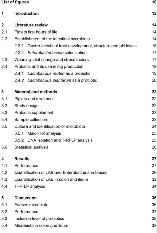
Figure 1. Structure of the small intestine. Most of the digestive tract in pigs are constructed in the same way (Sjaaastad et al., 2010). Permission given by the Scandinavian Veterinary Press. 16
Figure 2. Quantity of LAB in probiotic treated piglets and control piglets at 14, 28 and
42 days of age. Values shown are means for all tested piglets at the different ages and treatments, and the vertical bars show the positive
standard error of the mean. 29
Figure 3. Quantity of Enterobacteriaceae in probiotic treated piglets and control piglets at 14, 28 and 42 days of age. Values shown are means for all tested piglets at different ages and treatments, with the positive standard error of the mean indicated by vertical bars. 30
Figure 4. Proportion of LAB species detected with Maldi-Tof analysis, and how it
changes over time in the two treatment groups. n=number of colonies
tested. 31
Figure 5. Quantity of LAB detected in colon mucosa (CM), colon digesta (CD) and ileal mucosa (IM) of euthanized piglets. Quantity shown for probiotic and control are mean values of the piglets from the three litters (n=3). 32
Figure 6. LAB species identified by Maldi-tof analysis from three different samples in
colon and ileum from euthanized piglets at 5 weeks of age. The first row shows probiotic treated piglets (P) and the second row control piglets (C).
33
Figure 7. The LAB community profile* detected with T-RFLP analysis, shown in mean values for the probiotic (P) and control (C) group at 14, 28 and 42 days of age. *Values < 5% are not shown 34
Figure 8. The community profile of LAB* in six individual piglet (1-3=probiotic, 4-
6=control) and how it changes over the experimental period, identified with T-RFLP analysis. *Values < 1% is not shown. 35
A good animal welfare is the key to a successful animal production. Impaired wel-fare is closely related to health problems for the animals, and it can thereby affect animal performance (Jensen, 1998). Health problems around weaning is one of the most common problem in Swedish and European pig production, which can affect pigs growth and thereby have negative impact on the economy of the farm (De Angelis et al., 2007). The stressful time that occurs at weaning for the piglets, with diet change and removal of the sow, can affect the microbiota in the gut so it is more sensitive to infections. Weaning diarrhoea caused by pathogenic Escherichia coli (E. coli) are a common disease of piglets because the gastro intestinal tract is not fully developed yet, both regarding absorption of nutrients, a developed microbiota and the correct pH-level (Li et al., 2008). Around 30% of the piglet producing farms in Sweden have reported problems with weaning diarrhoea (Holmgren et al., 2005). This dilemma in the piglet production, have for a long time been solved by adding antibiotics or zinc oxide in the weaning diet (Milani et al., 2017).In Sweden 1986 and in Europe 2006, the use of preventive antibiotic in animal production was banned because of the rising bacterial resistance (Jordbruksverket, 2018a). In addi-tion, zinc oxide that has been used instead of antibiotics, have also been restricted and will be banned within a few years, since it showed to have negative impact on the environment (Buff et al., 2005; EMA, 2017).
Therefore, other ways must be tested in order to lower the incidence of diarrhoea in pig production worldwide and prevent this welfare problem. Lactic acid bacteria has been tested as an alternative, since it is a family of beneficial bacteria that al-ready is present in the piglets gastro intestinal tract, where it can compete with path-ogenic bacteria and thereby lower the incidence of infections (Liu et al., 2014).
The goal with this research is to find a supplement that can support the piglet to resist infections early in life. In this study the aim was to investigate if two Lacto-bacillus species (LactoLacto-bacillus plantarum and LactoLacto-bacillus reuteri) could work as a probiotic for piglets to prevent health problems around weaning.
13
The tested hypothesis was that piglets given probiotic feed supplement from the age of three days until weaning would have better growth, higher levels of lactoba-cilli and less enterobacteria in their intestinal microbiota and also better faeces con-sistency compared to piglets without this treatment.
2.1 Piglets first hours of life
The gestation period for a sow last for 115 days, and thereafter the piglets are born (Ewing, 2011). A piglet is born without subcutaneous fat and does not have any fur, and is therefore very sensitive to cold and great heat losses. However, piglets are born with a limited energy reserve containing glycogen stored in the liver (The Pig Site, 2011) to help keep up the body temperature and to provide enough energy for the piglet so it is able to get to a teat as soon as possible after birth (Ewing, 2011). In addition, piglets does not have a functional immune system at birth, and therefore the colostrum is of great importance so the piglet have a chance to initiate the estab-lishment of its own immune system (Ewing, 2011). Colostrum contains antibodies against different infectious agents that are present in a pigs environment, and it also contains vitamins and some extra energy. Antibodies are quite large, and the piglets intestine can only let through large molecules for a short period of time after birth, therefore it is important that the piglets receive colostrum as soon as possible for a sufficient uptake. In addition, the amount of antibodies in the colostrum decline rap-idly after farrowing, which adversely affects the last born piglet.
2.2 Establishment of the intestinal microbiota
The piglet is developed without an efficient immune system or a microbial ecosys-tem in the gastro-intestinal tract (GIT) (Ewing, 2011). As soon as the birth process starts, microbes will begin colonization of the gut and the establishment of this mi-crobial ecosystem is thought to be important for gut health. To maintain proper health and prevent infections, a well function intestinal homeostasis is essential (Zhang, 2014). An early development of the intestinal immune system combined with colonization of the GIT by microorganisms, is therefore of great significance.
15
At birth, the microbial establishment begins when the piglet is exposed to a com-plex of microorganisms mainly from the mother sow when passage through the birth canal and after that the surrounding environment (Zhang, 2014). The composition of microbiota is unstable during the first period of life, and is influenced mostly by environmental condition, combined with nutrition and exposure to stress (Schokker et al., 2014). For example, differences has been shown in piglets reared in groups compared to isolated- reared piglets in microbiota diversity, where better immune development and homeostasis were seen in piglets reared in groups (Mulder et al., 2009).
The microbiota that colonize the GIT, especially the beneficial microorganisms such as lactic acid bacteria (LAB), is crucial in order to maintain a good health (Li et al., 2008). Therefore a well function and developed microbiota early in life can benefit the piglet during critical periods later on, such as stress around weaning and change in diet which is well known to have an impact on the GIT microbiota.
2.2.1 Gastro-intestinal tract development, structure and pH levels
In piglets gut (Figure 1), the layers directly beneath the surface facing the lumen is called mucosa (Sjaastad et al., 2010). In the small intestine there are many folds (crypts) and extensions (villi), which makes the surface for absorption greater, since it is here most of nutrient absorption take place. The mucosa layer in the gut consist of three sections, epithelial layer, connective tissue and smooth muscles. One layer of epithelial cells line the gut, and it is mostly through those cells the absorption take place (Moeser et al., 2017; Sjaastad et al., 2010). These cells are connected with adjacent cells by tight junctions, so no intestinal fluid can pass in between the cells (Sjaastad et al., 2010). Therefore, this epithelial layer serves as a barrier against the exterior environment, so microorganisms that are present in the intestine does not translocate from the gut and enter the blood stream.
Figure 1. Structure of the small intestine. Most of the digestive tract in pigs are constructed in the same way (Sjaaastad et al., 2010). Permission given by the Scandinavian Veterinary Press.
Piglets GIT is matured during an extended period that starts before birth and continue until the post-weaning phase (Matton, 2018). During gestation the GIT is developed by hormonal and growth factors, that mostly changes its structure and function. Villi and crypts starts to develop in the small intestine, and thereafter the epithelial cells differentiate into enterocytes, which enables sugars, amino acids, li-pids and other molecules to be absorbed. Colon however, is mostly developed two weeks after gestation. Then, development and growth of the GIT is regulated by the intake of colostrum and after that milk by the piglet. (Matton, 2018)
For piglets, development of pH in the stomach is important for gut health (Mavromichalis, 2016). New born piglets have a rather high gastric pH (5-6) be-cause colostrum has strong buffering capacity (Manners, 1976; Mavromichalis, 2016). When the piglets starts to ingest milk, the gastric pH drops to 4 and stays at that level until weaning. However, at weaning, the pH stays relative high which makes it harder to digest plant- derived proteins, because the enzyme pepsin is most effective at pH level 2-3.5 (Mavromichalis, 2016). Also, this indigestion of protein gives rise to microorganisms that thrives on proteins and prefers a higher pH, for example E. coli, which then can result in diarrhea. It is therefore significant that weaner diets have low protein inclusion and is composed of highly digestible pro-tein, until the gut is mature and can produce enough hydrochloric acid so the pH level drops. A mature gastric pH is 2-3, and this level is reached about 4 weeks after weaning. A well establish gastric pH is essential for digestion, pathogen elimination and a good gut health in pigs.
17
2.2.2 Enterobacteriaceae colonisation
As described above, several microorganisms starts to colonise the GIT immediately after birth and among them bacteria from the Enterobacteriaceae family (Zhang, 2014). From this family, various species are present in a pigs environment, and in-cludes both pathogenic and non-pathogenic bacteria (Schierack et al., 2007). Bacte-ria from the EnterobacteBacte-riaceae family usually prefers an aerobic environments with temperature between 25-42 oC and a pH level between 5 -7.5 to grow (Tsuji et
al., 1982). In pig production, E. coli is one of the most important photogenic bacteria since it has a great economic impact on the production (Fairbrother et al., 2005). It is due to the fact that some E. coli strains can cause infections in piglets. For exam-ple, E. coli K88 is enterotoxigenic and can release enterotoxins in the piglets gut, which damage the barrier functions and can therefore induce losses of fluid. This occurs mostly after weaning when other factors effects the piglets stress levels, and can manifest as diarrhoea. This can be costly for the farmer because it often impair the piglets weight gain, and is also a common cause of death in weaned piglets. In a study by Tsuji et al. (1982), different gram- negative bacteria were compared, and it showed that E. coli was the bacteria that required the shortest time for growth to a certain population and also had the widest range of growth temperature, 18-47 oC.
Schierack et al. (2007) studied the composition of Enterobacteriaceae in the in-testine of healthy pigs, and showed that E. coli was the most dominant bacteria. Mucosa samples from jejunum and colon was cultured and analysed with Polymer-ase Chain Reaction (PCR), and the authors concluded that the intestinal population of Enterobacteriaceae is extremely individual and varied between pigs. Apart from E. coli, bacteria from the gen Klebsiella, Enterobacter and Citrobacter were present in the samples.
2.3 Weaning- diet change and stress factors
Weaning of piglets is a critical point in many pig herds because it is associated with stress for the piglets. The time of weaning is decided by the farmer and depends on the sows body condition and fitness, but regulations in Sweden states that the wean-ing age should not be earlier than 28 days after birth (Jordbruksverket, 2018b). A new regulation was introduced in Sweden on the 1st of December 2017 and state
that, if the farmer fulfil a list of demands regarding health and welfare, 10% of the piglets in a batch are allowed to be weaned at 21 days if the rest of the piglets in the batch are 26 days or older (Jordbruksverket, 2018c). However, according to EU reg-ulations, the weaning age in EU is set to 21 days at earliest.
At weaning, the sow is removed abruptly and the piglets are often moved to a different pen with a new environment (Göransson, 2009). In addition, the piglets
must survive on a diet based on less-digestible solid feed instead of highly- digesti-ble milk (Lalles et al., 2007). Most of the energy in sows milk consist of milk sugars, which is easy to digest for the piglets and favours the large amount of LAB in the gut (Göransson, 2009). LAB produces lactic acids in the gut which lowers the pH-level. When the diet is changed at weaning, the lactic acid production is reduced and the pH-levels increases. This can benefit undesirable bacteria to grow, for example bacteria from the Enterobacteriaceae family such as E. coli. These bacteria prefer an environment with higher pH but also nourish on proteins, which it is a surplus of in the intestine since piglets have a hard time digest all of the proteins in the diet at this stage (Göransson, 2009). In addition, the piglets immune system and GIT is not fully developed until five weeks post-weaning (Mavromichalis, 2016), which makes them vulnerable to invasion of pathogenic microorganisms at weaning (Hansen et al., 2012).
The change in diet and removal of the sow at weaning involve great stress for the piglets, which can have a negative impact on the immune system (Michiels et al., 2012). These changes combined, often results in lower feed intake with a tran-sient growth reduction as an outcome (Lalles et al., 2007). Also a shift in the bacte-rial balance in the intestine and in some cases even diarrhoea are consequences from a stressful period around weaning (Estrada et al., 2001; Suo et al., 2012).
In Sweden and Europe where preventive use of antibiotics is banned, the health problems around weaning is counteracted by well functioned management. This in-cludes good environmental hygiene and a well-adjusted feed that gives the best tran-sition possible from liquid to solid feed (SVA, 2017). During the last decades, sev-eral nutritional approaches has been examined to lower the incidence of health prob-lems around weaning (Lalles et al., 2007). For example, an inclusion of lactose in weaner diets have been shown effective, since it promote lactic acid production in the gut until the parietal cells can produce enough hydrochloric acid and establish the wanted pH-level (Göransson, 2009). Another management approach that is widely used in EU, is inclusion of zinc oxide in weaners diet to prevent diarrhoea (Milani et al., 2017). That is because zinc oxide has shown to prevent pathogenic bacteria in the gut (Milani et al., 2017) and also promote the immune system (Ou et al., 2007). However, due to the fact that only 20 % of zinc oxide is absorbed in the intestine for weaned piglets, the remaining 80% is excreted in the faeces (Buff et al., 2005). This has raised concerns for environmental pollution (Buff et al., 2005) but also that bacteria can become resistant against zinc oxide (Milani et al., 2017). Therefore, the European Commission banned zinc oxide for veterinary use in June 2017 (EMA, 2017). They concluded a transition time of five years, to farmers in EU could be able to phase out the product and find other alternatives.
19
2.4 Probiotic and its use in pig production
Probiotic is defined as live microorganisms which induce health benefits on the host when given in the right amount (Ou et al., 2007). Due to the fact that the problem regarding antibiotic resistance are increasing, probiotics have been considered an alternative to improve health and reduce infections.
In pigs, the GIT is already colonized by a wide range of bacteria just a few days after birth (Sears, 2005). The microorganisms used as probiotics, must therefore possess the property to colonize the GIT and compete with the unwanted microbes (Kenny et al., 2011). After ingestion of probiotic bacteria by the host, in this case the piglet, the probiotic encounters several stress factors such as low pH in the stom-ach and bile in the small intestine (Hou et al., 2015).
Probiotics used for pigs are therefore often LAB, since they can survive the gas-tric acid and are already a part of the pigs intestinal microbiota (Ericsson, 2009).It is a group of bacteria that is considered “good” for the piglet and have many bene-ficial properties. LAB is characterized as a group of bacteria that is gram-positive, non-sporulating and by fermentation of carbohydrates produce lactic acid (Meng et al., 2010). Supplement of LAB as probiotic can benefit the host through competition for binding sites on intestinal epithelium and for nutrient in the gut, which is factors that can inhibit colonization and growth of pathogenic bacteria (Malago, 2011). For example, it has been shown that supplement of LAB to neonatal piglets can benefit their health by regulating the formation of the microbiota in the gut (Siggers et al., 2008), and thereby decrease problems that can occur around weaning. However, probiotics have shown to have most effects for the host animal when the microbiota is unstable, like after birth, the time around weaning or after movement to a new environment (Jensen, 1998).
2.4.1 Lactobacillus reuteri as a probiotic
For pigs, Lactobacillus reuteri (L. reuteri) is one of the most dominant bacterial species in the GIT (Oh et al., 2010), and after several years of research there is now evidence that some strains possess probiotic characteristics (Hou et al., 2015).
In a study by Huang et al. (2004) weaned pigs was fed a basal diet with 0.1% supplement inclusion of a complex containing four lactobacilli strains (L. gasseri, L. reuteri, L. acidophilus and L. fermentum)with a content of 2.4x10⁵ colony
form-ing units (cfu)/g. The pigs were held in pens with three pigs in each pen, and the experiment lasted for 21 days from weaning. On day 18 to 21 of the experiment, faecal samples were collected from all pigs to be analysed for nutrient digestibility determination. The results showed that pigs fed a diet supplemented with probiotics had significant lower population of E. coli in the GIT than the control group, which
were treated with carbadox (an antibiotic). Additionally, the count for lactobacilli was significantly higher in pigs given the probiotic supplement and they also had a better digestibility of crude protein.
In a study by Liu et al. (2014), piglets was given an oral supplement containing probiotics from four days of age and continuously every day for 14 days. The LAB strain used as probiotic was L. reuteri I5007, initially known as L. fermentum I5007, which was fed orally once every day at the dose of 6x10⁹ cfu. Piglets fed the
probi-otic treatment had a higher average daily weight gain (ADG) the last eight days of the trial compared to the control group. The incidence for diarrhoea was also slightly lower for these piglets.
De Angelis et al. (2007) did an experiment were sows and piglets were fed a diet with LAB inclusion during 70 and 15 days respectively. The feed contained approx-imately 7.1 log cfu/g each of L. reuteri 3S7 and L. plantarum 4.1. The pigs were kept in individual pens to avoid contamination between pigs. Additionally, a control group of piglets and sows fed a basal diet was held under the same conditions as the treated animals. Results showed that LAB in piglets faeces increased during the 15 days of treatment, and was overall significant higher compared to the control group. However, after the end of treatment the bacterial count of LAB started to decrease. No difference was registered in LAB counts between treated and control sows, but the Enterobacteriaceae population in faeces were lower in treated sows compare to the control group. In treated piglets, the numbers of Enterobacteriaceae decreased linearly throughout the experimental time, but remained constant in the control group. However, six days after the treatment was finished, the number of Entero-bacteriaceae started to increase in treated pigs.
2.4.2 Lactobacillus plantarum as a probiotic
Lactobacillus plantarum (L. plantarum) is also a microorganism from the Lactoba-cillus family and is present in the GIT of pigs and humans (Suo et al., 2012). It has the ability to ferment various carbohydrates derived from plants, and can also, like most LAB, act as protection against pathogens. Nevertheless, experiments were L. plantarum is used as a probiotic in pig production is somewhat limited.
The preventive effect of L. plantarum on diarrhoea in relation to intestinal barrier function was tested by Yang et al. (2014) on piglets challenged with E. coli (ETEC) K88. Seventy-two male piglets, four days of age, were assigned to a diet with or without inclusion of L. plantarum (5x10¹⁰cfu/kg diet). On day 15, half of the piglets
on each assigned diet, were orally given one dose of ETEC K88 (1x10⁸ cfu/pig).
Piglets receiving the diet containing L. plantarum had enhanced body weight (BW), average daily gain (ADG) and average daily feed intake (ADFI) before day 15 com-pared to the piglets without supplement feeding. They also showed a trend towards
21
lower incidence of diarrhoea. Not surprisingly, greater incidence of diarrhoea could be seen in piglets after they were challenged with ETEC K88. However, piglets that were on the diet supplemented with L. plantarum before day 15 and that was chal-lenged with ETEC K88 on day 15, had greater BW, ADG and ADFI than piglets not receiving the feed supplement before challenged. When looking at intestinal segments using light microscopy, it was observed that ETEC K88 caused mucosal injuries such as lower villous height and deep crypts in duodenum and jejunum. Piglets on the diet supplemented with L. plantarum had however alleviated mucosal injuries caused by ETEC K88.
Lee et al. (2012) looked at the effect of L. plantarum CJLP243 on growth per-formance of weaned pigs that were challenged with ETEC. Piglets weaned at 20 days of age were assigned to one of six diets during a period of four weeks, with 18 pigs per treatment. The treatments were; one control diet, one control diet + ETEC challenge, one control diet with antibiotics and control diets with three different inclusion levels of L. plantarum (108, 109 and 1010 cfu/kg respectively). The pigs
were fed the assigned diet for 14 days and then all treatment groups except the pig-lets only fed the control diet were challenged with one dose of ETEC (5x10⁹ cfu).
Results showed that ADG was greater before compared to after the challenge for pigs fed the diet with antibiotic inclusion and the diet with the inclusion of 1010
cfu/kg L. plantarum. The ADFI was however similar between all treatment groups before the challenge, but was decreased in pigs fed only the control diet after the challenge. These results suggest that L. plantarum CJLP243 may, at higher inclu-sion levels (1010 cfu/kg diet), serve as an alternative to antibiotics to improve growth
3.1 Piglets and treatment
The study was carried out during the fall 2018 at the Swedish livestock research centre in Uppsala. Three litters were used in this study and included in total 30 pig-lets (3 litters x 10 pigpig-lets in each litter). The pigpig-lets used were crosses of Yorkshire sows and Hampshire boars. Three days after farrowing the piglets were enrolled to the experiment and continued until a week after weaning, which was approximately 6 weeks after farrowing. If a piglet needed to be medically treated for some reason, it was removed from the experiment. Only litters from multiparous sows with at least ten piglets, and without any medical treatment at farrowing was chosen.Five piglets in each litter received a supplement containing probiotics (treatment 1) and the other five piglets received a supplement only with placebo content (control group, treatment 2). The treatments were given orally, started 3 days after farrowing, and then continuously 3 times per week until weaning (33 ± 1 day of age).
The pigs was housed in conventional pens with partly slatted floor, enriched with straw bedding and together with the mother sow. They were held in intact litter groups, so the number of piglets in each pen differed a bit. The piglets were marked individually with ear tags, to be able to know which piglet that received which treat-ment.
From the Ethical committee of the Uppsala region, an approval was obtained for this study with reference number C54-16.
3.2 Study design
The field study was performed over a period of time that lasted for six weeks, where the piglets was given the same treatment during the whole experimental period. The
23
treatments was selected to the piglets in the litter based on gender and weight so these factors were as similar as possible between the control and probiotic piglets.
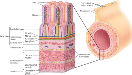
3.3 Probiotic supplement
The two treatments used was either a feed supplement containing 200 µL probiotics or a placebo containing 200 µL of a buffer solution for the control group (Table 1). Two LAB species was mixed and used as the probiotic supplement for this study, L. plantarum and L. reuteri. Each piglet in the probiotic group was orally supple-mented a dose of 8x10⁷ ± 3x10⁷ cfu L. reuteri and 2x10⁹ ± 5x10⁷ cfu L. plantarum
dissolved in sodium chloride (NaCl) and caramel colour at each supplementation day.
The supplement was prepared each supplementation day and delivered to the farm on ice. At each treatment day the piglets received a dose of 300 µL, containing the mixture seen in table 1. On the farm, the supplements was fed to the piglets by members of the staff within a few hours. Piglets received the supplement 15 times in total, between farrowing and weaning, approximately three times a week.
Table 1. Description of the feed supplements.
Probiotics (µL) Control (µL) L. reuteri (conc. 8x10⁷ ± 3x10⁷ cfu) 100 0 L. plantarum (conc. 2x10⁹ ± 5x10⁷ cfu) 100 0 NaCl 80 80 Buffer 0 200
Green caramel colour 20 20
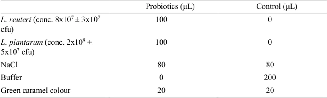
3.4 Sample collection
Fresh faecal samples was collected three times during the experimental period for each litter, at age 14, 28 and 42 days (Table 2). At the day for sampling, the faecal samples were placed in individual tubes and then held on ice until all the wanted samples had been collected, but no samples were stored on ice for more than five hours. Fresh faecal samples were not collected from all pigs of practical reasons due to differences in defecation frequencies, but equal number of samples were tried to be collected from both treatment groups. However, there were some variation in number of samples collected at the different ages of the piglets. The faecal con-sistency was visually assessed at the same time as the faecal samples were collected.
Scores were 0 = firm, normally shaped faeces; 1 = shapeless, pasty faeces; 2 = thick, liquid (soft) faeces; or 3 = thin, liquid faeces (Liu et al., 2010; Vente-Spreeuwenberg, 2003). When the faeces consistency was scored a 2 or 3, the piglets were considered to have diarrhoea. The piglets weight was registered individually at farrowing and then continually at the age of 14, 28 and 42 days.
In addition, samples were taken with a rectal swab on all animals included in the experiment at 14, 28 and 42 days of age to be used for a DNA analysis to character-ize the composition of the microbiota.
At weaning (33 ± 1 day), one pig from each treatment group and litter was eu-thanized for sampling of the gut. The abdominal cavity was opened and the gut was removed so samples from different parts of the intestine could be taken. The proxi-mal part of colon and ileum were opened lengthwise, and intestinal contents from colon was collected. A segment of colon and ileum were collected and rinsed with sterile NaCl and then mucus samples were collected by scraping the mucosa with a scalpel blade. The collected mucus samples were transferred to tubes which were immediately placed on ice. The samples were held on ice for approximately three hours before they were handled for bacterial culturing.
Table 2. Overview of the sample collection during the experimental period for each litter.
Sample 14 days 28 days 33± 1 days 42 days
Fresh faeces X X X
Rectal swab X X X
Mucosa and di-gesta sample
X
3.5 Culture and identification of microbiota
The fresh faecal samples were diluted in a 10- fold serial dilution with sodium chloride within five hours after collection, and then aliquots from the dilutions (10 -5, 10-6, 10-7 and 10-8) were cultured on both sterile de Man Rogosa Sharpe (MRS)
and Violet Red Bile Dextrose (VRBD) medium. The MRS agar was used to selec-tively culture bacteria from the Lactobacillus genus, and cfu counts were assessed after incubation in an anaerobic environment at 37 oC for 72 hours. To assess the
levels of Enterobacteriaceae in faeces, the VRBD agar plates was used for selective culture, and colonies were counted after incubation in aerobic environment at 37 oC
25
Samples collected from colon content and mucosa and ileum mucosa was also diluted and then cultured on MRS medium and kept at the same incubation condi-tions as described above. The cfu counts of LAB was then recorded for these sam-ples as well.
3.5.1 Maldi-Tof analysis
The cultured Lactobacillus from the fresh facal samples and the samples collected from colon and ileum, were analysed with matrix-assisted laser desorption ioniza-tion with time of flight (Maldi-Tof) method (Bruker, 2019) to be able to identify which species of lactobacilli that were present in the samples. Eight different colo-nies from each cultured sample was chosen based on colony morphology. These colonies needed to be fresh to get the best possible identification in the Maldi-Tof analysis, thus they were re-inoculated on new MRS medium before Maldi-Tof anal-ysis.
The isolated colonies were then added to the Maldi analysis plate, two tests per sample were analysed, to get more reliable results. Then 1 µL of 70% formic acid was added on top of each sample, to increase the permeability of the thick cell walls of gram positive bacteria. When dry, 1 µL of Bruker matrix was added to the sample. Then the Maldi-Tof analysis revealed the species identification of the tested colo-nies, which was completed within a few minutes.
3.5.2 DNA isolation and T-RFLP analysis
To analyse the community composition of lactobacilli in samples collected with rectal swabs, a molecular method called Terminal-restriction fragment length poly-morphism (T-RFLP) was used. This method provides a fingerprint of the microbial community, and can also provide a semi-quantitative measure of the microbial com-munity in the samples. This method was used in order to see if it was possible to identify differences in the Lactobacillus community composition between control and probiotic treated piglets, and also to see if the Lactobacillus community changed over time.
To be able to perform this analysis, preparations of the samples needed to be done. First, DNA was isolated from each swab sample, which was performed by using the QIAamp Fast DNA Stool Mini Kit (QIAGEN, 2018), according to the manufacturer’s instructions. 16S rRNA genes were amplified with PCR for each DNA isolate, with the use of the forward primer Bact 8F-FAM end labelled with a fluorescent dye and the reverse primer 677r for detection of lactobacilli.
The following PCR was used to amplify the 16S rRNA gene: the run started with an initial denaturing step at 94 oC for 3 minutes, and thereafter a total of 30 cycles
consisting of 40 s at 94 oC, 40 s at 55 oC, 60 s at 72 oC. It was then followed by a
final primer extension step at 72 oC for 7 minutes. The amount and size of the
PCR-amplified DNA product were confirmed by using agarose gel electrophoresis with the GeneRuler 100- bp Plus DNA Ladder as a size marker. A restriction enzyme (Bsul, Thermofisher Scentific) was used to cleave the PCR product at the wanted recognition site that was the sequence CCGG. The restriction digestion was per-formed by mixing the restriction enzyme, running buffer and water, 18 uL for each sample. Thereafter, 7uL of PCR-product sample was added to the restriction diges-tion and the PCR products were digested at 37 oC for 1 hour. When done, the
sam-ples were diluted and then submitted to the SciLifeLab in Uppsala for T-RFLP anal-ysis (Uppsala Genome Center, 2018).
3.6 Statistical analysis
To evaluate and compare the results from the collected samples between the two treatment groups, Microsoft Excel and the statistical program Past was used. For parametric testing, a Student’s t-test was performed and for nonparametric test, the Mann-Whitney U test was used.
The two treatments (probiotics and control) and age when the samples were col-lected (14, 28 and 42 days) was tested and compared regarding the parameters; av-erage daily weight gain, faeces score, quantity of Lactobacillus and Enterobacteri-aceae bacteria and proportion of different Lactobacillus in fresh faeces.
27
4.1 Performance
Piglet body weight at different ages can be seen in table 3 for the two treatment groups.
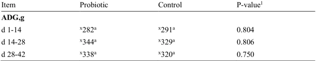
During the whole experimental period, no significant difference could be seen on average daily weight gain (ADG) between the piglets fed probiotics and the con-trol group (Table 4). Also, the ADG was similar within treatment groups during the entire study period (1-42 days).
Table 3. Body weight (BW) shown in mean values with the standard deviation (SD) in the probiotic group compared with control group over the entire study period.
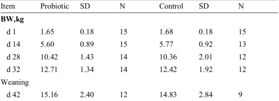
Item Probiotic SD N Control SD N
BW,kg d 1 1.65 0.18 15 1.68 0.18 15 d 14 5.60 0.89 15 5.77 0.92 13 d 28 10.42 1.43 14 10.36 2.01 12 d 32 12.71 1.34 14 12.42 1.92 12 Weaning d 42 15.16 2.40 12 14.83 2.84 9
P: Probiotic treatment with L. plantarum and L. reuteri C: Control piglets, placebo treatment
N: Number of piglets tested
4 Results
Table 4. Growth performance for piglets receiving probiotics compared with control shown as average daily weight gain (ADG) during the study period.
Item Probiotic Control P-value1
ADG,g
d 1-14 x282a x291a 0.804
d 14-28 x344a x329a 0.806
d 28-42 x338a x320a 0.750
1 P-value when comparing the treatments within different age spans.
a,b Means within a row with different superscripts are significantly different (P<0.05). x,y Means within a column with different superscripts are significantly different (P<0.05). Probiotic: Treatment with supplement containing L.plantarum and L.reuteri
Control: Placebo treatment
The faecal scores registered were very low throughout this study and no significant difference could be seen in the visual faecal scoring between the treatments groups at 14, 28 or 42 days of age (Table 5). However, within the probiotic and the control group, significantly higher faecal scores were registered at day 42 (P<0.001) com-pared to the rest of the study period.
Table 5. Effect of treatment with probiotics compared with control on faeces score* at different ages.
Item Probiotic Control P-value1
Faeces score
d 14 x0.166a x,y0.500a 0.363
d 28 x0.111a x0.125a 0.935
d 42 y1.166a y1.00a 0.363
*Faeces score= the mean value of the faeces consistency score of the piglets in each treatment.
(Score 0: firm, normally shaped faeces; 1: shapeless, pasty faeces; 2: thick, liquid faeces; or 3: thin, liquid faeces
1 P-value when comparing the treatments at different age.
a,b Means within a row with different superscripts are significantly different (P<0.05). x,y Means within a column with different superscripts are significantly different (P<0.05). Probiotic: Treatment with supplement containing L.plantarum and L.reuteri
29
4.2 Quantification of LAB and Enterobacteria in faeces
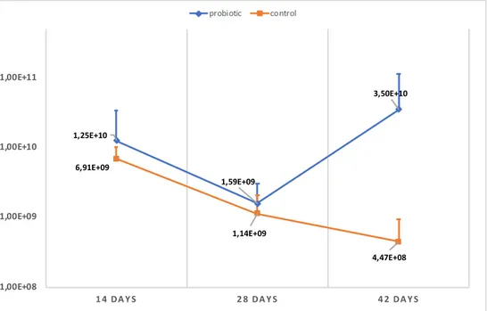
Over the whole experimental period, piglets treated with probiotic had higher quan-tity of LAB in their faeces than the control piglets (Figure 2). After weaning, at 42 days of age, a significantly higher (P=0.041) LAB count was registered in probiotic treated piglets compared to the control group. However, when comparing the bac-terial count of LAB in piglets faeces at 14 and 28 days of age, no significant differ-ence could be detected between probiotic and control treated piglets. When compar-ing the treatment groups mean values over the whole experimental period, no dif-ference could be seen in LAB count (P=0.40).
Within the group of probiotic treated piglets the bacterial count for LAB were more stable during the experimental period, and no significant differences could be seen when comparing pigs of different ages. It showed a small decrease between day 14 and 28, but thereafter an increase at day 42 which ended up with the highest detected value during the whole study period. The detected LAB count in the control group was highest at 14 days, thereafter the count decreased, to reach its lowest value at 42 days. This decrease showed to be significant (P=0.03) between day 14 and day 42.
Figure 2. Quantity of LAB in probiotic treated piglets and control piglets at 14, 28 and 42 days of age. Values shown are means for all tested piglets at the different ages and treatments, and the vertical bars show the positive standard error of the mean.
1,25E+10 1,59E+09 3,50E+10 6,91E+09 1,14E+09 4,47E+08 1,00E+08 1,00E+09 1,00E+10 1,00E+11
1 4 .DAYS 2 8 .DAYS 4 2 .DAYS
The registered count of bacteria from the Enterobacteriaceae family was similar between probiotic and control treated piglets (Figure 3). Over the whole experi-mental period, no significant difference (P=0.57) was detected between treatment groups, neither at day 14 (P=0.61), day 28 (P=0.88) or day 42 (P=0.44). The overall mean value of the bacterial count of Enterobacteriaceae were a bit lower for the probiotic group compared to the control group, with mean values at 7.29x10⁸ and
1.0x10⁹, respectively.
The bacterial count was unaffected by the age of the piglets (no significant dif-ference) within both treatment groups at 14, 28 and 42 days of age. However, both groups had the highest bacterial count at day 28.
Figure 3. Quantity of Enterobacteriaceae in probiotic treated piglets and control piglets at 14, 28 and 42 days of age. Values shown are means for all tested piglets at different ages and treatments, with the positive standard error of the mean indicated by vertical bars.
The proportion of different LAB species detected with Maldi-Tof analysis can be seen in figure 4. L. reuteri was the bacteria species that appeared at quite similar frequency for both the control and probiotic group at all ages (blue colour). L. am-ylovorus (green colour) was also a dominant species in both treatment groups, es-pecially at 28 and 42 days of age, compared to L. vaginalis (light grey colour) that was more frequent at 14 days of age. However, the distribution of L. reuteri and L. plantarum did not differ significantly between probiotic and control treated piglets during the experimental period.
3,85E+08 1,46E+09 3,40E+08 1,1E+09 1,20E+09 7,80E+08 1,00E+08 1,00E+09 1,00E+10
1 4 .DAYS 2 8 .DAYS 4 2 .DAYS
31 Figure 4. Proportion of LAB species detected with Maldi-Tof analysis, and how it changes over time in the two treatment groups. n=number of colonies tested.
4.3 Quantification of LAB in colon and ileum
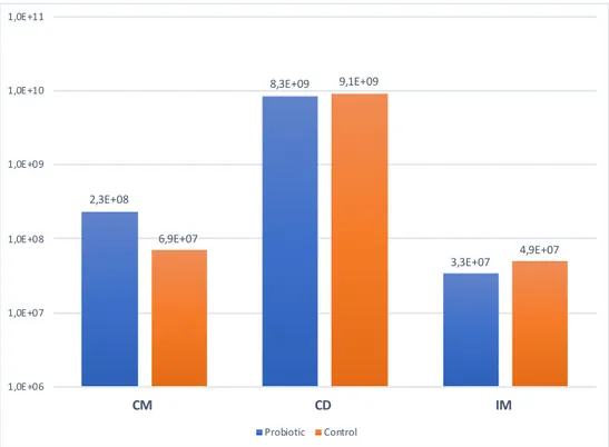
The piglets that were euthanized at weaning, showed the following quantity of LAB in the analysed samples from ileal mucosa (IM), colon mucosa (CM) and colon di-gesta (CD) when cultured (Figure 5). There were no significant difference regarding the quantity of LAB between probiotic and control fed animals in CM (P=0.20), CD (P=0.70) and IM (P=0.80).
Figure 5. Quantity of LAB detected in colon mucosa (CM), colon digesta (CD) and ileal mucosa (IM) of euthanized piglets. Quantity shown for probiotic and control are mean values of the piglets from the three litters (n=3).
The proportion of different LAB species in IM, CM and CD can be seen in figure 6. The distribution of LAB species was similar between the treatment groups, with L. reuteri and L. plantarum present in all samples. Also L. agilis (dark blue colour) appeared frequent in IM, whereas L. amylovorus (green colour) appeared most fre-quent in CM and CD for both treatment groups. The proportion of L. plantarum and L. reuteri in the different samples from the GIT, did not differ significantly in the group fed these bacteria in the probiotic supplement compared with the control group. 2,3E+08 8,3E+09 3,3E+07 6,9E+07 9,1E+09 4,9E+07 1,0E+06 1,0E+07 1,0E+08 1,0E+09 1,0E+10 1,0E+11 CM CD IM Probiotic Control
33 Figure 6. LAB species identified by Maldi-tof analysis from three different samples in colon and ileum from euthanized piglets at 5 weeks of age. The first row shows probiotic treated piglets (P) and the second row control piglets (C).
4.4 T-RFLP analysis
The T-RFLP analysis provides a molecular fingerprint of the LAB community com-position in the samples collected from the rectal swabs. The community composi-tion of LAB for all piglets fed probiotic was summarized and calculated to a mean value at day 14, 28 and 42, and the same thing was done for the control group (Fig-ure 7), to show the change over time in the LAB community profile. The T-RFLP data showed a quite similar community profile when looking at the mean values between treatment groups and age.
Figure 7. The LAB community profile* detected with T-RFLP analysis, shown in mean values for the probiotic (P) and control (C) group at 14, 28 and 42 days of age. *Values < 5% are not shown
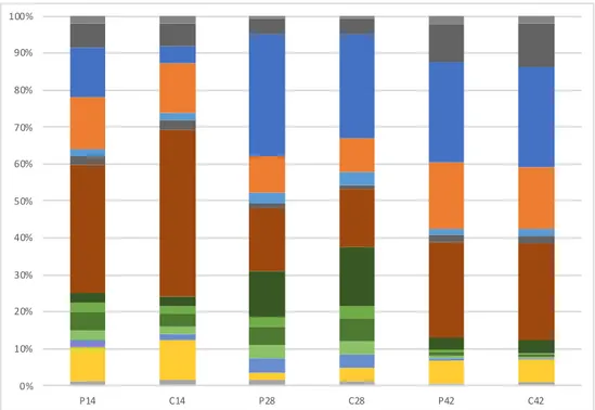
In addition, the community profile of LAB in six individual piglets were evaluated to show the change at 14, 28 and 42 days of age (Figure 8). When looking at indi-vidual animals, it shows quite a lot of difference between indiindi-vidual animals and also that the LAB community change over time. No clear differences or trends can be seen between probiotic and control piglets, but it seems like all piglets have somewhat less variety in LAB after weaning (day 42), since some segments of the bar are more prominent at that time.
0% 10% 20% 30% 40% 50% 60% 70% 80% 90% 100% P14 C14 P28 C28 P42 C42
35 Figure 8. The community profile of LAB* in six individual piglet (1-3=probiotic, 4-6=control) and how it changes over the experimental period, identified with T-RFLP analysis. *Values < 1% is not shown. 0% 10% 20% 30% 40% 50% 60% 70% 80% 90% 100% 1,P14 1,P28 1,P42 2,P14 2,P28 2,P42 3,P14 3,P28 3,P42 4,C14 4,C28 4,C42 5,C14 5,C28 5,C42 6,C14 6,C28 6,C42
5.1 Faeces microbiota
The results of this study indicated that piglets fed a supplement containing L. planta-rum and L. reuteri, had a somewhat higher bacterial count of LAB in their faeces than piglets without this treatment (Figure 2). This is consistent with previous stud-ies were piglets was fed probiotics in the form of LAB (De Angelis et al., 2007; Huang et al., 2004) and this increase in LAB count was detected in piglets both before and after weaning. In this study, the significantly higher LAB count in pro-biotic treated piglets compared to control piglets were detected after weaning (42 days of age), when the treatment with probiotics had been ended for over a week. This was somewhat unexpected, since previous studies showed that the bacterial count of LAB started to decrease when the supplement administration ended (De Angelis et al., 2007). However, it is well known that LAB compete with other bac-teria such as those from the Enterobacbac-teriaceae family in the GIT for binding sites and nutrients (Malago, 2011), and this can maybe explain the high LAB count at 42 days in probiotic treated piglets, since the Enterobacteriaceae count was at its low-est at this point (Figure 2 & 3). Another explanation could be that probiotic treated piglets may have lower pH in their gut compared to control treated piglets, and therefore the higher LAB count were detected at 42 days, since LAB thrives in lower pH. In addition, probiotics have shown to have greatest effect when piglets does not have a stable microbiota, for example directly after birth, around weaning (Jensen, 1998) or when challenged with pathogenic bacteria (Yang et al., 2014). This may also help explain the higher count for LAB in probiotic treated piglets compared to control piglets a week after weaning, and also the lower count for Enterobacteri-aceae bacteria.
When studying the results obtained from Maldi-Tof and T-RFLP analysis, it showed a quite similar distribution and community profile of LAB for probiotic and control treated animals (Figure 4 & 7). One could discuss why probiotic treated
37
piglets did not have a larger proportion of the LAB species (L. reuteri and L. planta-rum) given in the supplement compared to the control piglets, and no clear answer can be provided. None of the earlier published articles looked at how the proportion of the intestinal microbiota was effected by probiotics, so no relation can be made with previous studies. However, one idea is that the supplemented bacterial strains were outcompeted by the bacterial strains already present in the piglets GIT. In ad-dition, the values calculated and shown in the results are mean values, which means that some treatment effects can be misguided, since extreme values will affect the mean value. Therefore, when looking at individual animals (Figure 8) with the T-RFLP method, it is evident that the microbiota differs between individuals and over time, though no pattern could be seen between treatment groups. However, when looking at the proportion of LAB in individual animals (Figure 8), some pattern can be seen at different age. It seemed like there were higher diversity of LAB at 14 days compared to 42 days were some LAB species started to dominate, and thereby showed larger proportion.
5.2 Performance
It could be expected that the pigs fed the probiotic supplement should have a lower pH value in their gut compared to the control, since they had a bit higher LAB count, especially at 42 days age. This is because it is known that LAB produce lactic acid which lowers the pH in the gut (Göransson, 2009). The pH-value was not measured in this study, but would be of interest in future studies with probiotics, since it is an indicator for microbiota development and gut maturation. In addition, a low pH can suppress the growth of pathogenic bacteria which has a considerable effect to pre-vent diarrhoea. In this study, the faeces scoring that was performed showed overall low values (Table 5), with no incidence of diarrhoea in none of the treatment groups. This could probably be explained by the high level of biosecurity that is a routine at the Swedish livestock research centre in Uppsala were this study was performed, since environmental factors and overall hygiene have a large impact on diarrhoea (SVA, 2017). Higher faeces score can be expected on a commercial farm with poorer hygiene control and it is thereby also more likely to see difference between treatment groups (Liu et al., 2014); (Yang et al., 2014). However, many experi-mental studies have the piglets housed individually, which also can influence the results, because rearing environment have impact on microbiota development since bacteria is transferred between piglets (Mulder et al., 2009).
The body weight and average daily weight gain were, in this study, quite similar between treatment groups (Table 3 & 4). It could be discussed that this as well have a relation to the Swedish livestock research centre in Uppsala were the study was
conducted, because they already have quite high daily weight gain in their produc-tion stable. Previous studies have shown that LAB with probiotic properties have a positive impact on the ADG of the piglets (Lee et al., 2012; Liu et al., 2014; Yang et al., 2014). However, it is a possibility that this study would have seen an effect on the ADG if it were conducted on a commercial farm were the piglets had poorer growth in general.
5.3 Inclusion level of probiotics
The inclusion level of the LAB used as probiotic differs quite a lot between different studies, and there is no known upper or lower limits. In this study, the inclusion level per dose (each supplementation day) was 8x10⁷ ± 3x10⁷ cfu of L. reuteri and
2x10⁹ ± 5x10⁷ cfu for L. plantarum, and it was fed orally by the staff three
times/week. This was a quite high inclusion level compared to other studies that have been conducted in a similar way. However, the difference in supplementation compared to other studies were that they supplemented the pigs continuously every day for 14 days, instead of 3 times/week for five weeks as done in this study. Nev-ertheless, Lee et al. (2012) could only see an effect with the highest inclusion level of L. plantarum (1010) but not with lower inclusion levels, which indicates that some
limits exist for when the probiotic work. If LAB were to be used as an alternative to antibiotics and zinc oxide in piglet production, it is relevant to identify what the optimal inclusion level and bacterial strain is. However, it is difficult to answer this, since some studies have used only one species of LAB and some have used a com-plex of bacteria, and the comparison between inclusion level and results is therefore difficult.
5.4 Microbiota in colon and ileum
Cultured samples from the euthanized piglet at weaning did not indicated that there were any differences between probiotic and control animals regarding levels of LAB. This is in line with the other presented results of this study, with no significant difference between the two treatment groups for LAB count in fresh faeces at day 14 or 28. In addition, the distribution of LAB species detected with Maldi-Tof anal-ysis (Figure 6) in these samples were similar between probiotic and control animals at different ages. The identification of LAB species in fresh faeces was quite similar as the species identified in colon and ileum samples. L. reuteri was present in all samples and L. amylovorus was dominant in colon samples and fresh faeces. L. agilis did occur frequent in IM, but in very small proportion in CM, CD and not at all in fresh faeces.
39
The levels of LAB was a lot higher in CD (109 cfu) than in CM or IM (107 cfu).
It is, however, expected that the bacterial count is higher in digesta compared to mucosa. In addition, the large differences could also be due to difficulty when these samples were prepared for culture analysis, when samples from mucosa was very hard to dilute and get a uniform solution. This source of error should however only effect the bacterial count for these samples and not the distribution of bacteria, since it occurred similar in both treatment groups.
5.5 Method considerations
The large variation in data, can be an explanation for the high p- values calculated for bacterial count data and performance. In addition, the data set was quite small for this study, which require large differences in order to identify significant differ-ences between groups. As discussed above, many of the results had to be presented as mean values since the samples was not collected from the same piglets at each sampling, and therefore the calculated mean value can be affected by extreme values from some individuals. For this study, is was not possible to collect fresh faeces from all piglets at the different ages, since it was very time consuming combined with the time constrains put upon the sample collection for fresh faeces. That was because the samples should not be on ice for more than five hours before cultured, since it can have an effect on bacterial growth.
The Maldi-Tof analysis is a reliable method for species identification of LAB. The detected species are shown immediately together with a scale that present the certainty of the result. However, it is wise to consider in this study, that only eight colonies were tested from each cultured sample, and that these colonies were se-lected only by looking at the colony morphology (colour, shape and size). Therefore, it is a possibility that some other LAB was present in the faeces without being iden-tified using the selected approach. The distribution of LAB presented in figure 4 & 6 could therefore look somewhat different if all colonies were tested with Maldi-Tof analysis. However, this was not achievable from a work load perspective within this study period. Another consideration with this method is that some LAB species maybe tend to grow faster on the selected substrate, and will therefore suppress other more slow growing LAB.
T-RFLP analysis was used in addition to Maldi-Tof, since all animals in the three litters could be tested for their LAB- profile with this method. The samples needs to be prepared with both DNA-extraction and PCR amplification before ana-lysed with T-RFLP method. Considerations during DNA-extractions is the usage of bead beating which is applied to disrupt the cell walls of gram positive bacteria. The time for this needs to be optimized long enough so the cell walls breaks but
not too long so the DNA gets damaged. A critical step in PCR amplification, is the selection of primers. The selected primers must be specific enough to only target LAB, but at the same time not be too specific so some LAB are missed. Consider-ations with the T-RFLP analysis is also how the results are interpreted, since some species of LAB will have very similar fragment lengths, and thereby similar val-ues. The values are rounded off to the closest whole number, and it is therefore a risk that two different bacteria species will appear as the same species. In addition, something to keep in mind when using a DNA- method is that it can detect DNA from both living and dead bacteria.
5.6 Conclusion
The results from this experiment did not prove that supplement of L. reuteri and L. plantarum improved piglets performance or faecal consistency at the time before and after weaning. Neither did it affect the Enterobacteriaceae bacterial count in fresh faeces. However, significantly higher Lactobacillus counts were detected after weaning in piglets receiving this supplement, which is positive from a pathogen elimination perspective. From these results, it is not safe to state that probiotics can replace other commonly used supplements in piglet production to prevent problems that can occur around weaning. There is a lot more to explore in this field, to further investigate how lactic acid bacteria work as a feed supplement and what the optimal inclusion level should be to see an effect on piglets health and intestinal microbiota.
41 Buff, C.E., Bollinger, D.W., Ellersieck, M.R., Brommelsiek, W.A., Veum, T.L., 2005. Comparison
of growth performance and zinc absorption, retention, and excretion in weanling pigs fed diets supplemented with zinc-polysaccharide or zinc oxide. J. Anim. Sci. 83, 2380–2386.
https://doi.org/10.2527/2005.83102380x
De Angelis, M., Siragusa, S., Caputo, L., Ragni, A., Burzigotti, R., Gobbetti, M., 2007. Survival and persistence of Lactobacillus plantarum 4.1 and Lactobacillus reuteri 3S7 in the gastrointestinal tract of pigs. Vet. Microbiol. 123, 133–144. https://doi.org/10.1016/j.vetmic.2007.02.022 EMA, 2017. Zinc oxide | European Medicines Agency [WWW Document]. URL
https://www.ema.europa.eu/medicines/veterinary/referrals/zinc-oxide#all-documents-section (ac-cessed 11.9.18).
Ericsson, A., 2009. Effekten av ett probiotikum i en besättning med hög incidens spädgrisdiarré. Sve-riges lantbruksuniversitet. Veterinärprogrammet. (Examensarbete 2009: 49)
Estrada, A., Drew, M.D., Van Kessel, A., 2001. Effect of the dietary supplementation of fructooligo-saccharides and Bifidobacterium longum to early-weaned pigs on performance and fecal bacte-rial populations. Can. J. Anim. Sci. 81, 141–148. https://doi.org/10.4141/A00-037
Ewing, Kerstin, 2011. Grisar. Natur & Kultur.
Fairbrother, J.M., Nadeau, E., Gyles, C.L., 2005. Escherichia coli in postweaning diarrhea in pigs: an update on bacterial types, pathogenesis, and prevention strategies. Anim. Health Res. Rev. 6, 17– 39.
Genome Center, n.d. Deliver samples for CE - Uppsala Genome Center - Uppsala University, Swe-den [WWW Document]. URL http://ugc.igp.uu.se/sample-delivery/sample-delivery-ce/ (ac-cessed 12.11.18).
Göransson, L., 2009. Utfodring av smågrisar upp till 30 kg. Gård och Djurhälsan, pp. 2-4. Hansen, C.H.F., Nielsen, D.S., Kverka, M., Zakostelska, Z., Klimesova, K., Hudcovic, T.,
Tlaska-lova-Hogenova, H., Hansen, A.K., 2012. Patterns of Early Gut Colonization Shape Future Im-mune Responses of the Host. PLoS ONE 7. https://doi.org/10.1371/journal.pone.0034043 Holmgren, N., Mattson, B., 2005. Tvättning, desinfektion och tomtid i tillväxtstallar 4.
Hou, C., Zeng, X., Yang, F., Liu, H., Qiao, S., 2015. Study and use of the probiotic Lactobacillus reuteri in pigs: a review. J. Anim. Sci. Biotechnol. 6. https://doi.org/10.1186/s40104-015-0014-3 Huang, C., Qiao, S., Li, D., Piao, X., Ren, J., 2004. Effects of Lactobacilli on the Performance,
Diar-rhea Incidence, VFA Concentration and Gastrointestinal Microbial Flora of Weaning Pigs. Asian-Australas. J. Anim. Sci. 17, 401–409. https://doi.org/10.5713/ajas.2004.401
Jensen, B., 1998. The impact of feed additives on the microbial ecology of the gut in young pigs. J. Anim. Feed Sci. 7, 45–64. https://doi.org/10.22358/jafs/69955/1998