ACTA UNIVERSITATIS
UPSALIENSIS
Digital Comprehensive Summaries of Uppsala Dissertations from the Faculty of Medicine 1598
CT Guided Ablation of T1 Renal
Tumors
VANESSA ACOSTA RUIZ
Dissertation presented at Uppsala University to be publicly examined in Grönwallsalen, Akademiska sjukhuset, ingång 70, bv, Uppsala, Friday, 1 November 2019 at 09:15 for the degree of Doctor of Philosophy (Faculty of Medicine). The examination will be conducted in Swedish. Faculty examiner: Senior Professor Mikael Hellström (University of Gothenburg).
Abstract
Acosta Ruiz, V. 2019. CT Guided Ablation of T1 Renal Tumors. Digital Comprehensive
Summaries of Uppsala Dissertations from the Faculty of Medicine 1598. 71 pp. Uppsala: Acta
Universitatis Upsaliensis. ISBN 978-91-513-0750-3.
The widespread use of medical imaging contributes to the increased detection of incidentally detected small renal tumors, a majority which are often indolent masses found in elderly patients with preexisting chronic kidney disease. In Sweden, partial nephrectomy with minimal invasive surgical approach is the current standard for removing these tumors, although another option is percutaneous image-guided tumor ablation that allows treatment of elderly patients with comorbidities for who surgery is a risk. Due to the lack of long-term follow-up studies and prospective randomized trials, ablation is still considered an alternative option to surgery in Sweden. The aim of this thesis was to evaluate treatment of T1 renal tumors with CT guided radiofrequency (RFA) and microwave ablation (MWA).
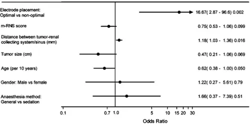
Factors affecting the efficacy rate of complete tumor ablation with RFA after a single session were evaluated (Paper I). Optimal electrode placement and a long tumor distance to the collecting system were associated with an increased primary efficacy. Renal tumor RFA was compared with laparoscopic partial nephrectomy (LPN: Papers II-III): both methods had comparable secondary efficacy rates, but RFA involved several treatment sessions. Total session times and hospitalization times were shorter and complications less frequent for RFA than for LPN (Paper II). After treatment, renal function impact was assessed by evaluation of both renal function quantity and quality through determination of the split renal function (SRF: Paper III). Standard renal function measurements were assessed and both RFA and LPN were nephron sparing when treating small renal tumors and did not affect creatinine or GFR. However, LPN involved greater SRF reduction in the affected kidney than RFA. Initial experience with microwave ablation was evaluated and this new ablation technique demonstrated high efficacy rates with fewer complications, and was comparable with the mid-term results of now established ablation techniques (Paper IV).
In conclusion, CT guided RFA and MWA are safe and effective treatments for the removal of T1 renal tumors. This thesis provides further insights into the field of thermal ablation of small renal masses, which can aid future treatment selection and patient management.
Keywords: Renal tumor, Ablation, Radiofrequency ablation, Microwave ablation Vanessa Acosta Ruiz, Department of Surgical Sciences, Radiology, Akademiska sjukhuset, Uppsala University, SE-75185 Uppsala, Sweden.
© Vanessa Acosta Ruiz 2019 ISSN 1651-6206
ISBN 978-91-513-0750-3
List of Papers
This thesis is based on the following papers, which are referred to in the text by their Roman numerals.
I. Acosta Ruiz, V., Lönnemark, M., Brekkan, E., Dahlman, P., Wernroth, L., Magnusson, A. (2016)
Predictive factors for complete renal tumor ablation using RFA.
Acta Radiologica, 57(7):886-93
II. Acosta Ruiz, V., Ladjevardi, S., Brekkan, E., Dahlman, P., Hägg-man, M., Lönnemark, M. Wernroth, L., Magnusson, A. (2018) Periprocedural outcome after laparoscopic partial nephrectomy ver-sus radiofrequency ablation for T1 renal tumors: a modified
R.E.N.A.L nephrometry score adjusted comparison.
Acta Radiologica, 60(2):260-8
III. Acosta Ruiz, V., Båtelsson, S., Onkamo, E., Wernroth, L., Nilsson, T., Lönnemark, M., Dahlman, P., Magnusson, A.
Split renal function after treatment of small renal masses: compari-son between Radiofrequency ablation and Laparoscopic partial ne-phrectomy.
In manuscript
IV. Acosta Ruiz, V., Dahlman, P., Brekkan, E., Lönnemark, M., Mag-nusson, A.
Percutaneous CT guided microwave ablation of 105 T1a-T1b renal tumors: technique efficacy with a mean 2-year follow-up.
In manuscript
Contents
1. Introduction ... 11
1.1 Renal Cell Carcinoma ... 11
1.1.1 Epidemiology and Etiology ... 11
1.1.2 RCC Subtypes and Benign Tumors ... 11
1.1.3 RCC Diagnostic Assessment ... 13
1.2 Small Renal Mass Management ... 13
1.2.1 The Clinical Dilemma of Small Renal Mass Management ... 13
1.2.2 Imaging Modalities for Small Renal Mass Evaluation ... 14
1.2.2.1 Computed Tomography (CT) ... 14
1.2.2.2 Magnetic Resonance Imaging (MRI) ... 15
1.2.2.3 Other Imaging Modalities ... 15
1.2.3 Renal Mass Biopsy ... 15
1.2.4 Anatomic Scoring Systems ... 16
1.2.4.1 Modified R.E.N.A.L Nephrometry Score (m-RNS) ... 16
1.3 Treatment Options for T1a Renal Tumors ... 19
1.3.1 Surgical Approaches ... 19
1.3.2 Thermal Ablation (TA) ... 20
1.3.3 Radiofrequency Ablation (RFA) ... 21
1.3.4 Microwave Ablation (MWA) ... 22
1.3.5 Cryoablation ... 23
1.3.6 Planning a Thermal Ablation Treatment ... 23
1.3.7 Active Surveillance ... 25
1.4 Follow-up after T1a Tumor Management ... 26
1.4.1 Follow-up with Imaging ... 26
1.4.2 Renal Function Assessment during T1a Renal Tumor Treatment ... 26
1.4.2.1 Evaluation of GFR, Creatinine and Cystatin C ... 27
1.4.2.2 Measuring Renal Function with Imaging ... 27
1.4.3 Evaluation of Post-treatment Renal Function ... 28
1.4.3.1 Renal Function after Partial Nephrectomy (PN): ... 28
1.4.3.2 Renal Function after Thermal Ablation (TA): ... 29
1.5 Guidelines on T1 Renal Tumor Management over Time ... 29
2. Aim of the Thesis ... 33
2.1 General Aim ... 33
2.2 Specific Aims ... 33
3. Materials and Methods ... 34
3.1 Patients ... 34
4. Methods ... 36
4.1 Patient Recruitment ... 36
4.2 Patient and Tumor Analysis ... 36
4.3 CT Image Analysis ... 37 4.4 Pre-procedural Analysis ... 37 4.5 Anesthesia ... 37 4.6 Pre-ablation Biopsy ... 38 4.7 RFA Technique ... 38 4.8 LPN Technique ... 39 4.9 MWA Technique ... 40 4.10 Follow-up Routine ... 40
4.11 Treatment and Image Analysis ... 41
4.12 Statistical Methods ... 43 5. Results ... 45 5.1 Paper I ... 45 5.2 Paper II ... 46 5.3 Paper III ... 48 5.4 Paper IV... 50 6. Discussion ... 51 7. Conclusions ... 54 7.1 General ... 54 7.2 Specific ... 54 8. Future Perspectives ... 55
9. Svensk Sammanfattning (Summary in Swedish) ... 57
9.1 Delarbete I ... 58 9.2 Delarbete II ... 58 9.3 Delarbete III ... 59 9.4 Delarbete IV ... 59 9.5 Konklusion ... 59 10. Acknowledgements ... 61 11. References ... 63
Abbreviations
AML Angiomyolipoma
ASCO American Society of Clinical Oncology
AUA American Urological Association
CKD Chronic Kidney Disease
CT Computed Tomography
EAU European Association of Urology
(e)GFR (estimated) Glomerular Filtration Rate
GEE Generalized Estimating Equations
HU Hounsfield Units
ICC Intra Class Coefficient
LPN Laparoscopic Partial Nephrectomy
m-RNS Modified R.E.N.A.L Nephrometry Score
MRI Magnetic Resonance Imaging
MWA Microwave Ablation
NSS Nephron Sparing Surgery
PN Partial Nephrectomy
RALPN Robot Assisted Partial Nephrectomy
RCC Renal Cell Carcinoma
RFA Radiofrequency Ablation
RMB Renal Mass Biopsy
ROI Region of Interest
RRF Relative Renal Function
SRF Split Renal Function
SRM(s) Small Renal Mass(es)
TA Thermal Ablation
1. Introduction
Kidney cancer is the fourteenth most common type of cancer worldwide and in adults it includes malignant tumors developing from the renal parenchyma or renal pelvis (1). Renal cell carcinoma (RCC) (i.e. adenocarcinoma arising from the renal parenchyma) accounts for 90% of kidney cancers. Cancer in the renal pelvis arising from transitional cells represent 10% of kidney cancers (2). The latter malignancy is not a focus in this thesis.
1.1 Renal Cell Carcinoma
1.1.1 Epidemiology and Etiology
The highest incidence of RCC occurs in Europe and North America, repre-senting 2-3% of all cancers (2, 3). In Sweden, RCC is the tenth most common cancer diagnosis affecting approximately 1000 people per year (4). The ma-jority of patients diagnosed with RCC are between 60-80 years (25% are <60 years), with a male predominance (incidence: 61% men, 39% women) (4). Established risk factors for RCC are obesity, hypertension and cigarette smok-ing. Familial or inherited predisposition to renal neoplasms accounts for <4% of renal tumors (5). Hereditary forms of RCC are caused by Von Hippel Lindau syndrome, hereditary papillary renal cell carcinoma, hereditary leio-myomatosis and RCC, and Birt-Hogg-Dubé syndromes (3) and tend to be bi-lateral and multiple, and occur at an earlier age than sporadic renal tumors (5).
1.1.2 RCC Subtypes and Benign Tumors
Renal tumors are subdivided according to their cell of origin and morphologic appearance (6). The World Health Organization’s (WHO) classification of adult renal epithelial neoplasms differentiates RCC types based on pathology, cytogenetic and genetic analyses (5). The differentiation of RCC from benign tumors makes it possible to avoid overtreatment of benign tumors. Therapeu-tic strategies and prognosis differ between RCC subtypes, as different sub-types have their specific pathological and imaging features.
The most common malignant subtype is clear cell carcinoma, which repre-sented 77% of RCC diagnosed in Sweden during 2010-2014 (4). On CT
im-ages, it appears as a highly heterogeneously hypervascular tumor in the corti-comedullary phase and presents some washout of contrast in the nephro-graphic and excretory phases (7). Five-year survival of between 43-89% is reported (7).
The second most common malignant subtype is papillary RCC, and repre-sents 13% of reported RCC in Sweden (4, 7). On CT, papillary RCC is more homogenously low-enhancing than the adjacent cortex and demonstrates a ho-mogeneous gradual enhancement when assessed in the corticomedullary- and nephrographic phase. The low attenuation and homogeneous enhancement patterns make papillary RCC distinguishable from clear cell RCC. Five-year survival is reported between 57-85% (7). There are two types of papillary RCC (8): Type 1 (low-grade neoplasm with better prognosis) and Type 2 (high-grade neoplasm with worse prognosis).
Chromophobe RCC represents approximately 6% of RCC. The degree of enhancement on CT imaging is intermediate between clear cell RCC and is hypovascular compared to the renal cortex (7). Five-year survival ranges be-tween 76-100% (9). Other unusual malignancies include carcinoma of the col-lecting ducts of Bellini (<1% of RCC) (9, 10).
Benign renal tumors account for 7-33% of all kidney masses (3, 11) and can appear similar to RCC on imaging and are thus challenging to differentiate from malignant tumors under pathological assessment (11-13).
Oncocytoma, a benign tumor, accounts for 8% of all renal cell neoplasms (14). On CT images, oncocytoma present similar enhancement to clear cell RCC and chromophobe RCC; therefore, CT cannot be solely used for diagno-sis. Histopathological assessment is used for diagnosing oncocytoma, but not all oncocytomas can be differentiated from malignant tumors with this method (7, 11, 15).
Angiomyolipoma (AML) represent 3% of all renal tumors (14). The ma-jority of angiomyolipoma are benign, except those with epithelioid features, which can have malignant potential. Large size AML (>4 cm) may cause hem-orrhage and local invasion. The presence of macroscopic fat (<10 HU) is suf-ficient for diagnosing AML on imaging. However, 10% of AML are fat-poor, containing <25% adipose tissue, making AML difficult to differentiate from RCC on CT images (7, 9). Normally, the presence of macroscopic fat in a renal mass is characteristic of an AML, but some rare cases of fat-containing RCC may be secondary to cholesterol necrosis or engulfment of adjacent fat (16). However, the presence of calcifications can increase the suspicion of RCC as calcifications are not present in AML (17). Other renal masses that can be differentiated are renal adenoma, urothelial carcinoma, metastatic tu-mor, infarct, abscess, pseudotumor or vascular malformation.
1.1.3 RCC Diagnostic Assessment
According to the 2019 European Association of Urology (EAU) guidelines (18), the work-up for RCC assessment includes the following recommenda-tions:
• Contrast-enhanced multi-phasic abdominal CT or MRI should be used for renal tumor characterization
• For determining staging, lungs and mediastinum should be evaluated with a chest CT.
• When in need to avoid ionizing modalities, contrast enhanced ultra-sound can be used for further characterization of small renal masses, evaluation of tumor thrombus or unclear renal masses
• Renal tumor biopsy with a coaxial technique is suggested before ab-lative treatment, systemic therapy and for selected patients considered for active surveillance.
• Percutaneous renal tumor biopsy is preferred over fine needle aspira-tion for characterizing of solid renal tumors.
The TNM classification system (19) is used to classify and stage RCC (3, 6). Tumor size, venous invasion, renal capsular invasion, adrenal or lymph node involvement and the presence of distant metastasis are evaluated for TNM classification. Survival rates are strongly correlated to TNM-stages; the 5-year cancer specific survival rates in patients with T1 tumors is 91% for grade 1, 83% for grade 2, 60% for grade 3, and 0% for grade 4 (3, 20). In this thesis, only T1N0M0 (≤7 cm tumors limited to the kidney) tumors will be discussed. T1a includes tumors 4 cm or less in the greatest dimension, whereas, T1b in-clude tumors more than 4 cm but not more than 7 cm in the greatest dimension.
1.2 Small Renal Mass Management
1.2.1 The Clinical Dilemma of Small Renal Mass Management
Although there are varying size limits exists in the literature for a small renal mass (SRM), in this thesis the SRM is defined as ‘an incidentally detected ≤4 cm in diameter contrast-enhancing renal tumor’, as used in the American So-ciety of Clinical Oncology (ASCO) guidelines (21).
Tumor characteristics of patients diagnosed with RCC have changed over time. Between 1986 and 2010, a Swedish study (22) reported a decrease in median tumor size from 70 mm to 50 mm. This is partially explained by the widespread use of medical imaging, which has contributed to the increased the detection of asymptomatic incidentally found renal masses (23). In Swe-den, the proportion of incidentally found renal cancer has increased from 43% in 2005 to 60% in 2014 (4). The incidence of T1a renal tumors has also in-creased, now representing up to 66% of all diagnosed renal tumors (24). Even
though kidney cancer is treated aggressively, mortality from RCC has not de-clined. Possible explanations are that a large proportion of small renal masses (SRMs) may be clinically benign or indolent and that their removal represents over diagnosis and overtreatment, suggesting that the current practice is insuf-ficient (11, 25).
SRMs are often found in elderly patients with preexisting chronic kidney disease. However, in this patient group, the high incidence of co-morbidity means patients are more likely to die with their renal tumor rather than because of it (23, 26).
The likelihood of malignancy in a solid renal lesion increases with size each 1 cm increase in tumor size is associated with a 17% increase in the odds of malignancy (27). Approximately 80% of SRMs are malignant, with 20% be-ing benign (27), and an average growth rate of <3 mm per year for SRMs is reported (28). As the growth rate of SRMs is slow (29, 30), it raises the ques-tions of whether treatment can be delayed or if older patients need to be treated at all. Nevertheless, prediction of growth rate is not possible and there is no established growth trajectory for intervention.
Despite a small size, 20-30% of renal masses <4 cm are reported as aggres-sive with high metastatic potential (31). Simultaneously, the amount of accu-mulating evidence supporting the indolent nature of SRMs has questioned whether these lesions should be treated at all (23). This raises the question as to whether the exceptionally good 95% five-year survival (11) of surgically resected SRMs is explained by a high presence of benign and indolent tumors rather than treatment effect. Not all small renal masses can be fully character-ized with a method. Imaging with CT has limitations and renal mass biopsies of these small lesions can lead to non-conclusive results, further questioning the type of disease is being handled.
Given this setting, less aggressive treatment alternatives have been devel-oped. Lesions, traditionally managed with an aggressive surgical approach, are now managed with nephron sparing surgical procedures, percutaneous ab-lative treatments or by active surveillance. The continuous development of treatment alternatives raises several clinical dilemmas, such as which SRM should be treated, how it should be treated, and when it should be treated.
1.2.2 Imaging Modalities for Small Renal Mass Evaluation
Several imaging modalities are used to assess SRMs, but there is no absolute reference method. CT is the main imaging modality able to detect SRMs. MRI is sensitive, but rarely used as a first step imaging alternative, except for high-risk patients (32).
1.2.2.1 Computed Tomography (CT)
CT is used for characterization, aiding diagnosis and staging of renal cancer and aids pre-operative planning. CT examination both prior and after contrast
medium injection should be included, as long as renal function allows contrast administration. Image findings help to divide renal masses into solid or cystic lesions by evaluating lesions based on, for example, density measurements, demonstration of fat or calcifications, and enhancement patterns (6, 9). The majority of solid malignant lesions have characteristic contrast enhancement, with a change of more than 15 HU between images before and after contrast administration (33). Four-phase CT-scans (unenhanced, corticomedullary-, nephrographic- and excretory phase) maximize the potential for differential diagnosis. CT has a median sensitivity of 88% and specificity of 75% in diag-nosing adult RCC (34), and is more easily available and quicker than MRI. Disadvantages include radiation and risk of contrast-induced nephropathy (3, 9).
The characterization of SRMs can be challenging. In very small tumors (i.e. ≤1.5 cm), the presence of enhancement can be difficult to determine due to volume-averaging (16). Renal masses ≤5 mm are too small to be characterized on CT reconstructions with 3 mm slices and thinner slices entail image-noise inhibiting analysis. As these “too-small to characterize” masses are likely to be benign, additional imaging is usually not recommended (23).
A minority of small RCC, especially papillary RCC, have a low level of enhancement, and near to significant enhancement, and hence can be mistaken as hyperdense cysts (16). Furthermore, CT and MRI cannot reliably differen-tiate oncocytoma and fat-poor AML from malignant neoplasms (18). Approx-imately 20% of small, solid renal masses suspected as RCC (enhancing on CT images) have been found to be benign oncocytoma or fat-poor AML after sur-gical resection (6).
1.2.2.2 Magnetic Resonance Imaging (MRI)
MRI is preferentially used for patients who are pregnant, have an allergy to intravenous contrast, possible venous involvement or locally advanced malig-nancy (6). Patients with hereditary forms of renal maligmalig-nancy can use MRI as an option to minimize radiation exposure from CT scans.
1.2.2.3 Other Imaging Modalities
Ultrasound may be sufficient for distinguishing a simple cyst that requires no follow-up. However, the use of ultrasound for characterizing SRM (beyond simple cysts) has yet to be evaluated (23). Other modalities e.g. Positron emis-sion tomography and angiography are also reported as an aid to RCC charac-terization (35, 36).
1.2.3 Renal Mass Biopsy
Renal mass biopsy (RMB) aims to obtain material for renal tumor histopatho-logical diagnosis (18). The four-tiered WHO/ISUP (World Health Organiza-tion/International Society of Urological Pathology) grading system based on
nucleolar features is used for histological assessment as the grading system correlates to tumor aggressiveness and prognosis (18).
Although RMB has an high sensitivity (97.5%) and specificity (96.2%) and positive predictive value (99.8%), it has a non-diagnostic rate of 14% (12). The accuracy of tumor grade (Fuhrman grading) diagnosis with RMB ranges between 52-76%, entailing a variability that potentially affects patient man-agement and prognosis (12). Histologic heterogenic tumors present a chal-lenge (i.e. hybrid oncocytic tumors with chromophobe RCC) as RMB may not include all cell types (12). Differentiation between oncocytoma and chromo-phobe RCC may also present a challenge and a non-malignant biopsy result does not imply the presence of a benign lesion. Among patients undergoing extirpation despite a negative biopsy, 37% had malignant disease on final sur-gery pathology (13).
The roll of RMB in characterizing SRMs has been discussed and interna-tional guidelines differ in their recommendations. Small tumor size is predic-tive for biopsy failure, however, repeat biopsy has a high (80%) diagnostic success rate (37). The EAU 2019 guidelines recommend RMB before ablative treatment, and active surveillance for selected patients, in order to avoid un-necessary surgery in the event of a benign tumor (18). Conversely, the AUA does not require RMB for older patients who will be managed independently of RMB findings, but do recommend RMB prior to ablation (12). Considering the indolent nature of SRMs and the competing risks of mortality from other diseases in patients presenting with incidental SRMs, the ASCO guidelines recommend RMB when results may alter management (21).
1.2.4 Anatomic Scoring Systems
Several anatomical classification systems have been proposed that objectively describe renal tumor complexity in a quantitative manner (38). Initially devel-oped to aid choice of surgical approach for treatment, these classification sys-tems serve to predict treatment outcome, risk of complications and renal tumor malignancy grade (39-44). The standardized reporting of tumor characteristics facilitates comparison between tumor populations and reduces selection bias.
In this thesis, the modified R.E.N.A.L nephrometry scoring system (m-RNS) was used, as it is one of the most reported systems and it has a greater predictive performance of ablation success with radiofrequency than the orig-inal scoring system (45).
1.2.4.1 Modified R.E.N.A.L Nephrometry Score (m-RNS)
Gahan et al. (45) proposed a modified scoring system adapted to the smaller renal masses treated with ablation. Four of the five included parameters are given 1-3 points depending on different criteria (Table 1). A suffix is assigned to the total sum (ranging between 4-12 points) to describe the tumors’ anterior (“a”), posterior (“p”), neither a/p (“x”) and/or hilar (“h”) location (Figure 1).
A limitation of the scoring system is that several parameters influence each other. Growth in tumor size increases the tumor’s possibility of expanding into the renal pelvis, acquiring a central location. Similarly, exophytic tumors will often be further away from the collecting system. In addition, the m-RNS does not consider other factors that may affect the ablation approach, e.g. the tu-mor’s relationship to bowel and ureter affect the ablation approach, e.g. the tumor’s relationship to bowel and ureter (46, 47).
Table 1. Points given according to criteria for each parameter of the modified R.E.N.A.L nephrometry score (45).
1 point 2 points 3 points Radius (maximal diameter
in cm) <3 3-4 >4
Exophytic/endophytic
prop-erties ≥50% <50% Entirely endophytic Nearness of the tumor to the
collecting system or sinus (mm)
≥7 >4 but <7 ≤4 Location relative to the
po-lar lines Entirely above the upper or be-low the be-lower polar line
Lesion crosses polar line
>50% of mass is across polar line; or mass crosses the axial renal midline; or mass is entirely between the polar lines
Figure 1: Example of a SRM categorized with the modified R.E.N.A.L nephrometry score. Evaluation of parameters Radius (a), Exophytic/endophytic properties (b), Nearness of the tumor to the collecting system or sinus (c), Anterior or Posterior lo-cation (suffix) (d) and Lolo-cation relative to polar lines (tumor within the dashed cir-cle) (e). Total RNS score: 8a.
1.3 Treatment Options for T1a Renal Tumors
Several aspects need to be considered when choosing treatment alternatives. Patient factors (age, co-morbidities, life expectancy, renal function, personal preference), tumor characteristics (size, location) and technical considerations for each treatment method must be assessed.
Since the 1990s, treatment of localized RCC has evolved. There are three main management alternatives for patients with T1 renal tumors: surgery, ab-lation or active surveillance. Depending on the individual setting, these alter-natives can aim to cure the patient from disease, minimize the tumor burden, or actively follow the patient to discuss possible future intervention, if neces-sary.
1.3.1 Surgical Approaches
Nephron-sparing surgery (NSS) implies the enucleation of the tumor (without concomitant adrenalectomy) by partial nephrectomy (PN). In the 1970’s, PN was introduced for high-risk patients where the alternative was total nephrec-tomy followed by renal replacement therapy (48). As studies began to report equivalent tumor-free survival rates after both PN and total nephrectomy, PN gained increased interest (49-51). In the 1980s, survival rates were improved and the development of NSS continued from imperative to elective treatment, parallel to the increase of incidentally found SRM (52). Later, further ad-vantages including renal function preservation, reduced patient morbidity and risk for chronic kidney disease development made partial nephrectomy (PN) standard in treating T1a tumors with a curative intent. The EAU includes T1b tumors for NSS treatment, whenever technically feasible (4).
An open, laparoscopic or robot-assisted approach can be used depending on the surgeon’s knowledge and skills (3). Small, peripheral exophytic lesions are less complicated to remove and make good candidates to start with. Com-plex tumors e.g. invading the collecting system, totally endophytic, tumor in a solitary kidney, are be feasible with increased surgical expertise (53). Sur-geons may choose a transperitoneal (for anterior or upper pole tumors) or a retroperitoneal approach (for posterior tumors). CT imaging is important for identifying renal tumor location for pre-operative planning (18).
After port placement, renal hilar dissection and mobilization of kidney fat, renal vessels are prepared for cross-clamping. To improve visualization of the kidney tumor and minimize blood loss, renal hilar clamping can be used; this minimizes or shuts down renal blood flow allowing work in a bloodless field, i.e. obtaining renal ischemia. For large, intra-renal or hilar tumors, temporary hilar cross-clamping is used (to reduce bleeding and facilitate dissection) i.e. warm ischemia can be induced. Renal hypothermia, i.e. cold ischemia, may be induced to lower ischemic damage to the kidney e.g. by retrograde cold perfusion or with intracorporeal ice slush. The tumor is then resected followed
by pelvicalyceal repair (if necessary). The renal defect is then reconstructed over a hemostatic agent, e.g. oxidized cellulose (48, 54). Other hemostatic techniques may be used to aid hemostasis (e.g. fibrin glue, gelatin sponges, argon beam coagulator, electrocautery, laser) (48).
Current advantages of laparoscopic partial nephrectomy (LPN) over open partial nephrectomy include shorter hospital stay, reduced blood loss and an-algesic requirement (55). Difficulties in ensuring renal hypothermia, paren-chymal hemostasis, pelvicalyceal reconstruction and renorrhaphy make LPN a more technically challenging procedure than open partial nephrectomy. This may result in longer warm ischemia time (entailing subsequent renal dysfunc-tion) and higher per operative complication rates (55, 56). Even so, the bene-fits of reducing morbidity of the flank incision are substantial, encouraging the use of less invasive laparoscopic or robot assisted techniques (56-62). Con-traindications for LPN are multiple renal tumors, locally advanced disease, morbid obesity, renal vein/inferior vena cava thrombus, bleeding diathesis or other general contraindications to laparoscopic surgery (48, 54).
Robot-assisted laparoscopic partial nephrectomy (RALPN) is a further de-velopment for NSS. A camera, controlled by the primary surgeon, adds a three-dimensional view and the instruments allow greater range of motion, facilitating tumor excision and suturing. RALPN and LPN have comparable oncological outcomes and postoperative morbidity, but shorter hospital stay and reduced blood loss are reported after RALPN. The increased costs of this approach are a disadvantage (61) and as with all new methods, further evalu-ation of RALPN is needed.
1.3.2 Thermal Ablation (TA)
For this thesis, the terminology suggested by the Society of Interventional Ra-diology guidelines (63) are used for reporting ablation techniques and results.
The term tumor ablation implies the local application of chemical or ther-mal therapies to a specific focal tumor(s) in an attempt to achieve partial or total tumor destruction (64). The local application of thermal therapies will be discussed in the thesis. Several thermal energy sources destroy a tumor through heat (e.g. RFA, laser) or cold (cryoablation). Common for these tech-niques is they constitute an energy source connected to an antenna/probe that can be placed intra-operatively (through an open or laparoscopic approach) or percutaneously (under imaging guidance with CT, MR or ultrasound) into the renal tumor.
Some of the benefits of thermal ablation (TA) include the possibility of treating patients who are unfit for surgery, as the procedures can be performed under conscious sedation by a percutaneous approach. One of the main disad-vantages is that the tumor is treated in-situ, so tumor characterization is de-pendent on CT and renal mass biopsy alone. This contrasts to tumor enuclea-tion after surgery that gives the entire tumor for evaluaenuclea-tion after treatment.
1.3.3 Radiofrequency Ablation (RFA)
The effects of alternating current causing thermal damage in a tissue was first described in 1891 (65). The first report of RFA for cancer treatment was in the early 1990’s for treatment of hepatic carcinoma (66). Zlotta et al (67) re-ported the first RFA renal tumor treatments in 1997.
RFA transmits a high-frequency (300-500 kHz) alternating electrical cur-rent into the targeted tissue via an electrode. The alternating curcur-rent induces ionic agitation within the tissue, leading to frictional heat. In other words, the RF electrode itself is not the source of heat – it is the vibrating molecules that are the source of heat. Direct heating caused by the RF electrode occurs only within <1 cm proximity to the electrode tip, the rest of the ablation zone is created by thermal conduction (68). Reliable tissue destruction at temperatures above 55°C (for at least 15 seconds) leads to local tissue destruction and co-agulation necrosis (69). Immediate cell death occurs at 60°C, but at tempera-tures above 100°C tissue vaporization, gas formation, tissue carbonization and eschar form around the electrode and act as an insulator, thereby, reducing the efficiency of the treatment. Thus, the goal is to achieve temperatures between 55-100°C in the targeted tissue (69).
Several RFA systems are available, but they do not all perform the same; therefore, effectiveness rates in different studies need to be interpreted appro-priately. The size of the ablation zone may differ due to the length, diameter, surface area and temperature of the electrode. RFA generators vary in how they deliver energy. In the monopolar systems, the electrical circuit is formed between the RF generator, electrode and dispersing pads (placed on the pa-tient’s thigh) (70). In bipolar systems, the current flows from the generator to the active electrode, through the targeted tissue to the second electrode and back to the generator. The generator in impedance-based systems has a feed-back system that adapts power delivery in order to avoid quick temperature increases that can char the tissue (70). Temperature-based systems are de-signed to reach a pre-set target temperature in the tissue around the electrode for a predetermined duration. Internally cooled electrodes (with continuous internal saline perfusion) can create larger ablation zones by minimizing tissue charring. Perfusion electrodes infuse saline into the tissue through the elec-trode tip to achieve larger ablation zones. Although there are variations in techniques, the different RFA systems are equally effective in achieving cell death (69).
With increasing distance from the tip of the RF electrode, the temperature drops exponentially. Hence, radiofrequency waves can cause thermal ablation within a defined volume of tissue. The RF electrode can be positioned within the kidney through ultrasound guidance and/or CT fluoroscopy. CT fluoros-copy allows visualization of organs adjacent to the renal tumor and an addi-tional temperature probe may be used to control the temperature at the border
of the ablation zone (71). Contrast medium may be used to visualize the direct effect after ablation (Figure 2).
Figure 2: Example of transverse sections of CT images during an RFA procedure: (a) Renal tumor before ablation (indicated by the yellow dotted line) (b) Tumor tar-geting during ablation and positioning of the RF electrode (blue arrow) and tempera-ture probe (red arrow) with CT fluoroscopy (c) CT image directly after ablation showing no contrast enhancement within the ablated zone (yellow arrow, i.e. tumor successfully treated).
RFA, together with cryoablation, is one of the most widely used ablation sys-tems, therefore, more data are available on this system. However, RFA is cheaper and treatment durations are considerably shorter than with cryoabla-tion (72). Disadvantages of RFA include the occasional need for repeat treat-ment (3, 73-75), it is less effective in tumors larger than 3 cm and in tumors with central location in the kidney (73), and, the RF current flow may be re-directed to the high electrolyte content in urine causing unwanted ablation (70).
1.3.4 Microwave Ablation (MWA)
Microwave ablation (MWA) is a heat-based system. By applying an oscillat-ing electromagnetic field (at 900-2500 MHz), polar molecules in a tissue (pri-marily water molecules) are forced to rotate billions of times per second, lead-ing to heat generation. Tumors (and solid organs) have a high water content, making them conducive to microwave heating (76). Irreversible tissue de-struction occurs at >55°C, and when tissue is heated to 100°C water starts to boil and escapes as gas, resulting in tissue dehydration (68).
The ablation system is composed of a generator, a power distribution sys-tem and an interstitial antenna that radiates the electromagnetic field, directly heating a volume around the antenna. Microwaves have the capacity to prop-agate through charred tissue, water vapor, dehydrated tissues and other high impedance tissues, allowing larger active zones of heating. This differs from RFA, which is dependent on good tissue electrical conductivity to heat the tissue immediately adjacent to the electrode. MWA can reach temperatures
over 100°C, as the microwaves can continue heating regions of low electrical conductivity. Chilled saline solution can be administered to circulate within the antenna, chilling the antenna shaft, to enable the delivery of higher powers for longer times and for producing larger ablation zones (68, 76, 77). Unlike RFA, MWA is less affected by the “heat sink effect” and does not require grounding pads (68, 76, 77). Although there are several manufacturers of MWA-systems, each with its’ positive and negative properties, the efficacy of MWA on renal tumors has not been fully evaluated and is the reason this ab-lation system is still considered as experimental for treating renal tumors (18).
1.3.5 Cryoablation
Cryoablation implies tissue destruction by freezing and thawing. Cryoprobes are inserted in the tumor and rapid cooling of the cryoprobe (by rapid expan-sion of argon gas) causes cellular necrosis and changes to the cellular micro-environment. Ice crystals are formed in the extracellular space, creating an osmotic tension that draws free intracellular water from the cells, which de-hydrates them (78). The increase in intracellular solute concentration results in protein denaturation and damages enzymes. Active thawing with helium gas or passive thawing results in endothelial damage, microvascular throm-bosis and ischemic cell. The rapid temperature changes to below -40°C cause immediate cell death in the majority of tissues (79).
Cryoablation is advantageous in that the limits of the expanding ice ball are visualized in direct real time with ultrasound, CT or magnetic resonance (MR) and multiple cryoprobes can be used simultaneously, which enables adaption of the ablation zone to the shape of the tumor (79).
However, there is an increased risk of bleeding, as cryoablation does not coagulate the needle path, which can be performed with RFA, and storage of the gases for cryoablation is expensive (79). With cryoablation, complication rates are low (between 6-10.4% in experienced centers) and the most common complications are minor hemorrhages (80, 81). Single-session complete tumor treatment rate (i.e. primary efficacy rate) is high in experienced centers (up to 92.4%; median follow-up 20.1 months (80)) and long-term efficacy is high (5-year efficacy, 97%; overall survival 97.8% for median 2.8 cm tumors (81)).
1.3.6 Planning a Thermal Ablation Treatment
To maximize chances of obtaining complete renal tumor ablation in a single session and avoiding complications, several factors need to be considered.
Preprocedural imaging is essential for procedure planning. On imaging, the feasibility of the procedure, site of access, number of probes needed, nearness of the tumor to adjacent structures, and the need for any ancillary procedures can be determined (82). CT is usually the modality of choice for procedural
planning and probe guidance (82); MR can be used but is not often as availa-ble. Ultrasound can be used to enable direct monitoring during probe place-ment without exposing the operator (and patient) to ionizing radiation. How-ever, as the visualization of adjacent structures is limited, ultrasound in com-bination with CT is preferred (83).
The effects of ablation on renal tissue are dependent on several factors. Small tumor size, exophytic location and far distance from the collecting sys-tem are examples of tumor properties that make ablation more likely to result in complete tumor ablation (70, 79, 84-87). Tumor biology and histopathology may also have an effect (70, 79, 88). The thermal effect of TA is affected by vascularity in a tissue. High blood flow may lead to heat loss for the RFA/MWA zone i.e. the “heat sink effect” and heating for cryoablation). Perirenal- and hilar fat have insulating properties that can lead to ablation fail-ure and probe properties, such as the use of single/multiple probes, internally cooled, mono-/bipolar probes, affect the size of the ablation zone (46, 70, 89).
The few complications reported (between 7.4-11%) include hemorrhage, uretral strictures, obstruction of the ureteropelvic junction, renocolic fistula and neuromuscular complications with paresthesia secondary to ablation in the proximity of the psoas muscle, where the genitofemoral nerve is situated (74, 90). Many of these complications are related to the tumor’s proximity to other structures or its’ central position in the kidney. Therefore, tumor location relative to the bowel, ureter, psoas muscle or other adjacent organs and struc-tures needs to be considered in pre-operative planning. Hydrodissection and/or changing patient positioning may be used to increase the distance be-tween e.g. colon and tumor to avoid colon perforation (Figure 3) (46). Selected renal pre-embolization may increase the ablation zone and minimize hemor-rhage (46, 91) and placement of a ureter stent and pyelo-perfusion protects the ureter from thermal injury when ablating centrally located tumors (46).
Figure 3: Example of a renal tumor treated with RFA that required hydrodissection prior to ablation (a) location of the tumor (marked by the dashed yellow line) rela-tive to the bowel before hydrodissection (b) placement of a needle used for hydro-dissection (arrow) (c) increasing distance between bowel and renal tumor by infu-sion of glucose (white arrows) (d) end result with RF electrode placed within the tu-mor with enough distance from the bowel to avoid unintended ablation.
1.3.7 Active Surveillance
Active surveillance implies continuous imaging to follow potential tumor growth that would require tumor removal. This management is based on data showing that incidentally detected SRMs have a low RCC-specific mortality and low (0.13 – 0.31 cm/year) growth rate (29, 30, 92, 93). This has been supported by data showing that metastatic disease is rare (1-2%) and 20% of SRMs are benign (29, 92, 93). However, these findings are based on small sample studies that do not include young patients without co-morbidities (92), have short follow-up periods (92, 93) or fail to report histopathology (93). Currently, there are no means of truly predicting whether a tumor will be in-dolent or potentially aggressive (30, 94). Even tumors with zero growth have been proven to be malignant (29). There is always the potential risk of losing the alternative of undergoing NSS or ablation therapy if the tumor progresses to higher TNM stages during active surveillance.
The main challenge is to effectively inform patients of the risk-benefit of this method. There are a wide range of surveillance protocols, making it diffi-cult to establish an optimal. The EAU and AUA guidelines recommend active surveillance for elderly patients with severe comorbidities (making them high
risk for intervention) and limited life expectancy (<5 years), as they are more likely to die with their tumor (than because of it) due to competing death causes (3, 6).
1.4 Follow-up after T1a Tumor Management
Post treatment surveillance aims to assess complications, renal function, local tumor recurrence and metastasis. There is no consensus on an optimal interval between follow-up assessment. Surveillance schemes should be adapted to the patient’s risk profile and current evidence of recurrence risk for each treatment method (3).
1.4.1 Follow-up with Imaging
For PN and ablation therapies, imaging should be annually during the initial 5 years post treatment. As surgery has low recurrence risk and high 5-year can-cer specific survival (3, 48), surveillance can be discharged (for patients with a low risk profile) (3). For ablative therapies, there is a lack of long-term ran-domized prospective studies evaluating recurrence risk, therefore, CT imaging is recommended every 2 years after the initial 5-year follow-up period (3).
On CT, a complete tumor treatment is expected to lack enhancement after contrast injection, indicating coagulative necrosis. If the first follow-up CT presents nodular enhancement of >15-20 HU, residual disease should be con-sidered. If enhancement is seen on the treated site, after at least one follow-up study showing absence of viable tissue, then local tumor progression should be considered (63, 82).
1.4.2 Renal Function Assessment during T1a Renal Tumor
Treatment
Current guidelines do not specify a treatment strategy based on renal function. Besides recommending PN as a nephron sparing procedure, the EAU guide-lines make no further recommendation on how renal function should, or could, affect management (18). The AUA guidelines recommend evaluation of rou-tine blood tests including the glomerular filtration rate (GFR), urinalysis, com-plete blood count and comcom-plete metabolic panel to assess renal function (12). Nevertheless, renal function preservation is paramount in patients treated for SRMs. Not only should renal function recovery after treatment be assessed, but also the risk of progression to chronic kidney disease (CKD). The potential need of renal replacement therapy and long-term overall survival implications need to be reviewed prior to treatment. The term “renal function” will be used henceforward to denote the kidney’s filtration properties.
1.4.2.1 Evaluation of GFR, Creatinine and Cystatin C
For routine clinical purposes, renal function is evaluated through the glomer-ular filtration rate (GFR), creatinine and cystatin C. The GFR represents the filtering capacity of the kidneys, showing at which rate substances are filtered from the blood at the nephrons. Creatinine is formed as a degradation product of muscle cells and most of it is cleared by glomerular filtration. A low renal function (i.e. low GFR) will imply a low filtration and therefore an accumula-tion of creatinine in the blood, resulting in high plasma creatinine values. This test is a cheap and quick way of measuring renal function. However, it is af-fected by the individual’s muscle mass, which varies with age, gender and diet amongst others. Cystatin C, a protein produced in almost all cells in the body, and is not affected by muscle mass, can be assessed by blood tests and used for GFR calculations, but is affected by medication with corticosteroids (95).
Several mathematical models have been developed to calculate GFR based on creatinine and cystatin C levels and demographic and anthropometric data. Creatinine-based formulas provide an estimation of renal function in adults that is sufficient for clinical practice. The estimated (also called the relative) GFR (eGFR) is correlated to a standard body surface area and height (corre-sponding to an individual of 170 cm and 63 kg). As GFR is reported relative to an area of 1.73 m2, GFR-values between groups of individuals based on
reference intervals for normal GFR values can be compared and are therefore applicable for research purposes. The absolute GRF is based on an individ-ual’s height and weight, therefore, preferably used prior to contrast medium administration (96).
A normal GFR for young healthy individuals is 100-130 mL/min/1.73 m2.
GFR decreases with age; a 10 ml/min/1.73 m2 decrease is expected per
10-year period between 40-50 10-years of age (96). Chronic kidney disease (CKD) implies decreased renal function with permanent albuminuria and/or hematu-ria and a GFR <60mL/min/1.73 m2. Common reasons leading to CKD are age,
general atherosclerosis, diabetes and chronic renal inflammatory disease. De-creased GFR increases the risk for complications after surgery, use of medi-cation and intravenous contrast administration. Decreased renal function in-creases the risk of developing and aggravating cardiovascular disease (96) and is also associated with increased risk of hospitalization, cardiovascular events and mortality from any cause (97).
1.4.2.2 Measuring Renal Function with Imaging
As GFR represents the combined renal function of both kidneys, this limits assessment of the individual kidney’s contribution to the total renal function. Prior to nephrectomy in patients with unilateral renal cancer, the individual kidney function needs to be assessed to predict post-operative renal function. The internal function ratio between the two kidneys, the split renal function (SRF), expresses the contribution of each kidney (in percent) to the total renal
function. Renal scintigraphy with radioactive tracers has long been the routine for this. The uptake of radioactive tracers in each kidney are visualized and quantified with a gamma camera. However, nuclear renography is a time-con-suming procedure and implies exposure to radioisotopes.
The intravenous contrast medium used in CT examinations is freely filtered through the glomeruli, is not bound by plasma proteins and is not reabsorbed or secreted in the tubuli. Therefore, the amount of contrast medium accumu-lating in the kidney is directly proportional to the GFR of that kidney. Meas-uring the mean attenuation of each kidney on contrast enhanced CT reflects the kidney’s uptake of contrast medium via renal perfusion (98). The calcu-lated result can be recalcu-lated to the volume of renal parenchyma to be able to assess GFR per volume unit of renal parenchyma. Björkman (99) calculates the SRF by multiplying the kidney’s volume with the mean contrast attenua-tion of the renal parenchyma, which provides sufficient accuracy for clinical routine purposes. As the RCC patient already undergoes a contrast enhanced CT for pre-treatment planning, the SRF can be assessed without adding any extra imaging.
However, the SRF is limited to an internal comparison of the two kidneys of an individual, presented in percentages (e.g. 45% right kidney; 55% left kidney) and will not reflect absolute measurements of renal function. A change in SRF will reflect a change in the internal ratio of function between the kidneys.
1.4.3 Evaluation of Post-treatment Renal Function
Non-modifiable factors in patient (baseline renal function, co-morbidities, age) and tumor characteristics (location, size) affect renal function after renal tumor surgical resection and thermal ablation (100).
Small renal masses are more commonly found in patients of high age and with several co-morbidities. The risk for developing CKD is higher in this patient category. The average age at RCC diagnosis is 64 years old and the incidence of CKD in the group ≥60 years is roughly 26% (101). Some of these patients (26%) already have pre-existing CKD (102). CKD is associated with risk of renal failure, cardiovascular events, hospitalization and premature death (97). This emphasizes how the sparing of nephrons is paramount in these patients in order for minimizing development of end-stage renal disease (3, 102).
1.4.3.1 Renal Function after Partial Nephrectomy (PN):
Long term GFR after PN depends on the amount of remaining renal paren-chyma (kidney quantity) and the preoperative function of the kidney (kidney quality) (21). There are non-modifiable factors that predict worse long-term GFR after surgery: higher body mass index, male sex, hypertension, diabetes
mellitus, increasing tumor size, age, higher Charlson comorbidity index, and lower baseline GFR.
Several groups have discussed modifiable procedure-related factors. Sur-gically induced CKD is associated with a lower risk of progressive renal func-tional decline than pharmacologically induced CKD: thus, the impact of sur-gery on postoperative renal function is dependent on pre-existing renal disease and function (103).
A warm ischemia time should be less than <30 min, as longer times results in diminished post-operative GFR and/or irreversible renal damage (21). Cold ischemia allows longer durations of ischemia and is related with improved renal functional results. However, ischemia times vary with tumor size and complexity and a safe lower limit has not been established (104).
Renal parenchymal volume preservation is one of the most important de-terminants of renal function, with limited ischemia time playing a secondary role (105, 106). Factors associated with increased renal parenchymal loss are large tumor size and central location (i.e. tumors of greater complexity) (107, 108). In summary, the factors affecting post-operative renal function after PN are complex and inter-related.
1.4.3.2 Renal Function after Thermal Ablation (TA):
TA can be performed with zero ischemia time, as it is in-situ and percutane-ous. However, the evaluations of renal function after TA present contradictory results for ablation in older patients with more co-morbidities and lower pre-operative GFR (109-111). A meta-analysis (112) reports similar changes in eGFR, incidence of CKD and rates of acute kidney injury for PN and TA, but data on how the affected kidney responds to treatment is limited. Further stud-ies assessing renal functional preservation after TA are needed, where both GFR change is considered and the quality and quantity of the remaining renal parenchyma is assessed.
1.5 Guidelines on T1 Renal Tumor Management over
Time
Initially only small renal tumors were treated by RFA with single electrodes. Early studies report a high efficacy rate after treating <3 cm tumors and a greater risk for residual tumor after treating >3 cm tumors (73, 85). Later, RFA techniques evolved introducing the possibility of treating larger tumors with saline perfused electrodes or multiple electrodes (73, 113, 114). Parallel to this, CA grew as a treatment alternative and development of thinner cry-oprobes reduced the risk of hemorrhage (115). Even though ablation has been performed on T1b tumors (80, 114, 116, 117), the role of ablation for these tumors is not well defined. As RFA and CA are the most studied ablation
techniques, they are considered in currents guidelines, but MWA is still con-sidered as experimental.
In 2009, the AUA were the first to include ablation as a treatment alterna-tive reserved for elderly, comorbid patients who are unfit for surgery (6). With more emerging data, TA now figures in several urology-, radiology- and on-cology guidelines, but recommendations among them differ.
The 2016 AUA and 2019 EAU guidelines restrict RFA and CA as alterna-tives to PN for T1a tumors due to the higher reported recurrence rates after TA (i.e. low primary efficacy rate) (118) and the scarce data on long-term oncological efficacy (12, 119). Conversely, the 2017 ASCO guidelines favor TA as an equivalent alternative to PN in cases where ablation can be reliably achieved, i.e. preferably tumors <3 cm, which are reported with the most avail-able data (21). The CIRSE (Cardiovascular and Interventional Radiology So-ciety of Europe) 2017 guidelines present TA as a valid treatment of T1a tu-mors with excellent long term (>5 years) outcomes (82). In the oncology com-munity, the ESMO (European Society for Medical Oncology) 2019 guidelines recommend RFA, MWA, and CA as treatment options for ≤3 cm tumors, highlighting the comparable oncological outcome between PN and TA, how-ever, reserving any definitive conclusions due to the quality of available data (120).
The reservation for recommending TA as an equivalent treatment alterna-tive to PN is often due to the selection bias in patient and tumor selection in TA studies and the lack of prospective randomized control studies comparing PN and TA (121). For partial nephrectomy of T1a tumors, the 5-year recur-rence-free survival is higher than 97% (122). For RFA and CA, there are sev-eral long-term ≥5-year follow-up studies, reporting comparable oncological outcome for PN, although TA may include several treatment sessions (74, 81, 116, 118, 123). Hence, future studies must be heavily powered to determine significant differences in cancer survival when treating this relatively indolent disease.
There are current ongoing attempts to recruit patients for prospective ran-domized trials aimed at comparing TA with surgery (124, 125). Even though a direct randomization of percutaneous ablation with partial nephrectomy would be highly desirable, it is not always feasible to recruit sufficient num-bers of patients for randomization (126). One group (126) attempted to com-pare active surveillance and TA in a randomized control trial, and screening of eight centers over 11 months found 154 patients, of whom only 36 patients were eligible for inclusion. Of these 36 patients, 7 patients agreed to partici-pate in the study, one patient was ineligible after biopsy results; thus, only 6 patients were available for randomization, demonstrating that the full trail was not feasible. Patient and clinician preferences, availability of different treat-ment alternatives and organizational factors affect the possibility of perform-ing these trials. Alternatives to randomized trials are raised by CIRSE (Cardi-ovascular and Interventional Society of Europe), which would consider robust
evidence from a multi-center registry with >1000 patients with biopsy-proven sporadic T1a RCC treated with TA and followed for a minimum of five years (82). Ongoing registries and databases will hopefully prove promising in aid-ing future treatment decisions.
1.6 Treatment of T1 Renal Tumors in Uppsala since
2007
In Sweden during 2014, the majority (60%) of T1 tumors (≤7 cm) were found in the Uppsala-Örebro region (4). In Sweden, there was an increase in the per-formance of NSS for T1a tumors (≤4 cm) from 43% in 2010 to 56% in 2014 (4). Centralization of surgical treatment centers has allowed specialization and increased learning experience with these methods.
At the Uppsala University Hospital, Uppsala, Sweden, both LPN and RFA were introduced in the autumn of 2007. The use of LPN has been stable from 2007 to 2015 (approximately 9 patients per year), in comparison to the use of ablative therapies, which increased exponentially during this period (Figure 4).
Figure 4: Number of thermal ablation sessions performed per year at Uppsala Uni-versity Hospital, Uppsala, Sweden.
This increase is partially explained by the fact that Uppsala University Hospi-tal has for a long time been the only center in the Uppsala-Örebro region per-forming ablative therapies on renal tumors. Patients from the entire region (with a population of approximately 2 million people (127)) could be referred if there was a necessity for ablative treatments. Approximately 75% of patients
0 10 20 30 40 50 60 70 80 90 100 2008 2009 2010 2011 2012 2013 2014 2015 2016 2017 2018
Thermal ablation sessions of renal tumors per year at Uppsala University Hospital
RFA MWA CA
treated with ablative therapies have been referral patients from outside Upp-sala County. In comparison, surgical tumor extirpation is performed at other hospitals in the same region. Since 2016, there has been a stable number of TA treatments of approximately 90 treatments per year.
At the beginning of the study, RFA was still considered as an alternative method for treating renal tumors due to the absence of long-term studies. Pa-tients chosen for ablation were high-risk paPa-tients where laparoscopic interven-tion was considered a high risk. As the RFA technique developed over time, larger tumors (>3 cm) were treated. In May 2014, MWA was introduced as a new technique that could potentially reduce ablation times and provide a safer and more predictable ablation zone. In 2017, the first cryoablations were per-formed on patients with centrally located tumors that were not technically fea-sible for surgery. In 2014, LPN was replaced by RALPN and since then, ap-proximately 100 patients have been treated with this technique.
Ablations are performed through a percutaneous approach under CT guid-ance. In Uppsala, both PN and TA patients are followed with contrast en-hanced CT images. Initially, RFA patients were followed at 3 months after treatment. With more data supporting comparative oncological results with PN being available, both TA and PN patients were later followed with the same follow-up CT protocol at 6 months and yearly after treatment (for up to 5 years for low risk patients).
2. Aim of the Thesis
2.1 General Aim
The general aim of this thesis was to evaluate treatment of T1 renal tumors with CT guided radiofrequency (RFA) and microwave ablation (MWA).
2.2 Specific Aims
1. To identify factors that may affect primary efficacy of complete renal tumor ablation with radiofrequency after a single session. (Paper I) 2. To compare percutaneous RFA and LPN for treating
small-interme-diate (≤5 cm) renal tumors through evaluating efficacy rates, peripro-cedural outcome (technical success, session and hospitalization time and complications), taking tumor complexity into account. (Paper II) 3. To compare the renal function preservative properties of RFA and LPN after SRM treatment, through evaluation of the split renal func-tion, creatinine and eGFR values. (Paper III)
4. To evaluate technique efficacy and complications of our initial expe-rience of percutaneous MWA-treated renal tumors for patients having a minimum follow-up interval of 12 months. (Paper IV)
3. Materials and Methods
3.1 Patients
The overall inclusion criteria for this thesis were patients treated for a renal tumor with RFA (Papers I-III), LPN (Papers II-III) or MWA (Paper IV) and contrast enhanced CT-scans from before and after treatment.
In a retrospective study (Paper I) of subjects undergoing percutaneous RFA ablation between October 2007 and May 2012, a total of 60 renal tumors in 51 patients were treated by percutaneous RFA. Only 44 patients (with 52 in-dex tumors in total) were included for assessment as seven patients did not follow the CT protocol required for inclusion into the study.
A consecutive subset of patients with index renal tumors (≤5 cm diameter) for whom RFA or LPN was chosen as primary treatment methods for treating the tumor with a curative intent were included in the study in Paper II. Both methods were introduced at Uppsala University Hospital in October 2007; be-tween October 2007 to May 2014 a total of 97 RFA (91 patients) and 57 LPN treatments (57 patients) were performed. Retrospective analysis included 84 tumors (in 82 patients) in the RFA group after excluding patients with multiple (n = 4) or large (n = 1) tumors and those not following the CT protocol (n = 4). In the LPN group 49 tumors (in 49 patients) were analyzed after exclusion of patients with multiple (n = 2) or large (n = 4) tumors, those who did not follow the CT protocol (n = 1) or had simultaneous laparoscopic cholecystec-tomy (n = 1).
The same patients were included in the study for Paper III, with the further addition of patients treated until to December 2016 (total of 166 patients treated with RFA and 92 patients treated with LPN). Only patients with single T1a, non-hereditary, renal tumors originating from the renal parenchyma treated with a curative intent by only percutaneous CT guided RFA or LPN as treatment methods were included. A 100% success rate (i.e. complete tumor treatment/absence of residual tumor or local tumor progression) during the follow-up time of the study (median 3.2 years) was a requirement. In total, 60 patients treated with RFA and 31 treated with LPN (total 91 patients) were included.
Patients treated with MWA were assessed in the study for Paper IV. Be-tween May 2014 and August 2017, a total of 156 patients with 173 tumors were treated with percutaneous CT guided MWA with a curative intent. Only patients with diagnostic biopsies, treated for ≤T1b renal tumors, either renal
cell carcinoma or oncocytoma, and who were only treated with MWA were included. After excluding patients who did not meet the criteria, a total of 93 patients (105 tumors) were followed.
4. Methods
The study was approved by the Uppsala regional ethical review board (Dnr 2012/518).
4.1 Patient Recruitment
Initial treatment decision was based on CT image findings alone (i.e. tumor appearance). Previous tumor biopsy was not a requirement for renal tumor treatment, as this did not alter management. Choice of treatment alternatives (RFA/LPN/MWA) was discussed at a joint meeting between the urologists, radiologists and pathologist. RFA/MWA was preferred for elderly patients with severe co-morbidities, with a solitary kidney, impaired renal function, multiple or bilateral tumors, and/or hereditary predisposition for developing renal tumors. LPN was considered for patients fit for surgery. Neither tumor location nor tumor size (≤5 cm) influenced treatment choice between RFA/LPN/MWA. However, tumors adjacent to the pelvo-ureteric junction were not considered for RFA/MWA treatment. LPN was preferred for patients who were fit for surgery. The final treatment decision was taken during an individual consultation between the urologist and patient.
4.2 Patient and Tumor Analysis
The patients’ medical records were assessed retrospectively. Data collection included patient age, gender, co-morbidity status (with the Charlson comor-bidity index), ASA score, weight, height, BMI and associated hospital (if the patient was a referral patient).
The parameters for calculating m-RNS (45) were assessed retrospectively by a medical student in consensus with an experienced radiologist. This was performed on pre-procedural CT images in the corticomedullary phase. The histopathological diagnosis was obtained from the pathology report.
4.3 CT Image Analysis
CT images served for pre-procedural planning, peri-procedural assessment and for post-treatment analysis. The technical development of CT reflects the use of different scanners in different parts of the study. A Somatom Definition scanner was used between October 2007 to January 2012 and Somatom Defi-nition Flash was used February 2012 to June 2015 (Siemens, Forchheim, Ger-many).
To obtain baseline images for pre-procedural planning, patients had CT scans the day before treatment. This included unenhanced and contrast en-hanced corticomedullary-, nephrographic, and excretory phase images with contrast medium (Iomeron 400 mg I/ml iomeprol, 1 ml/kg, maximum 80 ml, Bracco Imaging SpA, Milano, Italy).
4.4 Pre-procedural Analysis
Prior to RFA/MWA treatment the pre-procedural CT was reviewed to meas-ure tumor size, skin-tumor distance, assess the tumor’s position relative to ad-jacent organs, and possible RFA/MWA electrode/antenna entry points. The need for pyelo-perfusion of hydrodisecction was evaluated. For LPN patients, tumor size, tumor location and renal vascular anatomy was assessed before treatment. The m-RNS was evaluated for all renal tumors irrespective of treat-ment method.
4.5 Anesthesia
Before RFA/MWA, the anesthesiologist, referring urologist and the interven-tional radiologist discussed the method of anesthesia: age, health, co-morbid-ities and the patient’s own wishes were considered. Patients were treated pref-erably under conscious sedation, which allows patient interaction and collab-oration during treatment. For conscious sedation, Midazolam 1mg/ml (Mid-azolam Panpharma, Rotex Medica GmbH, Trittau, Germany) was administered intravenously at intervals of 0.5 mg until the desired effect was reached. In addition, Remifentanil 5 mg (Ultiva, GlaxoSmithKline, Solna, Sweden) was diluted to a concentration of 50 μg/ml and administered to an initial plasma target level of 0.5 ng/ml. If needed, a maximal plasma target level of 3 ng/ml was used. Vital parameters were monitored constantly by the anesthesiologist during the procedure. Full anesthesia was used when patients did not want the treatment under conscious sedation. All LPN patients were treated under general anesthesia.
4.6 Pre-ablation Biopsy
Directly before ablation, between one to three 1.2 mm core biopsies of the tumor were taken under CT fluoroscopy, unless diagnostic biopsy had been achieved before. A non-contrast enhanced CT scan was used to control the position of the tumors(s) in the kidney. The SeeGrid® position marker
(Apri-oMed, Uppsala, Sweden) was placed on the patient’s affected kidney side to estimate the biopsy needle’s entry position on an axial image and calculate the needle path. A mark was drawn on the skin with a laser slice indicator to assess the tumor’s projection relative to the skin. The SeeStarTM guiding device
(AprioMed, Uppsala, Sweden) was used to visualize and adjust the CT-guided biopsy needle track and the skin and needle track were infiltrated with 8-10 ml local anesthesia, 10 mg/ml Lidocaine with 5 μg/ml Epinephrine (Xylocain adrenalin, Astra Zeneca, Södertälje, Sweden) before a 2-5 mm incision was made. The percutaneous placement of the biopsy needle was done under CT fluoroscopy and biopsy samples were macroscopically evaluated. If the first biopsy yielded sufficient material, further biopsies were not taken.
4.7 RFA Technique
A single tumor was ablated per session. Patients with multiple tumors were treated successively with 1 electrode per tumor in one session, if their health condition permitted; otherwise, two sessions were planned. RFA treatments were performed by three radiologists experienced in CT guided interventions with 30-, 20- and 10-years of experience respectively.
Before tumor targeting, it was ensured that adjacent organs were at least 2.5 cm away from the intended ablation zone. When needed, hydrodissection was used to increase the distance between the structures. Glucose (100 to 500 ml of 50 mg/ml: Glucos, Fresenius Kabi AB, Uppsala Sweden) mixed with contrast agent (20 mL/L Omnipaque 300 mgI/mL) was continuously infused percutaneously under the control of CT fluoroscopy until organs were satis-factorily separated. A ureter catheter with continuous pyelo-perfusion was used to protect the pelvo-ureteric junction before ablating tumors close to the pelvo-ureteric junction or ureter (tumors with 3 points in “Nearness” parame-ter from the m-RNS and/or less than 1 cm away from the ureparame-ter).
The Cool-tipTM RF Ablation system E Series (Covidien, Boulder,
Colo-rado, USA) was used for RFA. Choice of electrode type was based upon the recommendations from the manufacturer. The aim was to cover the tumor with the ablation zone, achieving at least a 5 mm ablation margin beyond the tumor border. If the ablation zone from a single electrode could not achieve this, multiple or cluster electrodes were used. The SeeStarTM was used to position
single RFA electrodes of sizes 2 and 3 cm, as this design allows only these electrodes to be positioned.