Svensk Botanisk
Tidskrift
Utgiven av
SvensKa BotanisKa Föreningen
Redigerad av
STEN AHLNER
v
Svenska
Botaniska Föreningen
SVENSKA BOTANISKA FÖRENINGENS
styrelse och redaktionskommitté år 1969.
Styrelse:
J. A. NANNFELDT, ordförande; T. HEMBERG, v. ordförande; L. ELIASSON, sekreterare; S. AHLNER, redaktör och ansvarig utgivare av tidskriften; A. L. STORK, skattmästare; E. BJÖRKMAN, N. FRIES, G. HARLING, E. v. KRUSENSTJERNA, T. NORLINDH,
W. RASCH, H. WEIMARCK.
Redaktionskommitté:
F. FAGERLIND, N. FRIES, E. HULTÉN, J. A. NANNFELDT, B. PETTERSSON, C. O. TAMM.
SVENSK BOTANISK TIDSKRIFT utkommer med fyra häften årligen.
Prenumerationsavgiften (för personer, som ej tillhöra Svenska Botaniska Föreningen) är 45 kronor. Svenska och utländska bokhandlare kunna direkt hos föreningen erhålla tidskriften till samma pris.
Medlemsavgiften, för vilken även tidskriften erhålles, är 35 kronor för medlemmar, bosatta i Sverige, Danmark, Finland, Island och Norge, och kan insättas på föreningens postgirokonto 29 86 eller översändas på annat sätt. Giroblankett för inbetalning av påföljande års medlemsavgift åt följer häfte nr 4. Har inbetalning ej skett före utgivandet av häfte nr 1, utsändes detta mot postförskott, varvid porto debiteras. Medlemmar er hålla i mån av tillgång äldre årgångar av tidskriften till ett pris av 24 kronor per årgång. För årgångarna 1965-66 är dock priset 25 kronor och fr. o. m. årgång 1967 35 kronor per årgång.
Generalregister över de första 40 årgångarna finnas nu tillgängliga.
SVENSK BOTANISK TIDSKRIFT, edited by Svenska Botaniska Föreningen (The Swedish Botanical Society), is issued quarterly.
An annual fee of 45 Sw. Kr., which includes the journal, applies to mem bers outside Sweden, Denmark, Finland, Iceland and Norway. The jour nal is available to booksellers for the same amount. Back volumes are available to members at 24 Sw. Kr. according to supply. For the volumes 1965-66 the price is, however, 25 Sw. Kr. and from 1967 forth 35 Sw. Kr. A general index, in two parts, to Volumes 1-40 is now available.
Inträde i föreningen kan vinnas genom anmälan hos någon av styrelsens ledamöter eller direkt hos föreningen.
Svensk Botanisk Tidskrift. Bd
63, H. 3. 1969.
MICROLOMA HEREROENSE
WANNTORP (ASCLEPIADACEAE), A NEW
SPECIES FROM SOUTH WEST AFRICA.
BY
HANS-ERIK WANNTORP.
Institute of Botany, University of Stockholm.
Microloma hereroense Wanntorp spec. nov.
Fruticulus ad 1 m altus, ramis volubilibus. Caules circa 1.5 mm crassi,
teretes, virides, apicem versus puberuli, basi subere verrucosi-striati. Folia linearia, plana, apice acuta, basin versus angustata, 20-50 mm longa, 1—1.5 mm lata, petiolata; petioli circa 2 mm longi. Inflorescentiae umbellatae (floribus 5-10). Pedunculi circa 5 mm longi, patentes, minute puberuli.
Bracteae 2 mm longae. Pedicelli 5-9 mm longi, tenues. Sepala anguste
triangulata, 5-7 mm longa, basi 0.5 mm lata, extus sparsim puberula, vinosa.
Corolla tubulosa, calyce sesquilongiore. Tubus 6-8 mm longus subvinosus,
basi 1.5-2 mm diametro, apice 2.5-3 mm diametro, sursum 5-cristatus.
Lobi acuminati, lateralibus compressis, virides, marginibus coccineis, 2 mm
longi, apice conniventes. Gijnostegium 3.5-4 mm longum, stipitatum; stipite 1.5 mm longo. Antherarum appendices angulato-ovatae, ad apices hirsutae.
Folliculi fusiformes, laeves, murini, 60-70 mm longi, 4-6 mm crassi, leviter
curvati. Semina 6 mm longa, plano-ovata, minute tuberculata, penicillo 30 mm longo instructo.
Type: Henni and Hans-Erik Wanntorp839 a (Holotype in the Museum
of Natural History, Stockholm).
Collections: South West Africa, Omaruru District: Nordenstam 2801,
31.5.1963, Brandberg, Königsstein, summit, 2575 m s. m. (Botanical Museum, Lund); H. and H. E. Wanntorp 839 a and 839 b, 11.4.1968, table
mountain by Ozondati, 90 km along the road Omaruru-Fransfontein (Mus. Nat. History, Stockholm).
Microloma hereroense is a woody twiner, branches and leaves at
first slightly puberulous, soon becoming glabrous; leaves 20—50 mm long, 1—1.5 mm broad, linear, acute and tapering into the 1-2 mm long petiole. They are spreading or somewhat reflexed; inter nodes 50—70 mm long; cymes subaxillary, 5-10-flowered; peduncles 2—5 mm
Sv. Bot. Tidskr., 63 (1969): 3
338
HANS-ERIK WANNTORPlong, minutely puberulous, projecting perpendicularly from the stem; bracts 2 mm long, subulate; pedicels 5-9 mm long, minutely and sparingly puberulous; sepals ascending-spreading, narrowly lanceolate, usually § as long as the corolla, pale vinaceous within, more whitish without, sparsely and minutely puberulous; corolla tubular with 5 sharp longitudinal ridges, which are minutely asperu- lous; tube 5-8 mm long, pale vinaceous, widening towards the mouth. It is provided inside with 5 tufts of dellexed, fulvous hairs; lobes 2 mm long, pale green with purple margins, laterally compressed, so that the ridges of the tube turn into sharp crests on the lobes; staminal
column 4 mm long, spurred below the middle; staminal appendages
0.8 mm long, 0.4 mm broad, ovate-lanceolate, membranous and hyaline, apically provided with 0.5-1 mm long hairs; follicles 60-70 mm long, 4-6 mm thick, slightly curved, usually solitary; seeds 6 mm long, Hat, ovate, minutely tuberculate and furnished with a 30 mm long tuft of silvery hairs.
Microloma hereroense is a distinct species, easily distinguished from
all other South West African species by its long and slender leaves. Superficially it resembles M. tenuifolium K. Sciium., to which, how ever, it shows no close affinity. By its essential characters M. here
roense comes close to M. calycinum E. Mey., M. pennicillatum Schlech ter, and M. schaeferi Dinter, and also to the tomentose M. incanum
Done. This group of species is characterized by similar flower shape, and especially by the anthers. These are all slender and spurred as in M. hereroense (Fig. 1 D).
In floral parts M. hereroense differs from M. calycinum in the shorter sepals, the conspicuously 5-crested corolla-tube which widens towards the mouth, and especially in the lobes which are acuminate, crested and short (not more than i of the total corolla length). In
M. calycinum the corolla tube is bluntly 5-angled, the lobes taper
gradually towards the apex, they are evenly rounded on the back and are at least of the total corolla length.
The type of the new species was collected on ledges of a south facing precipice of the rhyolite plateau which projects southwards from the Okonjejc Mountain in the centre of the Otjihorongo Native Reserve. On this site it was locally abundant near the top (approxi mately 1400 m s.m.), twining in grass and shrubs. A white-flowered form (H. and H. E. Wanntorp839 b) occurred in the same locality. In all characters, except the flower-colour it was identical with the type described. A previous collection of M. hereroense was made by Sv. Bot. Tidslcr., 63 (1969): 3
MICROLOMA HEREROENSE
339
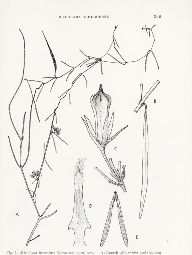
Fig. 1. Microloma hereroense Wanntorp spec. nov. — A: Branch with cymes and ripening
follicle, 0.5 x . — B: Leaf, 5 x. — C: Cyme, only one flower shown, 5 x . — D: Anther, 20 x . — E: Pollinium, 25 x. — H. and H. E. Wanntorp839 a.
340
HANS-ERIK VVANNTORPDr. B. Nordenstam on the Brandberg Mountain, about 80 km to the west of the locality described.
From a phytogeographical point of view these new finds of
Microloma are interesting, as they move the limit of the known dis
tribution of the genus more than 300 km northwards. As both collec tions of this new species were made on rather inaccessible mountain slopes, it is likely that further investigations of the little-known mountain flora of the Damaraland Region will show that Microloma
hereroense has a wider distribution there.
Svensk Botanisk Tidskrift. Bd
63, H. 3. 1969.
PHYSCIA KAIRAMOI IN SCANDINAVIA.
HY
ROLAND MOBERG.
Institute of Systematic Botany, Uppsala.
During my work on some taxonomic problems concerning the genus Physcia in the Scandinavian mountains, I found in the Uppsala herbarium two collections, one from Kongsvoll (Dovre, Norway) and one from Hamrafjället (Härjedalen, Sweden), not referring to any earlier known species from this region. The material was sparse and in summer 1968 1 visited the two places and collected additional material.
By the study of the description of some less known Physcia species, the new material was supposed to be Physcia kairamoi Vain. (Vainio
1921) and this was later verified by the study of Ihe type.
I he thallus is irregular in shape, up to 15 cm across with 1-4 mm broad lobes. The young lobe margins are isidious but the isidia develop later into erect-squarrose lobules (PI. 1:1, 2; compare also
Hale 1967 p. 22), sometimes covering small parts of the thallus. On old herbarium material these isidia-lobules may be broken (PI. 1:3) why the identification of such collections may be difficult.
1 he underside is black, usually with long, black and simple rhizinae which project beyond the lobe margins, but sometimes the rhizinae are short and white-tipped. Even on the isidia-lobules there are rhizinae, very typical for the species. The upper side is epruinose, usually ochraceous brown (Hair Brown in Ridgway 1912) but varies from greenish to brown-black. Upper cortex of the thallus para- plectenchymatous, 20-35 /a, medulla and algal layer 100-200 /u, and lower cortex paraplectenchymatous, 15-25 y.
Apothecia are fairly common, 1—3 mm across and usually with black epruinose discs. A “corona” of rhizinae projects from the underside of the apothecia. Hymenium hyaline, 85-110 y, with the upper part dark brown. Asci cylindrical, containing 8 two-celled
342
ROLAND MOBERGspores which have the wall thickened at the apices and the septum, forming round to angular lumina. Spore size 20-24(-27)x 10-12 (-15) y.
Pycnidia flask shaped, scattered over the thallus and except for the uppermost part totally immersed in thallus. Pycnoconidia ellipsoid, 2-4x1 /.i.
Chemical reactions: K=, C=, KC = , Pd=. Hymenium 1+ blue. No lichen substances found.
Physcia kairamoi seems to be closely related to Ph. sciastra (Acu.) Du Rietz, even if some old material has incorrectly been determined lo Ph. muscigena (Ach.) Nyl. These three species are compared in the following table.
Ph. kairamoi Ph. sciastra Ph. muscigena
brownish, no pruina greyish, no pruina dark brownish, white pruina narrow to broad lobes, irregular and isidious margins
narrow and linear lobes, no isidia on the margins
lobes broad with rounded tips and irregular nonisidious margins
black, simple1 and long rhizinae, projecting beyond the margins black, simple1 rhizinae, seldom projecting beyond the margins black, squarrose1 rhizinae, forming a dense felt isidia to isidia- like lobules,1 marginal isidia laminal or marginal no isidia apothecia with marginal rhizinae apothecia without marginal rhizinae apothecia without marginal rhizinae
Another species probably closely related to Ph. kairamoi but with more southern distribution is Ph. hispidula (Ach.) Frey. The latter has somewhat larger size and laminal soredia instead of marginal isidia.
Some material of Ph. kairamoi was incorrectly identified as Ph.
norrlinii Vain. (Vainio 1921). The type of this species seems to be
Ph. ciliata (Hoffm.) Du Rietz in very bad condition.
Physcia kairamoi was earlier known only from the type locality in
Orlow on the Kola peninsula (Kihlman 1891). The big distance to i
i Cf. Hale p. 22.
PHYSCIA KAIRAMOI IN SCANDINAVIA
343
the new localities, Oppland, Sör-Tröndelag and Härjedalen, may indi cate that the species has been overlooked.
In Härjedalen and Sör-Tröndelag (Dovre) the species seems to prefer calcareous schistose bedrock in the subalpine birch forest region. Either it grows directly on the rocks or on mosses on the rocks.
The locality in Härjedalen is a limited area at the base of the very steep and dry southwestern slope of Hamrafjället (PI. 11:4 and 5). Associated lichens: Buellia alboatra, Caloplaca obliterans, C. stil-
licidiorum, C. tiroliensis (=C. suboliuacea), Cladonia macrophyllodes, Collema tunaeforme, Lecanora albescens, Lobaria scrobiculata, Pannaria pityrea, Parmelia subargentifera, P. sulcata, Physcia ciipolia, Ph. caesia, Ph. constipata, Ph. dubia, Ph. endococcina, Ph. farrea, Ramalina pollinaria, Xanthoria elegans.
In Kongsvoll the species grows on steep W-exposed rocks both in the ravine of Driva and on the lower parts of the western slope of Mt. S. Knutshö. Associated lichens: Caloplaca stillicidiorum, C.
tiroliensis (=C. suboliuacea), C. tegularis, Candelariella vitellina, Collema nigrescens, C. tunaeforme, Lecanora albescens, L. arctica, Lecidea stigmatea, Pannaria pityrea, Parmelia glabratula v. fuliginosa, P. infumata, P. subargentifera, P. sulcata, Peltigera spuria, Physcia ascendens, Ph. caesia, Ph. constipata, Ph. dubia, Ph. endococcina, Ph. enteroxantha, Ph. farrea, Ph. wainioi, Rhizocarpon disporum, Xanthoria elegans, X. sorediata.
On the numerous Norwegian localities, visited by Dr. Sten Ahlner,
Ph. kairamoi was found on boulders in rather open Alnus incana, Betula or Pinus forests.
A name to be referred to Ph. kairamoi as a synonym is Ph. ulothrix ssp. pityrophylla Nyl. (in Norrlin, in Medd. Soc. F. Fl. Fenn., 1 :20. — Ph. obscura var. orbicularis f. pityrophylla Blomberg et Forss. in Enumer. Plant. Scandin. 1880 p. 66. — Ph. constipata ssp. pityro
phylla Räsänen in Ann. Bot. Soc. Zool.-Bot. Fenn. Vanamo, Tom 12, N:o 1 p. 98). The holotype (H) has been examined and found to be Ph. kairamoi. The collection is sparse and not in good condition.
Specimens examined.
USSR
Lapponia ponojensis. Orlow, 1889 Kihlman(H, holotype).
Karelia onegensis. Walkiamäki, 1870 Norrlin (H), “Ph. pityrophylla”. SWEDEN
Härjedalen. Hamrafjället, SW-slope, alt. 850-900 m, 1958 Santesson
344
ROLAND MOBERGn. 12.531 b (UPS), “Physcia sp.”; 1968 Moberg n. 986, 993, 996, 999, 1007 a,
1010, and 1012 (all in UPS). NORWAY
Sör-Tröndelag. Dovre, Kongsvoll, 1870 Zetterstedt (UPS) “Ph. pulve-
rulenla var. muscigena”; 1927 Nilsson (Degelius) (UPS) “Physcia sp.”; 1946 Frisendahl (UPS) “Ph. muscigena det. G. Degelius”. Dovre, Kongs voll, alt. 850-1100 m, 1964 Tibell n. 2209 j, 2213 b (UPS) “Ph. ciliata”;
1968 Moberg n. 1032, 1042, 1045 b, 1059 a, 1063, 1070-74, 1077, 1088 (all
in UPS).
Oppland. Nord-Fron, Kvarn, alt. c. 280 m, 1949 Ahlner(S) “Ph. norrlinii”.
Nord-Fron, Sjoa, alt. c. 280 m, 1958 Ahlner (S) “Physcia sp.”. Vågå, Tolstadskriu, alt. 340-370 m, 1937 and 1958 Ahlner (S) “Physcia sp. and
Ph. norrlinii”. Lom, Böverdalen, Hågå, alt. c. 440 m, 1948 Ahlner(S) “Ph.
norrlinii”; alt. c. 460 m, 1948 Ahlner(S) “Ph. norrlinii”. Lom, Böverdalen,
N. Bövertjern, alt. c. 950 m, 1948 Ahlner(S) “Ph. norrlinii”.
I wish to express my gratitude to Docent R. Santesson for valuable
help and stimulating discussions. For the loan of herbarium material from H, S, and UPS I want to thank the curators of the herbaria, especially Docent S. Ahlner. I also thank Mrs. G. Fällman, Husum, for correcting the English text.
REFERENCES.
Hale, M. E., Jr., 1967: The Biology of Lichens. — London.
Kihlman, A. Osw., 1891: Neue Beiträge zur Flechten-Flora der Halb-Insel Kola. — Medd. Soc. F. Fl. Fenn. 18: 41-59. Helsingfors.
Ridgway, R., 1912: Color standards and color nomenclature. — Washington DC.
Vainio, E. A., 1921: Physcia norrlini, Physcia kairamoi. Medd. Soc. F. FI. Fenn. 46:3. Helsingfors.
Explanation of the Plates. Plate ].
1. Physcia kairamoi. Part of the thallus with apothecia. Note the rhizinae of the apo-thecia and the marginal isidia. Moberg1012. 2 x .
2. Ph. kairamoi. Irregular lobes with rhizinae projecting beyond the margins. Moberg
999. 6 x .
3. Ph. kairamoi. Part of the holotype. Somewhat different appearance to this old specimen is due to the broken marginal isidia and rhizinae. 1889 Kihlman. 2 x.
Plate 2.
4. Mt. Hamrafjället from West. Physcia kairamoi was found at the base of the steep rock-wall.
5. Detail of the rock-wall seen from a distance of c. 2 m. The Physcia kairamoi com munity indicated by the arrow.
R. MOBERG! PHYSCIA KAIRAMOI IN SCANDINAVIA
Plate I
'■*1. jfP* ■££ råifc tiKa?s«*v
:»
zJ&K
a Sv. Bot. Tidskr., 63 (1969): 3Plate II
R. MOBERG: PHYSCIA KAI RAM Ol IN SCANDINAVIAsia®
Hi
L£;-f$VW:3^
'■Svensk Botanisk Tidskrift. Bd 63, H. 3. 1969.
XYLOGONE SPHAEROSPORA, A NEW
ASCOMYCETE FROM STORED PULPWOOD CHIPS.
HY
J. A. VON ARX and T. NILSSON.
Centraalbureau voor Schimmelcultures, Baarn, and Skogshögskolan, Stockholm 50.
A large number of fungal cultures isolated in Sweden from stored pulpwood chips by the second author have been sent to the Cen traalbureau voor Schimmelcultures for identification. Some of the strains representing a member of the Eurotiales (Ascomycetes) could not be identified with any of the genera or species known to the authors. Therefore the name Xylogone sphaerospora gen. & spec, nov. is proposed for this organism:
Xylogone gen. nov.
Ascomata globosa, obscura, parva, clausa, pariete e hyphis texto; asci in glomerulo centrali nati, obovati vet subglobosi, octoxpori, membrana evanescenti; ascosporae globosae, hyalinae, leves. Conidia (arthrosporae) hyalina, cylindracea, pariete inspissato, septata, ex erectis simplicibus conidiophoris oriunda.
Species typica: Xylogone sphaerospora v. Arx& Nilsson.
Ascomata spherical, dark, small, astomous, with a wall composed of hyphal cells; asci borne in a central cluster, obovate or nearly spherical, 8-spored, with a thin, evanescent membrane; ascospores spherical, hyaline, smooth. Conidia (arthrospores) hyaline, cylin drical, thick-walled, septate, arising by septation and breaking up of simple, erect conidiophores.
Type species: Xylogone sphaerospora v. Arx & Nilsson.
Xylogone sphaerospora spec. nov.
Coloniae celeriter crescentes, appressae, primum hyalinae, deinde obscurae ascomatibus maturescentibus. Hyphae radiales regulariter septatae et rami- ficatae, tenuitunicatae, hyalinae, 4-7 y latae; hyphae aeriae lateraliter natae,
346
.1. A. VON ARX AND T. NILSSONFig. 1. Electronmicrograph of a radial longitudinal wood section showing lumenal erosion in a birch fibre-tracheid attacked by Xylogone spliaerospora. Shadowed with Au.
(Cambridge Stereoscan Electron Microscope.)
wgggam 1If.
Jx* *
-' -, t å \ ■tjS&Lhyalinae vel brunneolae, 2-4 latae, hyphae submersae plerumque tenues et hyalinae.
Conidiophora lateraliter e hyphis radiantibus vel e liyphis aereis cras- sioribus orta, plerumque simplicia, erecta, deorsum 2-2.5 /i lata, sursum expansa, primum oblonge clavata, deinde cylindracea, apice rotundato, usque ad 150 n longa; mox crassitunicata numerosis septis divisa in ar- throsporas secedunt, quae sunt cylindraceae, septatae, usque ad 4-cellulares, 8-18 /x longae, 3-4 fi latae (cellulae quaeque 3-6 /i longae), pariete crasso duplici circumdatae.
Ascomata e primordiis spiralibus lateralibus prodeunt; maturitate glo- bosa vel subglobosa, superficialia, brunnescentia, clausa, usque ad 50-90 in diametro; paries 4-7 n crassus e hyphis 1.5-2.5 /x latis, brunneo-tunicatis dense textus. Asci in glomerulo centrali irregulari oriuntur e hyphis ascogenis ramificatis; obpyriformes vel subglobosi, pariete mox evanescenti, octospori, 8-12 /x in diametro; ascosporae globosae, hyalinae, leves, crassitunicatae, refringentes, 3-4.5 n in diametro.
Holotypus: Suecia, Ångermanland, Örnsköldsvik, isolatus e ligno pineo dirupto. 1966 T. Nilsson(herb. S).
Cult. CBS 186.69 =E 7-2 (Baarn).
Colonies fast growing (on corn meal agar 9-11 mm in 24 h. at 24°C), appressed to the substrate, at first colourless, becoming dark with age due to the ascomata which ripen within 2 weeks. Main
XYLOGONE SPHAEROSPORA
347
hyphae radially spreading, regularly septate and branched, thin- walled, hyaline, 4-7 p broad; aerial hyphae borne as side branches, hyaline or brownish, 2-4 p broad, intramatrical hyphae mostly thin and hyaline.
Conidiophores borne as side branches of the main hyphae or of thicker aerial hyphae, mostly simple, erect, at the base about 2-2.5 p broad, becoming broader above, when young elongated clavate, later cylindrical, rounded at tip, up to 150 [x long. They soon become thick-walled, abundantly septate and break up into arthrospores, which are cylindrical, 2-4 celled, 8-18 fx long and 3-4 [x broad (single cells 3-6 p long) and have a thick, double wall.
The ascomata develop from side branches of hyphae which be come coiled. The filaments of the coils are 1.5-2.5 p broad, but become broader in the central part of the developing ascoma. Mature ascomata are spherical or nearly so, superficial, at first bright, becoming brown or black when ripe, astomous, reaching a diameter of 50-90 ix. The wall of the ascomata is 4-7 p thick and composed of 1.5—2.5 [x broad, brown-walled, densely woven hyphal cells. rl he asci are borne from the central part in an irregular cluster on a different level lo the branched ascogenous hyphae. They are obpyri- forrn or nearly spherical, thin-walled, evanescent when mature, 8-spored and 8-12 fx in diameter. The ascospores are spherical, hya line, glabrous, thick-walled, refractive, 3-4.5 ix in size.
Holotype: Sweden, Ångermanland, Örnsköldsvik, isolated from pine chips. 1966 T. Nilsson (herb. S).
Cult, in Baarn as CBS 186.69 = E 7-2.
Other strains studied were CBS 187.69 = D 25—17, isolated from spruce chips from Böksholm (Sweden) and CBS 188.69= K 103—3, isolated from birch chips from Jössefors (Sweden).
A number of other strains have been isolated by the second author from stored pulpwood chips of different trees.
Monosporic cultures from ascospores of Xylogone sphaerospora again produced ascomata with ripe ascospores. Therefore the fungus can be considered as being homothallic. Suitable media for the production of ascomata are malt-, cornmeal- or oatmeal-agars. Conidiophors and conidia are observed especially on media devoid of sugars, especially on potato-carrot-agar.
This fast growing fungus is typically cellulolytic; cellulose agar is cleared. The fungus is able to attack different kinds of hardwoods (e.g. aspen, ash, birch, and beech). The attack starts as a lumenal
348
J. A. VON ARX AND T. NILSSONerosion which ultimately leads to a general dissolution of the cell walls. In a decay experiment the fungus was cultured on birch wood blocks that had been impregnated with a solution of ammonium nitrate. After 3 months’ incubation at 35°C the loss of wood substance was 11%.
The taxonomical position of the genus Xylogone within the Euro- tiales is uncertain. Conidia borne as arthrospores in this order are known only in the Gymnoascaceae. Barron and Booth (1966)
described a new species with an Oidiodendron conidial state as
Arachniotns striatisporus. Oidiodendron-conidia are further known
for Myxotrichum cancellatum Phillips and Myxotrichum ochraceum
Berk. & Br. (Apinis, 1964). The conidial state of Xylogone sphaero-
spora however is not related to Oidiodendron, to Sporendonema or to
another genus of the imperfect fungi with arthrospores.
1 he ascigerous state has to be compared with genera such as
Pseudeurotium van Beyma, Emericellopsis van Beyma, Anixiopsis
Hansen, Aphanoascus Zukal or Allescheria Sacc. In Emericellopsis the ascospores are provided with winged appendages and conidia are borne on phialides when present. In Anixiopsis llie ascomata are bright brownish and the ascospores are reticulate (de Vries, 1969). In Pseudeurotium and Allescheria the ascospores are smooth. In the former genus the wall of the ascomata is composed of flattened cells, in the latter of hyphal elements. In both genera the ascospores are not strictly hyaline and conidia are aleuriospores (chlamydospores) or blastospores (radulaspores) when present.
REFERENCES.
Apinis, A. E., 1964: Revision of British Gymnoascaceae. — Commonwealth Mycol. Inst. Kew, Mycol. papers no. 96.
Barron, G. L. and C. Booth, 1966: A new species of Arachniotus with an Oidiodendron conidial state. — Can. J. Bot. 44: 1057-1061.
de Vries, G. A., 1969: Das Problem: Aphanoascus Zukal oder Anixiopsis Hansen. — Mykosen 12: 111-122.
Explanation of the Plate.
Xylogone sphaerospora.
1. Conidial state. 2-5. Ascogonial coils.
6. Cross-section of a ripe ascoma, x 1400. Sv. Bot. Tidskr., 63 (1969) : 3
J. A. VON ARX AND T. NILSSON: XYLOGONE SPHAEROSPORA
Plate I
Su. Bot. Tidskr., 63 (1969): 3
#7# •
■* é *
7s//
k
Svensk Botanisk Tidskrift. Bd 63, H. 3. 1969.
ÉTUDES URÉDINOLOGIQUES.
PAR
LENNART HOLM.
9. Sur 1’urédo des Gymnosporangium.
Un trait caractéristique du genre Gymnosporangium est 1’absence presque totale d’un stade urédien. D’apres Kern (1964) 55 espéces en seraient connues au stade télien, dont trois seulement pourvues d’un stade urédo, å savoir Gymnosporangium Gaeumannii, G. noot-
katense et G. paraphysatum. Quant å la premiere espéce nous en
avons fait une investigation, publiée dans l’étude précédente de cette serie (Holm 1968). Le prétendu urédo s’en est révélé si extraordi naire, que nous avons étendu nos recherches å toutes les espéces de
Gymnosporangium auxquelles on avait attribué un stade urédien. Les
résultats en sont présentés ci-aprés. Nous croyons qu’ils sont aussi de conséquence plus générale comme référant au probléme principal si le manque d’urédo chez le genre est en somme un phénoméne de reduction ou non.
Cette question fut aussi traitée par Kern, dans sa grande mono graphic (1911) et ii y répondit par 1’affirmative. A ce temps-lå le stade urédo était connu chez un seul Gymnosporangium, å savoir
G. nootkatense. Puisque cette espéce a des écidies cupulées — condi
tion primitive selon Kern — le maitre américain supposa selon ses premisses, que 1’urédo représente un trait primitif de Gymnosporang
ium et que l’absence de ce stade est un phénoméne de reduction.
Cette doctrine n’a guére été contestée mais eile mérite certainement d’etre discutée. Avant d’aborder ce probléme nous présenterons les cas prétendus des Gymnosporangium urédiniféres.
Gymnosporangium nootkatense Arthur.
/
Cette espéce semblait longtemps le seul Gymnosporangium urédo- sporé. Il doit étre observe que des téliosores n’y sont pas connus, mais que les téliospores prennent naissance dans les urédosores, ce qui
350
LENNART HOLMest évidemment un trait avancé. Cependant, il n’y a pas de télio- spores dans le seul matériel que nous ayons examiné (USA, Oregon, Mt Jefferson, sur Chamaecyparis nootkatensis, 15.VIII.1914, hérb. Jackson no. 1394, S). Les urédospores — qu’il s’agisse de telles semble incontestable — possédent une paroi hyaline, épaisse de 2.5 p environ, finement verruculeuse, et percée de nombreux pores ger- minatifs épars (Pl. 1:2). (D’apres Arthur 1934, p. 358, la paroi est
épaisse de 3-4 g et pourvue de deux pores équatoriaux. La derniére indication, du moins, doit étre erronée.) En somme les urédospores chez G. nootkatense présentent une tres grande ressemblance avec les écidiospores (PI. 1:1). Nous reviendrons ci-aprés å cette condition remar quable.
Pendant ces derniéres années, un stade urédo a été rapporté chez encore quatre espéces de Gymnosporangium (sensu lato), ä savoir G. Gaeumannii (Zogg 1948), G. paraphysatum (Viennot-Bourgin
1960), Uredo cupressicola et Uredo apacheca (Peterson 1967 a, b).
Cependant, il faut observer que le terme « urédo » y est employé pour des structures fort differentes, comme nous allons le démontrer. Quant å G. Gaeumannii nous renvoyons ä notre étude récente (Holm
1968) ou est émise la supposition que les urédospores prétendues sont en réalité des téliospores unicellulaires. Méme si elles peuvent fonctionner comme des urédospores, leur parenté morphologique avec les téliospores est manifeste.
Gymnosporangium paraphysatum Viennot-Bourgin.
G. paraphysatum n’est connu que de la récolte originale (Viet-Nam, Dalat, sur les feuilles de Heyderia (Calocedrus) macrolepis, I. 1959, leg. P. Tixier), déposée å 1’Institut National Agronomique de Paris. Gråce ä Pobligeance de M. Viennot-Bourginnous avons pu examiner
ce matériel, ce qui nous permet de confirmer dans 1’essentiel la description détaillée fournie par le savant francais (Viennot- Bourgin 1960).
Aucun stade écidien n’est connu. L’espece a des urédosores et des téliosores séparés. Ceux-lå contiennent et des urédospores et de nombreuses « paraphyses », les unes comme les autres d’un type extraordinaire (PI. II). A ce que nous sachions, les urédospores ne ressemblent pas å d’autres spores, rencontrées parmi les Rouilles, mais il est probablement juste de les qualifier d’urédospores. Elles ont å peu pres la forme de cylindres courts, en moyenne de 30 x 18 g environ, å paroi hyaline, verruculeuse, et pourvue de pores proémi- Sv. Bot. Tidsltr., 63 (1969): 3
ÉTUDES URÉDINOLOGIQUES
351
nents et bien distincts. Il y en a 8, le pore apical excepté, disposés å deux niveaux, c.-å-d. 4 pores sous-apicaux et 4 pores supra-basals.
(Viennot-Bourgin n’indique que 2 pores å chaque étage.) Les
urédospores se détachent tres facilement de leur pédicelle.
Dans les urédosores on trouve en plus d’abondantes structures singuliéres, désignées « paraphrases » par Viennot-Bourgin. Elles sont renflées en forme de massue, å paroi extrémement épaissie, et deviennent de bonne heure fortement pigmentées. La pigmentation n’en est pas toujours uniforme; une partie sous-apicale, intérieure, plus ou moins sémi-globuleuse, est souvent plus noircie (PI. II: 2, 3). Cette partie semble solide mais représente probablement une löge supérieure rabougrie et engorgée. Il parait évident que les paraphyses sont des téliospores métamorphosées.
Les téliosores, rares dans ce matériel, hébergent également des structures différentes (PI. III). La plupart sont des téliospores å paroi mince, du type Gymnosporangium normal, c.-å-d. å deux cellules, dont chacune å un pore. On trouve aussi des téliospores å trois cellules et, plus rarement, celles å quatre cellules. Mais, en plus, il y a des téliospores unicellulaires assez fréquentes, dont la paroi est fort épaissie, jusqu’a 10 [i å l’apex. Celles-ci deviennent fort colorées, de teinte presque brun-noir. Nous ne croyons guére qu’elles fonction- nent comme des téliospores. Certaines d’entre elles ont aussi une loge apicale rabougrie. II semble que ces structures exceptionelles furent septées å un stade tres jeune, apres quoi la cellule supérieure est presque disparue å cause de l’épaissisement de la paroi. Leur ressemblance avec les paraphyses des urédosores est manifeste. Il est remarquable que Viennot-Bourgin ne fasse pas allusion å ces
structures; pourtant elles doivent étre représentées dans la fig. 2 de 1’ouvrage cité.
Uredo cupressicola Peterson.
Cette Bouille est certainement un Gymnosporangium. Elle engendre des galles sur les rameaux de Cupressus pygmaea et jusqu’a ce jour eile n’est connue que de cinq collections de Californie et de Basse Californie. Peterson (1967 a) en a donné une bonne description.
Ces matériaux sont préservés å l’herbier de « Intermountain Forest and Range Experiment Station », Ogden, Utah, USA, etM. Richard
Krebill de cet Institut les a aimablement mis å notre disposition, ce qui nous permet de confirmer la description originale (Peterson
1967 a) dans l’essentiel.
352
LENNART HOLMComme indiqué par Peterson les spores de YUredo cupressicola (PI. IV), le pédicelle å part, sont du type écidiosporé caractéristique de Gymnosporangium. Elles sont d’une couleur brun-roux, å paroi forte, de 2.5 g environ, finement verruculeuse, mais percée en général de 6 pores germinatifs seulement; ceux-ci sont entourés d’un renforcement annulaire ä 1’intérieur.
Les spores sont attachées å un long pédicelle hyalin, atteignant au moins 125 g (selon Petersonjusqu’a 145 g). Un fait non remarqué par Pauteur américain est que le pédicelle est trés souvent septé, parfois méme bisepté (PL IV: 1, 3). Une cloison est généralement formée å une distance de 15-30 g du point d’insertion sporée. Les pédicelles septés sont quelquechose de trés remarquable chez les Roubles — å ce que nous sachions on les connait seulement chez les téliospores de Tranzschelia. Le pédicelle sporé de YUredo cupressicola a ainsi une cellule apicale, longue de 15-30 g, et celle-ci devient le plus souvent nettement renflée. Par exception cette cellule peut se transformer en une spore inférieure ce qui donne deux spores en chaine (PI. IV: 4). Peterson a également remarqué ce phénoméne: « Urediniospores are occasionally borne in tandem on a single pedicel and then appear somewhat like teliospores » (op. cit., p. 50 et fig. 2). Un phénoméne encore plus singulier doit aussi étre observé å litre exceptionnel: deux spores aux extrémités du méme pédicelle (voir PI. IV: 2 et Peterson op. eil. fig. 3). Cela peut å peine s’ex- pliquer autrement que par ce que le pédicelle fut bisepté de bonne heure et que la cellule sous-apicale y fut transformée en une spore secondaire.
De quelle sorte de spores s’agit-il ici? Peterson les nomine, sans hésiter, semble-t-il, des urédospores. Des données cytologiques, en état d’élucider ce probléme, nous font défaut, malheureusement. Cependant, Peterson a observé des spores germantes et il en repro- duit une microphotographie, ou l’on voit un tube de germination, se ramifiant au sommet. En effet, la figure n’est pas trop dissemblable de deux épibasides malformées. Mais 1’auteur cité n’a vu aucune formation de sporidies et e’est probablement pourquoi il qualifie les spores comme des urédospores. En tout cas, Uredo cupressicola est une Rouble remarquable, dont les ressemblances de Gymnosporan
gium Gaeumannii sont évidentes. Ainsi, nous tenons å indiquer la
possibilité que ces spores sont, en réalité, des téliospores. Quoi qu’il en soit, leur homologie avec les téliospores est incontestable.
L. iiolm: études urédinologiques
Planche I
f " \ /
) -
j
H
f 1^1 * f W[ - - .» Xi —") /^*\
. i! •■' I v '
Planche 11
L. holm: etudes urédinologiquesL. holm: ETUDES urédinologiques
Planche III
C Cb
Planche IV
L. holm: études urédinologiquesÉTUDES URÉDINOLOGIQUES
353
Uredo apacheca Peterson.
Uredo apacheca, enfin, provoque également la formation de galles,
sur les rameaus de Juniperus deppeana, étant connu jusqu’ä ce jour seulement de trois récoltes, du Nouveau-Mexique. Nous avons eu l’occasion d’étudier aussi ces matériaux, grace a l’obligeance de
M. Krebill, de Ogden, et nous pouvons attester l’essentiel de la description originale; on peut ajouter, pourtant, que les spores possédent 10 pores germinatifs épars, environ.
Comme il ressort de la description, et comine Peterson (1967 b)
l’indique lui-meme, ce champignon est de point de vue morpholo- gique un Caeoma. Tant que les téliospores sont inconnues l’espece doit indubitablement étre classée dans le genre d’attente Caeoma (si l’on ne veut la placer directement dans les Gymnosporangium, ou eile appartient sans aucun doute). Peterson l’a qualifiée de
Uredo, vu que les sores évidemment prennent naissance d’un mycé-
lium dicaryotique — 1’auteur américan en a fait une investigation cytologique qu’il référe. Cette condition singuliére est d’ailleurs indiquée déjå par 1’absence de pycnides, et, conformément å la terminologie d’Arthur, Peterson regarde cette Rouille comme un
stade Uredo. Certes, nous avons ici affaire ä des urédies au sens d’Arthur, mais il n’est pas pour cela justifié de placer 1’espéce dans le genre imparfait Uredo, qui est établi sur des critériums non cyto- logiques mais strictement morphologiques.
Une espéce parallele en est, en quelque mesure, le singulier Gymno
sporangium bermudianum, espéce autoique, également dépourvue
de pycnides, formant seulement les stades I et III, qui évidemment sont engendrés du méme mycélium. Ainsi il y a toutes les raisons de croire que les écidies y prennent naissance d’un mycélium dica ryotique et, en conséquence, qu’elles représentent un stade urédo, au sens d’ARTHUR. De telles conditions mettent en évidence les conséquences singuliéres d’une adhérence stricte å la terminologie arthurienne (voir aussi la critique de Savile, 1968).
Comme résumé de 1’exposé précédent il convient de souligner:
Gymnosporangium nootkatense a un stade urédo dont les spores sont
fort ressemblantes aux écidiospores de 1’espéce — qui sont assez differentes du type écidiosporé normal du genre. G. paraphysatum est également pourvu d’urédo dont les spores sont d’un type unique; en plus 1’espéce est le seul Gymnosporangium aux paraphyses. Quant å G. Gaeumannii les urédospores prétendues sont å notre avis plutötdes
354
LENNART HOLMtéliospores unicellulaires. En tout cas leur homologie avec les télio- spores est manifeste. Uredo cupressicola en est assez proche. Uredo
apacheca, enfin, est un Caeoma.
Discussion.
Apres cet apercu succinct des cas reels ou prétendus d’uredo chez
Gymnosporangium nous sommes å meine de prendre position å
1’opinion soutenue par Kern qui dit que la présence d’uredo est un
Irait primitif chez ce genre et que 1’absence générale en est un phénoméne de réduction.
La suppression du stade uredo est en soi un phénoméne assez commun parmi les Urédinales. 11 est ä remarquer, cependant, que cela se rapporte en premier lieu aux Micro-formes, dont le manque d’uredo est la conséquence du raccourcissement du cycle évolutif, réduit å une seide génération. Pour élucider le probléme qui nous concerne, il faut examiner les Opsi-Urédinales. On y observe que presque toutes ces Rouilles, les Gymnosporangium å part, sont des formes autoiques. En eilet, les seules exceptions sont Puccinia inter-
veniens et Pucciniastrum goeppertianum. Il est interessant que la
premiere espéce est evidemment apparentée aux Gymnosporangium. Quant a la seconde, il n’est guére douteux que la suppression d’uredo en soit un trait avance comme chez les Opsi-formes autoiques.
(Puccinia e/J'usa, P. Podophylli etc.). L’hétéroicité est considérée
comme une condition fort ancienne chez les Urédinales, et chez le genre Puccinia eile est certainement originaire. Par consequent il n est pas surprenant que la suppression d’uredo ait eu lieu chez les
Puccinia autoiques seulement, abstraction faite de P. interveniens
ou cependant 1'absence d’urédo peut étre primitif. Sur ce fond il semble en soi fort singulier qu’une telle réduction ait frappé si complétement le genre voisin Gymnosporangium, également hété- roique.
L’explication la plus simple de cette condition est naturcllemcnt que Gymnosporangium primitivement a manqué d’urédo. Mais, si c’est le cas, on ne peut plus regarder la présence d’urédo chez G.
nootkatense et d’autres comme un trait d’atavisme; au contraire
1’urédo y représente une néoformation. Il est manifeste que, en ce cas, cet urédo doit étre d’un type tres primitif, tandisqu’un urédo en train de disparaitre devrait presenter certains traits avancés. Comment en est-il dans le cas actuel?
Pour répondre å cette question il faut sürement avoir certains Sv. Bot. Tidskr., 63 (1969): 3
ÉTUDES URÉDINOLOGIQUES
355
critériums sur les urédospores primitives et avancées respectivement. Ce probléme fut cliscuté å fond par Cummins (1936). Sur la base
d’une statistique comparée, celui-ci conclut qu’une paroi sporale hyaline, pourvue de nombreux pores épars, y est å compter comme primitive. 11 ne discute pas 1’épaisseur de la paroi urédosporée, mais regarde les paraphyses comme des structures primitives. Si l’on part des critériums de Cumminson est sans aucun doute obligé
de qualifier l’urédo de G. nootkatense comme primitif, et peut-étre aussi l’urédo de G. paraphysatum. Les soi-disant urédospores de
G. Gaeumannii et de Uredo cupressicola sont pourtant d’un tout autre
type: un trait primitif est 1’existence de pores nombreux mais la forte pigmentation de la paroi serait, d’apres Cummins, un trait avancé chez les urédospores.
On peut discuter le probléme aussi en partant des considerations plus générales. Il n’est pas douteux que le stade urédo est relative- ment secondaire. Selon une opinion unanime le stade téleuto est le plus ancien — encore plus vieux que les Urédinales peut-étre — tandisque les stades 0, I et II se sont dévéloppés pendant Involution des Rouilles. Il y a des raisons de croire que l’urédo en. est le plus jeune, puisqu’il n’est pas un membre nécessaire du cycle évolutif. Les urédospores sont de leur nature des diaspores végétatives et de telles structures ne représentent pas, en général, un trait archalque.
C’est un fait intéressant qu’il existe une ressemblance remarquable entre les écidiospores et les urédospores de plusieurs Rouilles.
Arthur (1929, p. 143) l’a fait remarquer. On peut encore rappeier
les genres Chrysomyxa et Coleosporium, dont les urédospores mémes sont formées en chaines avec des cellules intercalaires. Ces ressem- blances ne peuvent guére étre accidentelles et, en effet, ne sont pas du tout surprenantes; ce sont plutöt les dissemblances entre des spores de la méme fonction qui sont de nature å étonner. La proche parenté entre 1’écidium et Furédo est illustrée aussi par la condition bien connue que l’un peut remplacer l’autre dans le cycle évolutif (1’urédo primaire et les écidies secondaires, respectivement). En réalité le stade urédo est en principe une répétition du stade écidien. La supposition nous semble done plausible que 1’urédo des Rouilles avait å Forigine de grandes ressemblances du stade écidien, et en outre que les écidiospores étaient le « modele » å Forigine des urédo spores. Il convient de remarquer cette ressemblance chez G. nootka-
tensel
Pourtant, il ne parait pas impossible que 1’urédo pourrait étre
356
LENNART HOLMdéveloppé aussi d’une autre maniére, å savoir du stade télien. Des téliospores germant sans aucune fusion nucléaire pourraient fonc- tionner comme des urédospores, semble-t-il. Une forme transitionelle entre les urédo- et les téleutospores est d’ailleurs représentée par les « amphispores », trouvées chez Hyalopsora, entre autres.) Quant å
Uredo cupressicola cette hypothése doit étre envisagée. Si les spores en
fonctionnent réellement comme des urédospores — et non pas comme des téliospores ce que nous sommes enclins ä croire — nous croyons quand méme qu’elles sont dérivées de téliospores.
Dans une étude antérieure (Holm 1968) nous avons déjå développé des raisons de croire que les écidiospores des Rouilles avaient pri- mitivement une grande ressemblance des téliospores unicellulaires. La conséquence de notre argumentation ci-dessus est done que les trois types sporés — télien, urédien et écidien — sont dérivés d’un méme type original.
Pour conclure cet exposé sur 1’urédo de Gymnosporangium, nous n’avons trouvé aucun support réel de la doctrine de Kern, selon laquelle les cas tres peu nombreux d’urédo représenteraient le reste d’une condition antérieurement commune, et que le genre a ainsi donné lieu å une suppression presque totale de 1’urédo. Les cas incontestables d’urédo — Gymnosporangium nootkatense et G.
paraphysatum — ne montrent pas d’indices d’une dégénération de la
phase urédienne — au contraire, celle-ci est rnieux développée que la phase télienne, et G. nootkatense, du moins, nous montre des caractéres urédiniens, manifestement primitifs.
Summary.
The genus Gymnosporangium is generally devoid of uredia, this stage so far being reported in three species only, viz. G. Gaeumantiii,
G. nootkatense, and G. paraphysatum. It is also reported in two
species formally referred to the imperfect genus Uredo, viz. Uredo
cupressicola and U. apacheca.
The present paper gives an account of the alleged uredo of these Rusts. A great diversity is met with, and morphologically quite different structures have been designated as uredospores. According to the author an indubitable uredo stage is found in Gymnosporan
gium nootkatense and G. paraphysatum only. As to G. Gaeumannii he
has elsewhere (Holm 1968) put forth the view that the “uredo
spores” probably are unicellular teleutospores. The supposed uredo- Su. Bot. Tidskr., 63 (1969): 3
ÉTUDES URÉDINOLOGIQUES
357
spores of Uredo cupressicola are rather similar to those of G. Gaea-
mannii, and the author suggests that they may be teliospores, too.
Irrespective of their function, they are, however, morphologically of the type characteristic for the aecidiospores of Gymnosporangium.
Uredo apacheca, finally, is a Caeoma.
G. nootkatense is the only species, of those discussed here, which
is known to possess all spore forms. Its uredospores are remarkably like the aecidiospores, differing mainly by their thinner spore wall with less distinct germ pores. The peculiar G. paraphysatum, is known from the type collection only, which has uredia and sparse telia. In the uredia are found numerous “paraphyses” (Viennot-Bourgin).
The author reports similar structures in the telia and suggests that they all represent metamorphosed teliospores. The uredospores of this species are of a unique type, with prominent, mostly bizonate germ pores.
The existence of at least two truly uredosporiferous members of
Gymnosporangium is of considerable phylogenetic interest. In his
classical monograph Kern (1911) advocated the view that the gene
ral lack of an uredostage in the genus is due to reduction, and that
Gymnosporangium on the whole is an advanced group. Within the
genus, G. nootkatense was regarded as primitive by Kern (at this
time it was the only member of the genus claimed to have an uredo). The author points out that the uredia of G. nootkatense and G.
paraphysatum show no signs of being vestigial structures — on the
contrary they are abundant and much better developed than the te- lial stage, especially in G. nootkatense. If applying Cummins’s crite
ria we must admit that the uredia of these species are of a decidedly primitive type. Per se, it is obviously a plausible assumption that the uredo represents an innovation in Gymnosporangium and that the genus originally was devoid of this stage. The author also empha sizes the remarkable similarity between the uredospores and the aecidiospores of G. nootkatense, and suggests that this reflects an original resemblance between aecidiospores and primitive uredo spores. In a previous paper (Holm 1968) the author derived aecidio
spores and teliospores from a common ancestral “Bauplan” (sit venia
verbo Trolliano). Now this hypothesis is generalized to include the
uredospores, too.
The evolutionary trends within Gymnosporangium will be further discussed in the next paper in this series.
358
LENNART HOLM BIBLIOGRAPHIE.Arthur, J. C., 1929: The Plant Rusts. — Boston.
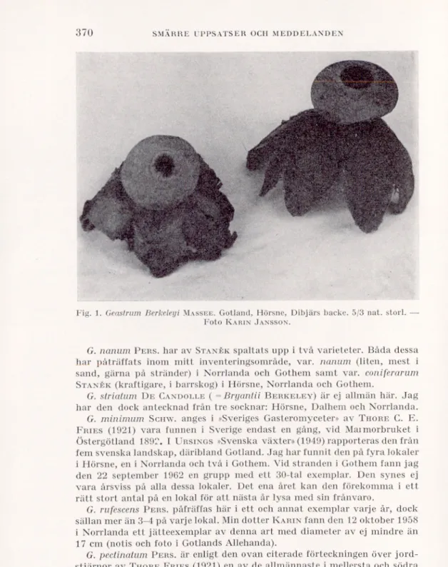
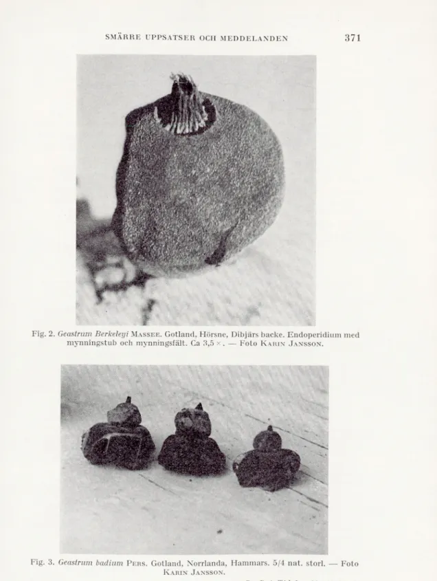
—»—, 1934: Manual of the Rusts in United States and Canada. — Lafayette.
Cummins, G. B., 1936: Phylogenetic significance of the pores in urediospores. — Mycologia, 28.
Holm, L., 1968: Etudes urédinologiques 8. — Sv. Bot. Tidskr., 62.
Kern, F. D., 1911: A Biologic and Taxonomic Study of the Genus Gymno- sporangium. — Bull. New York Bot. Gard., 7.
—»—, 1964: Lists and Keys of the Cedar Rusts of the World. — Memoirs New York Bot. Gard., 10 No. 5.
Peterson, R. S., 1967 a: Cypress Rusts in California and Baja California. — Madrono, 19 No. 2.
—»—, 1967 b: Studies of Juniper Rusts in the West. — Ibid., No. 3.
Savile, D. B. O., 1968: The case against “uredium”. — Mycologia, 60.
Viennot-Bourgin, G., 1960: Gymnosporangium paraphysatum sp. nov. sur le Heyderia macrolepis au Viet-Nam. — Rev. Myc., 25.
Zogg, H., 1949: Über ein neues, Uredo-bildendes Gymnosporangium: Gymnosporangium Gaeumanni n. sp. — Ber. Schweiz. Bot. Ges., 59.
Légende des planches. Planche I. Gymnosporangium nootkatense. 1. Écidiospores. — x 480. 2. Urédospores.—x 480. Planche II. Gymnosporangium paraphysatum. 1. Urédospores et paraphyses. — x 200.
2. Paraphyses å corps sous-apical noirci. — x 480. 3. Idem.—x 200.
Planche III. Gymnosporangium paraphysatum. 1. Téliospores.—x 200.
2. Téliospores de types diflérents; observez la spore unicellulaire å paroi épaissie et, au milieu, deux spores similaires, avec une loge supérieure rabougrie. — x 480.
Planche IV. Uredo cupressicola.
1. Spores å pédicelie septé, dont la cellule apicale est renflée. — x 480. 2. Deux spores aux extrémités du méme pédicelie. — x 480.
3. Spores aux pédicelles ramiliés; septa aux lléches. -— x 480. 4. Deux spores en chaine, portées par le meine pédicelie. -—• x 480.
Svensk Botanisk Tidskrift. Bd 63, H. 3. 1969.
STUDIES TO FIND THE EFFECT OF
TEMPORARY REMOVAL OF BELL JARS FROM
SEED BEDS ON JACOBSEN APPARATUS ON THE
GERMINATION OF PINES SILVESTRIS
AND PICEA ABIES SEED.
S. K. KAMRA.
Department of Forest Genetics, Royal College of Forestry, Stockholm 50, and Institute of Botany, University of Stockholm, Stockholm 50.
Introduction.
On Jacobsen apparatus, the bell jars have to be removed from the seed beds in order to count the number of germinated seeds. The func tion of the bell jars is to maintain a constantly high relative humidity of the air around the seeds. In those experiments of seed testing and research where the rate of germination is studied frequently (e.g. every day or every other day), it is of special interest to know, whether the temporary removal of the bell jars from the seed beds has any effect on the germination of the seed. An investigation was undertaken to study this problem and the results are reported here.
Material.
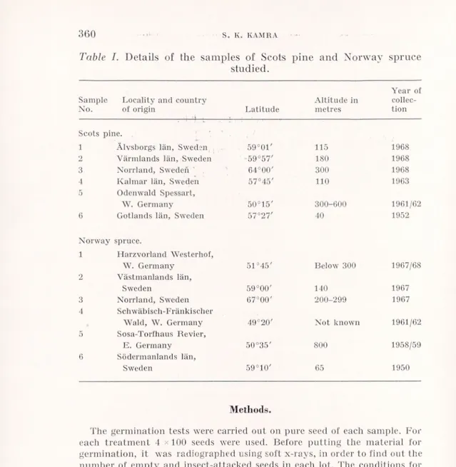
Six samples each of Scots pine (Pinus silvestris L.) and Norway spruce (Picea abies (L.) Karst.) were used for the experiments. They included seed with high viability as well as that with reduced viabi lity due to age or due to incomplete development of the embryo and/ or the endosperm. The details of the locality and the country of origin, latitude, altitude and the year of collection of each sample are given in Table I.
S. K. KAMRA
360
Table I. Details of the sain pies of Scots
studied.
pine and Norway spruce
Year of Sample Locality and country Altitude in collec-No. of origin Latitude metres tion
Scots pine. 1 Älvsborgs län, Sweden 59°0t' 115 1968 2 Värmlands län, Sweden 59°57' 180 1968 3 Norrland, Sweden ' 64°00' 300 1968 1 Kalmar län, Sweden 57°45' 110 1963 5 Odenwald Spessart, W. Germany 50°15' 300-600 1961/62 6 Gotlands län, Sweden 57°27' 40 1952 Norway spruce. 1 Harzvorland Westerhof, W. Germany 51°45' Below 300 1967/68 2 Västmanlands län, Sweden 59°00' 140 1967 3 Norrland, Sweden 67°00' 200-299 1967 4 Schwäbisch-Fränkischer
Wald, W. Germany 49°20' Not known 1961/62 5 Sosa-Torfhaus Revier,
E. Germany 50°35' 800 1958/59 6 Södermanlands län,
Sweden 59°10' 65 1950
Methods.
The germination tests were carried out on pure seed of each sample. For each treatment 4 x 100 seeds were used. Before putting the material for germination, it was radiographed using soft x-rays, in order to find out the number of empty and insect-attacked seeds in each lot. The conditions for radiography were: kV 14, mA =5, focus-film distance 50 cm, time of exposure 3 seconds. The X-Ray Industrial Film Type “L” (“Low speed”) manufactured by CEA Works, Strängnäs, Sweden was used. For further details of the procedure, see Kamra (1967).
Two Jacobsen apparatuses of stainless steel of the same model were used for the experiments. They were placed in a climate chamber, the tempera ture (20°C) and the relative humidity (60%) of which were regulated auto matically by special devices. The maximum deviation of the temperature was 1°C. The light was switched on and off automatically by an electrical watch at the fixed hours simultaneously for both the apparatuses. The following conditions were used for the germination tests:
GERMINATION OF PINUS SILVESTRIS AND PICEA ABIES SEED
361
Temperature =20°C (constant).
Light: About 1000 lux artificial light from day-light tubes for 8 hours daily.
Distance between water level and seed bed on Jacobsen apparatus: 13 cm. Duration of the germination tests: 21 days.
The germinated seeds were counted and removed from the tests every day up to the tenth day and every other day thereafter up to the twenty- first day. A seed was considered as germinated when the length of the root was at least equal to that of the seed itself. This criterion has been found to be reliable in comparative germination studies (cf. Kamra1968, 1969 a and
b). The advantages of this criterion have been discussed by Kamra(1969 c).
The germination percentages of all the samples were calculated uniformly on the basis of the number of filled seeds only in each lot.
Although the usual time for which the bell jars are removed from the seed beds for counting the germinated seeds is very short, in this investigation three series were used from which the bell jars were removed for 5, 30 and 60 minutes respectively at each occasion of counting the germinated seeds. The control series was kept covered continuously, except for counting it on the
7th, the 14th and the 21st day.
Results. (a) Germination percentage.
As may be observed from Figures 1-12, the fresh samples (Nos. 1-3) of both Scots pine and Norway spruce show practically the same germination percentages under the different treatments as in the control series. However, in the case of old samples, there are some variations in the germination percentages of the different treat ments as compared with the controls. But these fluctuations do not show any consistent trend, except in sample 4 of Norway spruce, where the germination percentage increases continuously with increase in the time of removal of the bell jars. Even if the germina tion conditions of the treatments are different, the variations in the germination percentages of the treatments and the controls are not large. In most cases, for instance, they do not exceed the tolerance limits for the germination percentages prescribed in the International Rules for Seed Testing (1966).
(b) Germination rate.
The germination rates of the samples of Scots pine are shown in Figures 1-6 and those of Norway spruce in Figures 7-12. As may be seen, the germination rates of the samples under the various treat ments do not show any appreciable differences.
362
S. K. KAM RA Germinationpercentaqe G. %-a<^e
Pinus sil vest ris
Fic^. 1 Sample Control — 5 min. — 30 min. -• 60 min. • 60-0123456789 10 12 14 16 18 20 21 0123456789 10 12 16 18 20 21
Figures 1-6. Germination of the Scots pine samples studied. (S. = Sample. G. = Germination.)
GERMINATION OF PINUS SILVESTRIS AND PICEA ABIES SEED
363
Germination percentage Sample 1 a Control --- 5 min. ---30 min. --- 60 mi.n. G. %-aqe Fiq. 10 Fiq. 11 S. 5 .0-0 _____ 0-0 18 20 21 12 14 0 1 23456789 10 12 14 16 18 20 21 23456789 10Figures 7-12. Germination of the Norway spruce samples studied. (S. = Sample. G. Germination.)
364
S. K. KAMRADiscussion.
From the results described above, it may be observed that the differences between the germination values of the controls and of the treatments of removing the bell jars from the seed beds for various periods are small and practically of no importance. This may perhaps seem surprizing, because the lifting away of the bell jars tends to reduce the relative humidity of the air on the seed beds. The reason why such a reduction of the relative humidity did not produce any appreciable effect on the germination is the fact, that the present investigation was carried out in a climate chamber where both the temperature and the relative humidity were constant (20°C and 60% respectively). This relative humidity is fairly high, in order to prevent the desiccation of seeds on uncovered seed beds for short periods. Thus the effect of the removal of bell jars was not well-marked in this study. Of course, it is possible that if the experiments are carried out in a room with lower relative humidity, the lifting away of the bell jars might affect the germination results (cf. Simancik et al. 1966, Table 2 and the note below the Table). However, the present inves tigation shows that under the conditions used here, the removal of the bell jars from the seed beds for a short time for counting the germinated seeds does not seem to affect the germination results of a sample.
Summary.
The present investigation was undertaken to find out, if the tem porary removal of the bell jars from the seed beds on Jacobsen appa ratus has any effect on the germination of Scots pine and Norway spruce seed. Six samples each of Pinus silveslris L. and Picea abies (L.) Karst, from areas lying between 50°-64° and 49°-67° Northern latitudes respectively were used for the experiments. The material included seed with high viability as well as that with reduced viabil ity due to age or due to incomplete development of the embryo and/or the endosperm (cf. Table I). The bell jars were removed from the seed beds for 5, 30 and 60 minutes at each occasion of counting the germinated seeds. This counting was done every day during the first ten days and every other day thereafter up to twenty-one days. The controls were kept continuously covered, except for a short time for counting the germinated seeds on the 7th, the 14th and the 21st day. There were practically no differences in the germination results Su. Bot. Tidskr., 63 (1969): 3
GERMINATION OF PINUS SILVESTRIS AND PICEA ABIES SEED
365
of the controls and of the series from which the bell jars were re moved for the various periods. The investigation thus showed that under the conditions used here, the removal of the bell jars from the seed beds for a short time for counting the germinated seeds does not seem to affect the germination results of a sample.
LITERATURE CITED.
I. S. T. A., 1966: International Rules for Seed Testing. — Proc. Intern. Seed Test. Assoc., 31 (1): 1-152.
Kamra, S. K., 1967: Comparative studies on the germination of Scots pine and Norway spruce seed under different temperatures and photo periods. — Studia Forestalia Suecica, No. 51: 1-16.
—»—, 1968: Effect of different distances between water level and seed bed on Jacobsen apparatus on the germination of Pinus silvestris L. seed. — Studia Forestalia Suecica, No. 65: 1-18.
—»—, 1969 a: Studies on the effect of different distances between water level and seed bed on Jacobsen apparatus on the germination of
Picea abies (L.) Karst, seed. — Sv. Bot. Tidskr., 63 (1): 72-80.
—»—, 1969 b: Further studies on the effect of different distances between water level and seed bed on Jacobsen apparatus on the germination of Pinus silvestris and Picea abies seed. — Sv. Bot. Tidskr., 63 (2): 265-274.
—»—, 1969 c: Investigations on the suitable germination duration for Pinus
silvestris and Picea abies seed. — Studia Forestalia Suecica, No. 73:
1-16.
Simanöik, F., Bevilacqua, B., und Simak, M. 1966: Bekämpfung von Schimmelpilzen im Jacobsenapparat durch ultraviolettes Licht. — Proc. Intern. Seed Test. Assoc., 31 (5): 731-739.
Svensk Botanisk Tidskrift. Bd 63, H. 3. 1969.
SMÄRRE UPPSATSER OCH MEDDELANDEN.
Föreningens medlemmar uppmanas att till denna avdelning insända meddelanden om märkliga växtfynd o. d.
Some new localities of rare plants in Lule Lappmark in northern Sweden.
In the summer of 1968 I was making a field study of the flora on ledges and cracks of the cliffs of mount Skerfe and mount Nammatj in the Sarek National Park, and on the uppermost parts of the talus slopes of the same mountains. The cliff of mount Skerfe (1206 m) and adjacent lower mountains faces south, towards the Rapa river and the delta landscape which has been built up by this river. The delta is 494.3 m above sea level. Mount Nammatj (826 m) stands alone, west of the delta, and has cliffs facing north, east and south.
On a small ledge only two meters from the base of the south-facing cliff on mount Nammatj, I found six small specimens of Poientilla multifida. The ledge appeared to be of sandstone, which was probably deposited directly upon the archaic rocks which are reported to be found at the bottom of the valley, only about 75 m vertically below the cliff. This ledge was very dry and there was almost no soil at all in which the plants could grow. The six specimens I found were small; the biggest collected had only two stems with a total of seven flowers, and there was only one more with buds or flowers. The other four had only a few small leaves.
Potentilla mullifida has been found on this mountain before, but the previously found locality (not published) is on a ledge which is very difficult to reach, high up on the eastern cliff (only one small specimen found by fil. stud. Mats Nettelblad in 1964; personal communication).
The next nearest locality is on mount Kebnats on the northern shore of lake Langas, just opposite Saltoluokta Tourist Station and about 33 km north of mount Nammatj.
As this new locality is so small it could easily be distroyed by even a minor rockfall from the cliff, and such rockfalls do occur now and then.
On the same mountain, but on the east-facing cliff and somewhat higher up, about 100 m vertically above the valley floor, I found some small specimens of Potentilla norvegica, only about 7 cm high. They were growing on a ledge near the base of the cliff and near the south-east corner of the mountain. This ledge was not quite so dry as the one where I found P.
multifida. The rock was some sort of shale.
The collected specimens have been studied by Professor N. Hylander,
Inst, of Syst. Botany, Uppsala. As far as Professor Hylander and I can
find out, the nearest localities are one near lakes Saggat and Skalka to the
SMÄRRE UPPSATSER OCH MEDDELANDEN
367
south, and around the Suorva reservoir in the north (G. Björkman: Supple ment to the flora of Stora Sjöfallets Nat. Park. Found at Suorva 1942-43, 1961 and at St. Sjöfallet 1943 and 1961) both about 35 km from the east facing cliff of mount Nammatj.
Normally P. norvegica is found on ruderate places, near roads and places where construction works are in progress. Here it is found on a steep cliff 7 km from the nearest human habitation, at Aktse, which is a very isolated place that can be reached only on foot or by air, as the nearest road, finished in 1968, stops about 15 km from Aktse.
As the locality is about 100 m above the track that goes into the Sarek National Park, and can be reached only by climbing the slope of boulders below the cliff, it is a place likely to be visited only by botanists and possibly some ornithologists and geologists. Therefore I can be fairly sure that it is a spontaneus locality and one not brought about by human beings.
Lektor Gunnar Wistrand has advanced a theory that P. norvegica is
colonizing the mountains from such bases as work camps, perhaps being brought to the new localities by birds (Wistrand1962 p. 116).
In the central part of the middle cliff of the low mountain immediately east of mount Skerfe, I found some flowering specimens and about 100 leafy specimens of Tussilago farfara, growing on a slope with seepage water, together with a lot of other moisture-demanding plants, e.g., Carex atrata,
Dadylorhiza maculata, Gymnadenia conopsea, Rumex acetosa and others.
One more locality for Tussilago farfara was found below the lower cliff of mount Skerfe in a place where there was some water from small springs in the cliff, which here consisted of shale with a rather high content of clay and graphite. This second locality is much more open and almost devoid of any vegetation, but in this one place I could count more than a hundred specimens of T. farfara and also specimens of other plants such as Tricho-
phorum caespitosum, Saxifraga oppositifolia and S. aizoides, Saussurea alpina
and Taraxacum croceum.
From July 17th to August 10th, 1968, I was a member of the botanical expedition to the Kaitum lake system. There is a plan to dam the Kaitum lakes and divert the water through a tunnel from the western part of the lower lake, Vuolep Kaitumjaure (575.8 m), to the water reservoir of Vietas power station in the Stora Lule älv river system. The water level of the lower lake will be raised about 15 m, and that of the middle lake, Kaska- Kaitumjaure (581.5 m), about 8 m. The aim of the expedition was to make an inventory of the flora below the future water level of the reservoir, at 590 m, and particularly to study the area around the site of the dam and places where mountain streams enter the lakes and also some of the shores. We also aimed to make comparable studies of some places above the 590 m level, to show any differences in flora and vegetation. (More of this in a special report to The National Nature Conservancy Office; summarized extracts will soon be published by the Bureau, editor Jim Lundqvist.)
One day I climbed the south-east-facing talus slope of mount Råvve (960 m) in order to get a better view of the area around Tjuonajokk and the planned dam site. It turned out to be a perfect habitat for Potentilla multi-
fida. When I reached the cliff and started climbing up to some wide